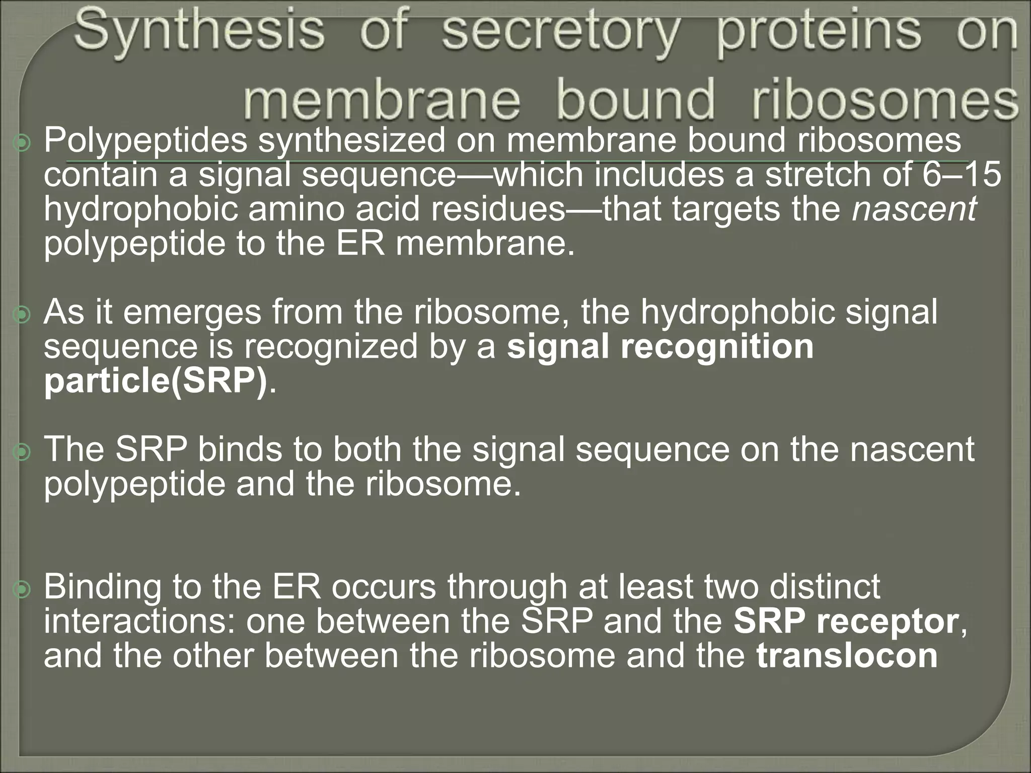
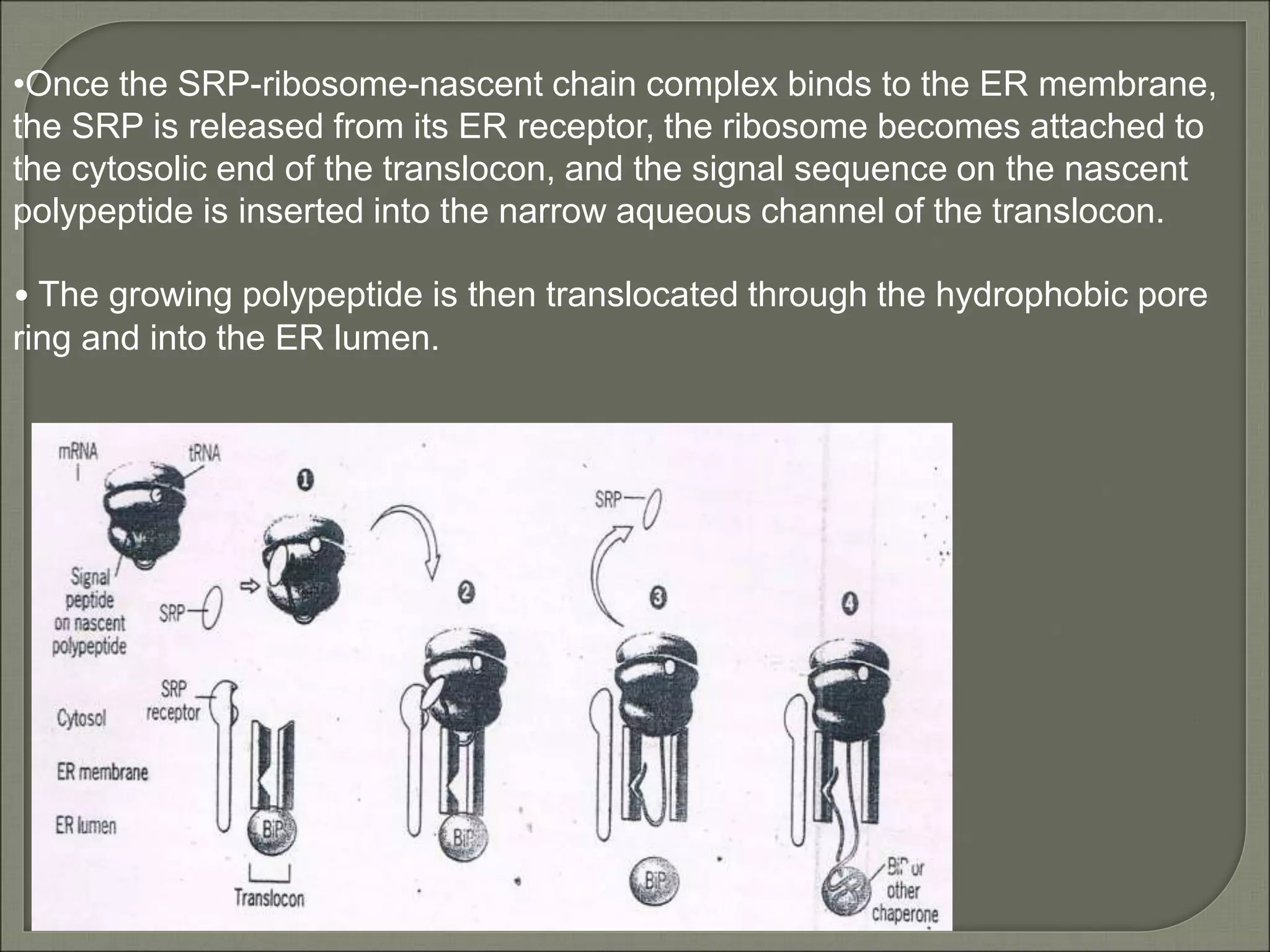
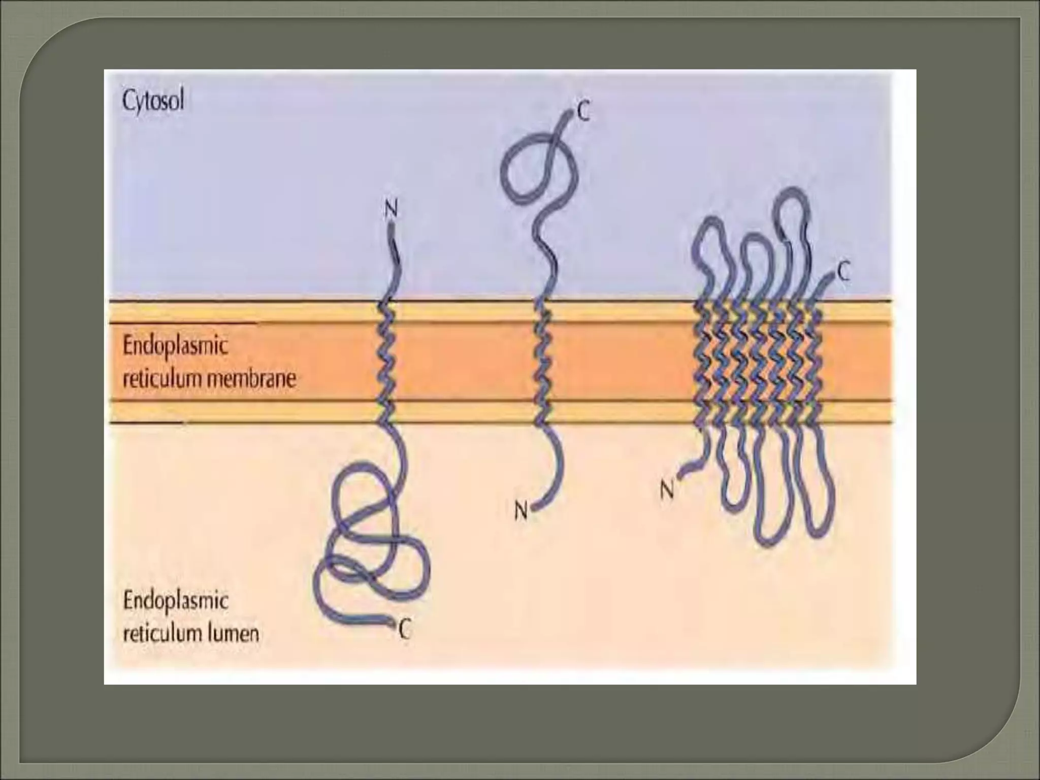
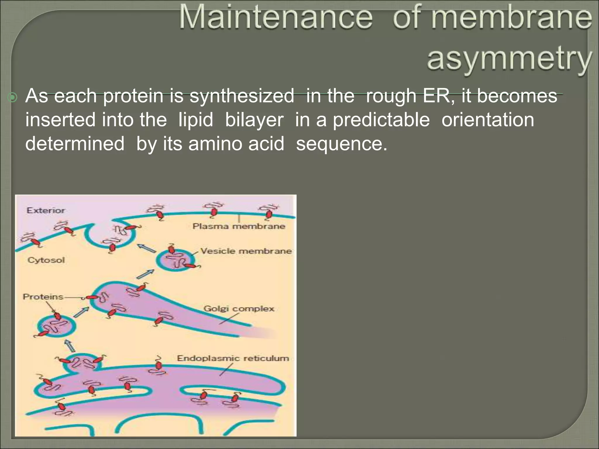
By
KAUSHAL KUMAR SAHU
Assistant Professor (Ad Hoc)
Department of Biotechnology
Govt. Digvijay Autonomous P. G. College
Raj-Nandgaon ( C. G. )
 Introduction
 Protein synthesis
 Synthesis of secretory proteins on membrane-bound ribosomes
 Processing of newly synthesized proteins in the ER
 Synthesis of integral membrane protein on membrane bound
ribosomes
 Maintenance of membrane asymmetry
 Conclusion
 Reference
 The rough ER is the starting point of the
biosynthetic pathway: it is the site of synthesis of
the proteins, carbohydrate chains, and
phospholipids.
 Certain polypeptides are synthesized on ribosomes
attached to the cytosolic surface of the RER
membranes.
 Other polypeptides are synthesized on “free”
ribosomes.
It includes: transcription and translation.
 Transcription- In transcription an mRNA chain is
generated, with one strand of the DNA double helix
in the genome as template.
 Translation- The synthesis of proteins is
known as translation. In translation, mRNA is
decoded to produce a specific polypeptide. This
uses an mRNA sequence as a template to guide
the synthesis of a chain of amino acids that form a
protein.
•Ribosomes synthesizing nascent proteins in the secretory pathway are
directed to the RER.
•After translation is completed on the ER, these proteins can move via
transport vesicles to the Golgi complex.
• Further sorting delivers proteins either to the plasma membrane or to
the lysosomes.
 Polypeptides synthesized on membrane bound ribosomes
contain a signal sequence—which includes a stretch of 6–15
hydrophobic amino acid residues—that targets the nascent
polypeptide to the ER membrane.
 As it emerges from the ribosome, the hydrophobic signal
sequence is recognized by a signal recognition
particle(SRP).
 The SRP binds to both the signal sequence on the nascent
polypeptide and the ribosome.
 Binding to the ER occurs through at least two distinct
interactions: one between the SRP and the SRP receptor,
and the other between the ribosome and the translocon
•Once the SRP-ribosome-nascent chain complex binds to the ER membrane,
the SRP is released from its ER receptor, the ribosome becomes attached to
the cytosolic end of the translocon, and the signal sequence on the nascent
polypeptide is inserted into the narrow aqueous channel of the translocon.
• The growing polypeptide is then translocated through the hydrophobic pore
ring and into the ER lumen.
 The N-terminal portion containing the signal peptide is removed from
most nascent polypeptides by a proteolytic enzyme, the signal
peptidase.
 Carbohydrates are added to the nascent protein by the enzyme
oligosaccharyltransferase.
 The RER lumen is packed with molecular chaperones that recognize
and bind to unfolded or misfolded proteins and give them the
opportunity to attain their correct (native) three-dimensional structure .
 The ER lumen also contains a number of protein-processing
enzymes,such as protein disulfide isomerase (PDI ).
 The formation (and rearrangement) of disulfide bonds is catalyzed by
PDI.
 Integral membrane proteins are also synthesized on membrane-bound
ribosomes of the ER.
 Integral proteins contain one or more hydrophobic transmembrane segments
that block further movement of protein into the ER lumen.
 These segments,called stop-transfer sequences,typically include at least
15 continous hydrophobic or uncharged amino acid residues that allow
stable integration into the lipid bilayer of the ER membrane.
 During the synthesis of membrane proteins, the inner lining of the
translocon is thought to orient the nascent polypeptide so that the more
positive end faces the cytosol.
 As a polypeptide passes through the translocon, it is proposed that this
lateral “gate” in the channel continually opens and closes, which gives each
segment of the nascent polypeptide an opportunity to partition itself
according to its solubility properties into either the aqueous compartment
within the translocon channel or the surrounding hydrophobic core of the lipid
bilayer.
•The nascent polypeptide enters the translocon just as if it were a secretory
protein (step 1-3).
•However, the entry of the hydrophobic stop-transfer sequence into the pore
blocks further translocation of nascent polypeptide through the channel(step 4).
•The ribosomes remain attached to the translocon, but is “tipped” so as to allow
the remaining portion of polypeptide ( the C-terminal portion) to be synthesized
in the cytosol. (step 5).
•Because ribosome is no longer tightly attached to the cytosolic end of the
translocon, a protein must bind on the luminal side of the channel to maintain
the membranes permeability barrier.
• At some point , the channel opens laterally, and the hydrophobic helix is
inserted into the bilayer.(step 6).
•Step 6 shows the final deposition of the protein.
 As each protein is synthesized in the rough ER, it becomes
inserted into the lipid bilayer in a predictable orientation
determined by its amino acid sequence.
Most membrane proteins function in the
following capacities: as receptors, as channels
or transporters, or as agents that transfer
electrons during the processes of
photosynthesis and respiration, and provides
strength.
Cell & Molecular Biology 5th edition Gerald
Karp
Molecular Cell Biology 6th edition Harvey
Lodish
The Cell A Molecular Approach 4th edition
Geoffrey M Cooper
Internet sources

membrane protein, synthesis by

  • 1.
    By KAUSHAL KUMAR SAHU AssistantProfessor (Ad Hoc) Department of Biotechnology Govt. Digvijay Autonomous P. G. College Raj-Nandgaon ( C. G. )
  • 2.
     Introduction  Proteinsynthesis  Synthesis of secretory proteins on membrane-bound ribosomes  Processing of newly synthesized proteins in the ER  Synthesis of integral membrane protein on membrane bound ribosomes  Maintenance of membrane asymmetry  Conclusion  Reference
  • 3.
     The roughER is the starting point of the biosynthetic pathway: it is the site of synthesis of the proteins, carbohydrate chains, and phospholipids.  Certain polypeptides are synthesized on ribosomes attached to the cytosolic surface of the RER membranes.  Other polypeptides are synthesized on “free” ribosomes.
  • 4.
    It includes: transcriptionand translation.  Transcription- In transcription an mRNA chain is generated, with one strand of the DNA double helix in the genome as template.  Translation- The synthesis of proteins is known as translation. In translation, mRNA is decoded to produce a specific polypeptide. This uses an mRNA sequence as a template to guide the synthesis of a chain of amino acids that form a protein.
  • 5.
    •Ribosomes synthesizing nascentproteins in the secretory pathway are directed to the RER. •After translation is completed on the ER, these proteins can move via transport vesicles to the Golgi complex. • Further sorting delivers proteins either to the plasma membrane or to the lysosomes.
  • 6.
     Polypeptides synthesizedon membrane bound ribosomes contain a signal sequence—which includes a stretch of 6–15 hydrophobic amino acid residues—that targets the nascent polypeptide to the ER membrane.  As it emerges from the ribosome, the hydrophobic signal sequence is recognized by a signal recognition particle(SRP).  The SRP binds to both the signal sequence on the nascent polypeptide and the ribosome.  Binding to the ER occurs through at least two distinct interactions: one between the SRP and the SRP receptor, and the other between the ribosome and the translocon
  • 7.
    •Once the SRP-ribosome-nascentchain complex binds to the ER membrane, the SRP is released from its ER receptor, the ribosome becomes attached to the cytosolic end of the translocon, and the signal sequence on the nascent polypeptide is inserted into the narrow aqueous channel of the translocon. • The growing polypeptide is then translocated through the hydrophobic pore ring and into the ER lumen.
  • 8.
     The N-terminalportion containing the signal peptide is removed from most nascent polypeptides by a proteolytic enzyme, the signal peptidase.  Carbohydrates are added to the nascent protein by the enzyme oligosaccharyltransferase.  The RER lumen is packed with molecular chaperones that recognize and bind to unfolded or misfolded proteins and give them the opportunity to attain their correct (native) three-dimensional structure .  The ER lumen also contains a number of protein-processing enzymes,such as protein disulfide isomerase (PDI ).  The formation (and rearrangement) of disulfide bonds is catalyzed by PDI.
  • 9.
     Integral membraneproteins are also synthesized on membrane-bound ribosomes of the ER.  Integral proteins contain one or more hydrophobic transmembrane segments that block further movement of protein into the ER lumen.  These segments,called stop-transfer sequences,typically include at least 15 continous hydrophobic or uncharged amino acid residues that allow stable integration into the lipid bilayer of the ER membrane.  During the synthesis of membrane proteins, the inner lining of the translocon is thought to orient the nascent polypeptide so that the more positive end faces the cytosol.  As a polypeptide passes through the translocon, it is proposed that this lateral “gate” in the channel continually opens and closes, which gives each segment of the nascent polypeptide an opportunity to partition itself according to its solubility properties into either the aqueous compartment within the translocon channel or the surrounding hydrophobic core of the lipid bilayer.
  • 10.
    •The nascent polypeptideenters the translocon just as if it were a secretory protein (step 1-3). •However, the entry of the hydrophobic stop-transfer sequence into the pore blocks further translocation of nascent polypeptide through the channel(step 4). •The ribosomes remain attached to the translocon, but is “tipped” so as to allow the remaining portion of polypeptide ( the C-terminal portion) to be synthesized in the cytosol. (step 5). •Because ribosome is no longer tightly attached to the cytosolic end of the translocon, a protein must bind on the luminal side of the channel to maintain the membranes permeability barrier. • At some point , the channel opens laterally, and the hydrophobic helix is inserted into the bilayer.(step 6). •Step 6 shows the final deposition of the protein.
  • 13.
     As eachprotein is synthesized in the rough ER, it becomes inserted into the lipid bilayer in a predictable orientation determined by its amino acid sequence.
  • 14.
    Most membrane proteinsfunction in the following capacities: as receptors, as channels or transporters, or as agents that transfer electrons during the processes of photosynthesis and respiration, and provides strength.
  • 15.
    Cell & MolecularBiology 5th edition Gerald Karp Molecular Cell Biology 6th edition Harvey Lodish The Cell A Molecular Approach 4th edition Geoffrey M Cooper Internet sources