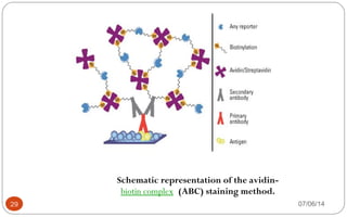
GEETHU
S NAI R
2011-09-104
1 07/06/14
Immunochemistry is the identification of a
certain antigen in a histological tissue
section or cytological preparation via an
antibody specific to the antigen
The localization of the primary antibody (and
therefore the target antigen) is then
visualized microscopically via an appropriate
enzymatic or fluorescent detection system
INTRODUCTION
2 07/06/14
Immunohistochemistry (IHC) combines
histological, immunological and biochemical
techniques for the identification of specific
tissue components by means of a specific
antigen/antibody reaction tagged with a
visible label. IHC makes it possible to
visualize the distribution and localization
of specific cellular components within a cell
or tissue.
INTRODUCTION
3 07/06/14
The use of immunohistochemistry to study
cellular markers that define specific phenotypes
has provided important diagnostic, prognostic,
and predictive information relative to disease
status and biology.
4 07/06/14
Cells to be stained can be attached to a solid
support to allow easy handling in subsequent
procedures. This can be achieved by several
methods: adherent cells may be grown on
microscope slides, coverslips, or an optically
suitable plastic support. Suspension cells can
be centrifuged onto glass slides (cytospin),
bound to solid support using chemical linkers, or
5 07/06/14
I n 1981 a new generat ion of
immunohist ochemical met hods
emerged wit h t he advent of t he
avidin-biot in met hods, which
remains widely used t oday .
 All avidin-biot in met hods rely on6 07/06/14
7 07/06/14
Other names
• Vitamin B7;
• Vitamin H;
• Coenzyme R;
• Biopeiderm
• The molecular formula for biotin is
CC1010 HH1616 NN22 OO33 S.S.
• 807/06/14
Chemical structure of biotin.
• Biotin is
a coenzyme for carboxylase enzymes,
involved in the synthesis of fattY
acids, isoleucine, and valine, and
in gluconeogenesis.
1007/06/14
• D-(+)-Biotin is a cofactor responsible for carbon
dioxide transfer in several carboxylase enzymes:
– Acetyl-CoA carboxylase alpha
– Acetyl-CoA carboxylase beta
– Methylcrotonyl-CoA carboxylase
– Propionyl-CoA carboxylase
– Pyruvate carboxylase
1107/06/14
Def iciency sympt oms
include:
• Hair loss (alopecia)
• Conjunctivitis
• Dermatitis in the form of a scaly, red rash around the
eyes, nose, mouth, and genital area.
• Neurological symptoms in adults, such as depression,
lethargy, hallucination, and numbness and tingling of
the extremities.
1207/06/14
13 07/06/14
• Avidin is a tetrameric or dimeric biotin-
binding protein produced in
the oviducts of birds, reptiles and amphibia
ns and deposited in the whites of their egg
• Functional avidin is found only in raw egg, as
the biotin avidity of the protein is destroyed by
cooking
• Avidin was first discovered by Esmond
Emerson Snell (1914–2003)
1407/06/14
15 07/06/14
Streptavidin
 Streptavidin is isolated from Streptomyces avidinii, and is similar in size and
affinity for biotin. In contrast to avidin, though, streptavidin is not glycosylated,
which makes the protein less-prone to nonspecific binding in IHC applications.
 There are considerable differences in the composition of avidin and streptavidin,
but they are remarkably similar in other respects. Streptavidin is also a tetrameric
protein, with each subunit binding one molecule of biotin with affinity similar to
that of avidin. Streptavidin is much less soluble in water than avidin.
Guanidinium chloride at pH 1.5 will dissociate avidin and streptavidin into
subunits, but streptavidin is more resistant to dissociation.
16 07/06/14
17 07/06/14
deglycoSylated avidin
 Thermo Scientific NeutrAvidin Protein is a specially deglycosylated version of
avidin, with a mass of approximately 60kDa.
 As a result of carbohydrate removal, lectin-binding is reduced to undetectable
levels, yet the biotin-binding affinity is retained because the carbohydrate is not
necessary for this activity.
 NeutrAvidin Protein offers the advantages of a near-neutral pH(6.3) to minimize
nonspecific adsorption, along with lysine residues that remain available for
derivatization or conjugation.
 NeutrAvidin Protein yields the lowest nonspecific binding among the known
biotin-binding proteins due to its near-neutral pH and lack of carbohydrate
group.
18 07/06/14
19 07/06/14
• The interaction of biotin and avidin or streptavidin has been
exploited for use in many protein and nucleic acid detection and
purification methods.
• Because the biotin label is stable and small,
• It rarely interferes with the function of labeled molecules enabling
the avidin-biotin interaction to be used for the development of
robust and
• Highly sensitive assays.
Schematic of the avidin-biotin
interaction. Avidin, streptavidin or
NeutrAvidin Protein can bind up to four biotin
molecules, which are normally conjugated to an
enzyme, antibody or target protein to form an
avidin-biotin complex. † denotes that avidin is
also often conjugated to an antibody, target
protein or immobilized support
22 07/06/14
• Immunohistochemical staining intensity is a function of the enzyme
activity, and improved sensitivity can be achieved by increasing the
number of enzyme molecules bound to the tissue.
• The multiple binding sites between the tetravalent avidin and
biotinylated antibodies (bound to the antigen) are ideal for achieving
this amplification.
• The two most common for amplifying the target antigen signal in
IHC are called avidin-biotin complex (ABC) and labeled streptavidin
binding (LSAB)
MethodS
• The Avidin-Biotin Complex (ABC) Staining MethodThe Avidin-Biotin Complex (ABC) Staining Method
• The Labeled Streptavidin Biotin (LSAB) Staining MethodThe Labeled Streptavidin Biotin (LSAB) Staining Method
the avidin-Biotin coMpleX (aBc)
Staining Method
The Avidin- Biotin Complex (ABC)
Staining Method
• Reporter intensity is a function of the localized enzyme activity, and
improved sensitivity can be achieved by increasing the number of
enzyme molecules bound to the target antigen.
• The multiple biotin binding sites in each tetravalent avidin molecule
are ideal for achieving this amplification.
28 07/06/14
Schematic representation of the avidin-
biotin complex (ABC) staining method.
29 07/06/14
The result is a greater concentration of enzyme (three
enzyme molecules to one avidin molecule) at the
antigenic site and therefore an increase in signal intensity
and sensitivity upon addition of substrate.
30 07/06/14
Advantages Disadvantages
Increased enzyme reporter
localized to the target antigen
Some tissue may require endogenous
biotin blocking to avoid nonspecific
labeling
Increased detection efficiency The ABC complex is large, which
hinders tissue penetration in some
applications
Requires less primary antibody
than direct methods of detection
Reduced assay time compared to
the PAP method
Important features of the Avidin-Biotin Complex (ABC) staining method.
THE LABELED STREPTAVIDIN BIOTIN (LSAB)
STAINING METHOD
• This method employs a streptavidin-enzyme conjugate to
detect the bound biotinylated-primary antibody on the
tissue section, and can be used if the avidin-biotin-
enzyme complex in the ABC method becomes too large
to penetrate the tissue.
• This smaller complex allows better tissue penetration,
has been reported to improve the sensitivy of detection
by 8-fold, and can be used with superior alternatives to
avidin to reduce background and improve sensitivity
even further.
• The information below describes the general staining
procedure and a diagram of the formed complex.
36 07/06/14
37 07/06/14
Advantages of using Avidin-
Biotin Systems
38 07/06/14
The avidin-biotin complex is the strongest known non-
covalent interaction (Kd = 10-15
M) between a protein and
ligand.
The bond formation between biotin and avidin is very
rapid, and once formed, is unaffected by extremes of pH,
temperature, organic solvents and other denaturing
agents.
These features of biotin and avidin – features that are
shared by streptavidin and NeutrAvidin Protein – are
useful for purifying or detecting proteins conjugated to
either component of the interaction.
39 07/06/14
Like secondary antibodies, the avidin-biotin detection
system allows an almost unlimited number of primary
detection reagents (i.e., antibodies, nucleic acids probes
and ligands) to be easily captured, recovered, immobilized
or detected with a very small number of secondary
detection reagents generated by modifying avidin or
streptavidin.
 Furthermore, if a specific biotinylated molecule is not
available, there are many commercially available reagents
to facilitate biotinylation in the lab.
 Likewise, avidin and streptavidin can also be modified as
needed
40 07/06/14
Disadvantages of using the
Avidin-Biotin System
41 07/06/14
Although the avidin-biotin system is simple to set up and
use, it does have certain limitations.
Because any biotinylated molecule will bind to any biotin-
binding protein, these reagents must be used in
combination with other detection-probe systems (i.e.,
primary-secondary antibodies) for multiplex experiments.
Also, because biotin is a biological molecule, endogenous
biotin can cause background and specificity issues when
performing assays with certain biotin-rich tissues and
extracts (i.e., brain, liver, milk, eggs, corn).
This also applies to samples containing endogenous biotin-
binding proteins such as eggs (source of avidin) or bacteria
likeStreptomyces avidinii (source of streptavidin).
42 07/06/14
For purification applications, the strength of the binding
interaction between biotin and avidin is a factor that
limits its utility. This is because harsh conditions are
required to break the avidin-biotin bonds (i.e., to
dissociate and elute), and these may denature target
proteins. To overcome this limitation, modified versions
of avidin resins and modified forms of biotin labeling
reagents are commercially available which make the
interaction readily reversible. These include monomeric
avidin, cleavable disulfide biotin reagents, and
iminobiotin and desthiobiotin derivatives (see discussion
of Protein Isolation and Enrichment below).
43 07/06/14
Applications for Avidin-Biotin Probes
44 07/06/14
Applications for which the avidin-biotin interaction is
used include:
Enzyme linked immunosorbent assay (ELISA)
Immunohistochemistry (IHC)
Western, Northern and Southern blotting
Immunoprecipitation
Cell-surface labeling
Affinity purification
Fluorescence-activated cell sorting (FACS)
Electromobility shift assays (EMSA)
45 07/06/14
The avidin-biotin system can be used for numerous
laboratory methods. The most common methods use
avidin or streptavidin forthe detection of biotinylated
probes. The following is an overview of some of the major
laboratory methods effectively using this system.
Protein Detection
Nucleic Acid Detection
Protein Isolation and EnrichmenT
46 07/06/14
07/06/1447
07/06/1448
07/06/1449
07/06/1450
07/06/1451

Immunocytochemical staining

  • 1.
  • 2.
    Immunochemistry is theidentification of a certain antigen in a histological tissue section or cytological preparation via an antibody specific to the antigen The localization of the primary antibody (and therefore the target antigen) is then visualized microscopically via an appropriate enzymatic or fluorescent detection system INTRODUCTION 2 07/06/14
  • 3.
    Immunohistochemistry (IHC) combines histological,immunological and biochemical techniques for the identification of specific tissue components by means of a specific antigen/antibody reaction tagged with a visible label. IHC makes it possible to visualize the distribution and localization of specific cellular components within a cell or tissue. INTRODUCTION 3 07/06/14
  • 4.
    The use ofimmunohistochemistry to study cellular markers that define specific phenotypes has provided important diagnostic, prognostic, and predictive information relative to disease status and biology. 4 07/06/14
  • 5.
    Cells to bestained can be attached to a solid support to allow easy handling in subsequent procedures. This can be achieved by several methods: adherent cells may be grown on microscope slides, coverslips, or an optically suitable plastic support. Suspension cells can be centrifuged onto glass slides (cytospin), bound to solid support using chemical linkers, or 5 07/06/14
  • 6.
    I n 1981a new generat ion of immunohist ochemical met hods emerged wit h t he advent of t he avidin-biot in met hods, which remains widely used t oday .  All avidin-biot in met hods rely on6 07/06/14
  • 7.
  • 8.
    Other names • VitaminB7; • Vitamin H; • Coenzyme R; • Biopeiderm • The molecular formula for biotin is CC1010 HH1616 NN22 OO33 S.S. • 807/06/14
  • 9.
  • 10.
    • Biotin is a coenzyme for carboxylase enzymes, involvedin the synthesis of fattY acids, isoleucine, and valine, and in gluconeogenesis. 1007/06/14
  • 11.
    • D-(+)-Biotin isa cofactor responsible for carbon dioxide transfer in several carboxylase enzymes: – Acetyl-CoA carboxylase alpha – Acetyl-CoA carboxylase beta – Methylcrotonyl-CoA carboxylase – Propionyl-CoA carboxylase – Pyruvate carboxylase 1107/06/14
  • 12.
    Def iciency symptoms include: • Hair loss (alopecia) • Conjunctivitis • Dermatitis in the form of a scaly, red rash around the eyes, nose, mouth, and genital area. • Neurological symptoms in adults, such as depression, lethargy, hallucination, and numbness and tingling of the extremities. 1207/06/14
  • 13.
  • 14.
    • Avidin is a tetrameric or dimeric biotin- binding protein producedin the oviducts of birds, reptiles and amphibia ns and deposited in the whites of their egg • Functional avidin is found only in raw egg, as the biotin avidity of the protein is destroyed by cooking • Avidin was first discovered by Esmond Emerson Snell (1914–2003) 1407/06/14
  • 15.
  • 16.
    Streptavidin  Streptavidin isisolated from Streptomyces avidinii, and is similar in size and affinity for biotin. In contrast to avidin, though, streptavidin is not glycosylated, which makes the protein less-prone to nonspecific binding in IHC applications.  There are considerable differences in the composition of avidin and streptavidin, but they are remarkably similar in other respects. Streptavidin is also a tetrameric protein, with each subunit binding one molecule of biotin with affinity similar to that of avidin. Streptavidin is much less soluble in water than avidin. Guanidinium chloride at pH 1.5 will dissociate avidin and streptavidin into subunits, but streptavidin is more resistant to dissociation. 16 07/06/14
  • 17.
  • 18.
    deglycoSylated avidin  ThermoScientific NeutrAvidin Protein is a specially deglycosylated version of avidin, with a mass of approximately 60kDa.  As a result of carbohydrate removal, lectin-binding is reduced to undetectable levels, yet the biotin-binding affinity is retained because the carbohydrate is not necessary for this activity.  NeutrAvidin Protein offers the advantages of a near-neutral pH(6.3) to minimize nonspecific adsorption, along with lysine residues that remain available for derivatization or conjugation.  NeutrAvidin Protein yields the lowest nonspecific binding among the known biotin-binding proteins due to its near-neutral pH and lack of carbohydrate group. 18 07/06/14
  • 19.
  • 20.
    • The interactionof biotin and avidin or streptavidin has been exploited for use in many protein and nucleic acid detection and purification methods. • Because the biotin label is stable and small, • It rarely interferes with the function of labeled molecules enabling the avidin-biotin interaction to be used for the development of robust and • Highly sensitive assays.
  • 21.
    Schematic of theavidin-biotin interaction. Avidin, streptavidin or NeutrAvidin Protein can bind up to four biotin molecules, which are normally conjugated to an enzyme, antibody or target protein to form an avidin-biotin complex. † denotes that avidin is also often conjugated to an antibody, target protein or immobilized support
  • 22.
  • 23.
    • Immunohistochemical stainingintensity is a function of the enzyme activity, and improved sensitivity can be achieved by increasing the number of enzyme molecules bound to the tissue. • The multiple binding sites between the tetravalent avidin and biotinylated antibodies (bound to the antigen) are ideal for achieving this amplification. • The two most common for amplifying the target antigen signal in IHC are called avidin-biotin complex (ABC) and labeled streptavidin binding (LSAB)
  • 24.
    MethodS • The Avidin-BiotinComplex (ABC) Staining MethodThe Avidin-Biotin Complex (ABC) Staining Method • The Labeled Streptavidin Biotin (LSAB) Staining MethodThe Labeled Streptavidin Biotin (LSAB) Staining Method
  • 25.
    the avidin-Biotin coMpleX(aBc) Staining Method
  • 26.
    The Avidin- BiotinComplex (ABC) Staining Method • Reporter intensity is a function of the localized enzyme activity, and improved sensitivity can be achieved by increasing the number of enzyme molecules bound to the target antigen. • The multiple biotin binding sites in each tetravalent avidin molecule are ideal for achieving this amplification.
  • 28.
  • 29.
    Schematic representation ofthe avidin- biotin complex (ABC) staining method. 29 07/06/14
  • 30.
    The result isa greater concentration of enzyme (three enzyme molecules to one avidin molecule) at the antigenic site and therefore an increase in signal intensity and sensitivity upon addition of substrate. 30 07/06/14
  • 31.
    Advantages Disadvantages Increased enzymereporter localized to the target antigen Some tissue may require endogenous biotin blocking to avoid nonspecific labeling Increased detection efficiency The ABC complex is large, which hinders tissue penetration in some applications Requires less primary antibody than direct methods of detection Reduced assay time compared to the PAP method Important features of the Avidin-Biotin Complex (ABC) staining method.
  • 33.
    THE LABELED STREPTAVIDINBIOTIN (LSAB) STAINING METHOD
  • 34.
    • This methodemploys a streptavidin-enzyme conjugate to detect the bound biotinylated-primary antibody on the tissue section, and can be used if the avidin-biotin- enzyme complex in the ABC method becomes too large to penetrate the tissue. • This smaller complex allows better tissue penetration, has been reported to improve the sensitivy of detection by 8-fold, and can be used with superior alternatives to avidin to reduce background and improve sensitivity even further. • The information below describes the general staining procedure and a diagram of the formed complex.
  • 36.
  • 37.
  • 38.
    Advantages of usingAvidin- Biotin Systems 38 07/06/14
  • 39.
    The avidin-biotin complexis the strongest known non- covalent interaction (Kd = 10-15 M) between a protein and ligand. The bond formation between biotin and avidin is very rapid, and once formed, is unaffected by extremes of pH, temperature, organic solvents and other denaturing agents. These features of biotin and avidin – features that are shared by streptavidin and NeutrAvidin Protein – are useful for purifying or detecting proteins conjugated to either component of the interaction. 39 07/06/14
  • 40.
    Like secondary antibodies,the avidin-biotin detection system allows an almost unlimited number of primary detection reagents (i.e., antibodies, nucleic acids probes and ligands) to be easily captured, recovered, immobilized or detected with a very small number of secondary detection reagents generated by modifying avidin or streptavidin.  Furthermore, if a specific biotinylated molecule is not available, there are many commercially available reagents to facilitate biotinylation in the lab.  Likewise, avidin and streptavidin can also be modified as needed 40 07/06/14
  • 41.
    Disadvantages of usingthe Avidin-Biotin System 41 07/06/14
  • 42.
    Although the avidin-biotinsystem is simple to set up and use, it does have certain limitations. Because any biotinylated molecule will bind to any biotin- binding protein, these reagents must be used in combination with other detection-probe systems (i.e., primary-secondary antibodies) for multiplex experiments. Also, because biotin is a biological molecule, endogenous biotin can cause background and specificity issues when performing assays with certain biotin-rich tissues and extracts (i.e., brain, liver, milk, eggs, corn). This also applies to samples containing endogenous biotin- binding proteins such as eggs (source of avidin) or bacteria likeStreptomyces avidinii (source of streptavidin). 42 07/06/14
  • 43.
    For purification applications,the strength of the binding interaction between biotin and avidin is a factor that limits its utility. This is because harsh conditions are required to break the avidin-biotin bonds (i.e., to dissociate and elute), and these may denature target proteins. To overcome this limitation, modified versions of avidin resins and modified forms of biotin labeling reagents are commercially available which make the interaction readily reversible. These include monomeric avidin, cleavable disulfide biotin reagents, and iminobiotin and desthiobiotin derivatives (see discussion of Protein Isolation and Enrichment below). 43 07/06/14
  • 44.
  • 45.
    Applications for whichthe avidin-biotin interaction is used include: Enzyme linked immunosorbent assay (ELISA) Immunohistochemistry (IHC) Western, Northern and Southern blotting Immunoprecipitation Cell-surface labeling Affinity purification Fluorescence-activated cell sorting (FACS) Electromobility shift assays (EMSA) 45 07/06/14
  • 46.
    The avidin-biotin systemcan be used for numerous laboratory methods. The most common methods use avidin or streptavidin forthe detection of biotinylated probes. The following is an overview of some of the major laboratory methods effectively using this system. Protein Detection Nucleic Acid Detection Protein Isolation and EnrichmenT 46 07/06/14
  • 47.
  • 48.
  • 49.
  • 50.
  • 51.

Editor's Notes

  • #10 Biotin Biotin, also known as vitamin H, is a small molecule (MW 244.3) that is present in tiny amounts in all living cells and is critical for a number of biological processes. The valeric acid side chain of the biotin molecule can be derivatized in order to incorporate various reactive groups that are used to attach biotin to other molecules. In the context of IHC, biotin is conjugated to antibodies or to the enzyme reporters used to detect target antigens.
  • #11 Once biotin is attached to a molecule, the biotin tag can be used to facilitate affinity purification of that molecule using an immobilized biotin-binding protein. Alternatively, a biotinylated molecule can be immobilized through interaction with a biotin-binding protein, and then used to affinity purify other molecules that specifically interact with it (i.e., co-immunoprecipitation or pull-down assays). In the context of immunohistochemistry and immunoblotting, biotin is most often conjugated to primary or secondary antibodies, and the biotin tag is then detected with a biotin-binding protein that is conjugated to an enzyme, fluorophore or other reporter molecule. Many proteins, such as antibodies, can be labeled with several biotin tags, each able to be bound by a biotin-binding protein. An optimized biotin-to-probe ratio can greatly increase the signal output of a detection system making it possible to create very sensitive assays. Biotin-labeled antibodies and other molecules are readily available from commercial suppliers making assay development routine for many applications. For assays in which a biotinylated probe is not available, there are many biotinylation reagents that enable researchers to chemically label proteins, nucleic acids and surface materials to make custom assay reagents.
  • #15 Avidin The extraordinary affinity of avidin for biotin allows biotin-containing molecules in a complex mixture to be specifically bound to avidin. Avidin is a glycoprotein found in the egg white and tissues of birds, reptiles and amphibia. It contains four identical subunits having a combined mass of 67 to 68kDa. Each subunit consists of 128 amino acids and binds one molecule of biotin; thus, a total of four biotin molecules can bind to a single avidin molecule. The extent of glycosylation on avidin is very high; carbohydrates account for about 10% of the total mass of the tetramer. Avidin has a basic isoelectric point (pI) of 10 to 10.5 and is stable over a wide range of pH and temperatures. Extensive chemical modification has little effect on the activity of avidin, making it especially useful for protein purification. However, because of its carbohydrate content and basic pI, avidin exhibits relatively high nonspecific binding properties. Avidin-biotin binding is the strongest known non-covalent interaction between a protein and ligand. The bond formation between biotin and avidin is very rapid, and once formed, is unaffected by extremes in pH, temperature, organic solvents and other denaturing agents. These features of avidin make detecting or purifying biotin-labeled proteins or other molecules particularly useful for a number of biomedical applications.