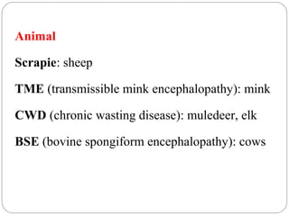
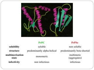
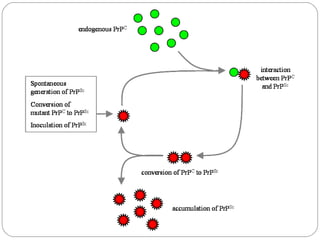
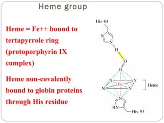
3-D Structure / Function
Animal
Scrapie: sheep
TME (transmissible mink encephalopathy): mink
CWD (chronic wasting disease): muledeer, elk
BSE (bovine spongiform encephalopathy): cows
Human
CJD: Creutzfeld-Jacob Disease
FFI: Fatal familial Insomnia
Kuru
  

PrPC 

PrPSc 

solubility 

soluble

non soluble

structure 

predominantly alpha-helical

predominantly beta-sheeted

multimerisation
state 

monomeric

multimeric
(aggregates)

infectivity 

non infectious

infectious
Susan W. Liebman, and James A. Mastrianni (2005) Trends in Molecular Medicine
11: 439-441
Myoglobin/Hemoglobin
First protein structures 
determined
Oxygen carriers
Hemoglobin transport O2 from 
lungs to tissues
Myoglobin O2 storage protein
Mb and Hb subunits structurally similar
8 alpha-helices
Contain heme group

Myoglobin

Mb monomeric protein
Hb heterotetramer (a2b2)

Hemoglobin
Heme group
Heme = Fe++ bound to
tertapyrrole ring
(protoporphyrin IX
complex)
Heme non-covalently
bound to globin proteins
through His residue
O2 binds non-covalently
to heme Fe++, stabilized
through H-bonding with
another His residue
Heme group in
hydrophobic crevice of
globin protein
Oxygen Binding Curves
•Mb has hyberbolic O2
binding curve
•Mb binds O2 tightly.
Releases at very low pO2
•Hb has sigmoidal O2
binding curve
•Hb high affinity for O2
at high pO2 (lungs)
•Hb low affinity for O2 at
low pO2 (tissues)
Oxygen Binding Curve
O 2 Binding to Hb shows positive
cooperativity
Hb binds four O2 molecules
O2 affinity increases as each O2 molecule binds
Increased affinity due to conformation change
Deoxygenated form = T (tense) form = low

affinity
Oxygenated form = R (relaxed) form = high

affinity
O 2 Binding to Hb shows positive
cooperativity
O 2 Binding
induces
conformation
change

T-conformation

R-conformation

Show Movie
Heme moves 0.34 nm
Exposing crystal of
deoxy-form to air cause
crystal to crack
Allosteric Interactions
Allosteric interaction occur when specific

molecules bind a protein and modulates activity
Allosteric modulators or allosteric effectors
Bind reversibly to site separate from functional

binding or active site
Modulation of activity occurs through change in

protein conformation
2,3 bisphosphoglycerate (BPG), CO2 and protons are

allosteric effectors of Hb binding of O2
Bohr Effect
Increased CO2 leads to decreased pH

CO2 + H2O <-> HCO3- + H+
At decreased pH several key AA’s protonated, causes Hb to take

on T-conformation (low affinty)
In R-form same AA’s deprotonated, form charge charge

interactions with positive groups, stabilize R-conformation (High
affinity)
HCO3- combines with N-terminal alpha-amino group to form

carbamate group.

--N3H+ + HCO3- 

--NHCOOCarbamation stabilizes T-conformation
Bisphosphoglycerate (BPG)
BPG involved acclimation to high altitude
Binding of BPG to Hb causes low O2 affinity
BPG binds in the cavity between beta-Hb subunits
Stabilizes T-conformation
Feta Hb (a2g2) has low affinity for BPG, allows fetus to

compete for O2 with mother’s Hb (a2b2) in placenta.
Mutations in a- or b-globin genes
can cause disease state
Sickle cell anemia – E6 to V6
Causes V6 to bind to hydrophobic pocket in deoxy-

Hb
Polymerizes to form long filaments
Cause sickling of cells
Sickle cell trait offers advantage against malaria
Fragile sickle cells can not support parasite
Fourteen-strand fibers of Hb S and the
interactions involving beta 6 Val

Protein structure 3 d