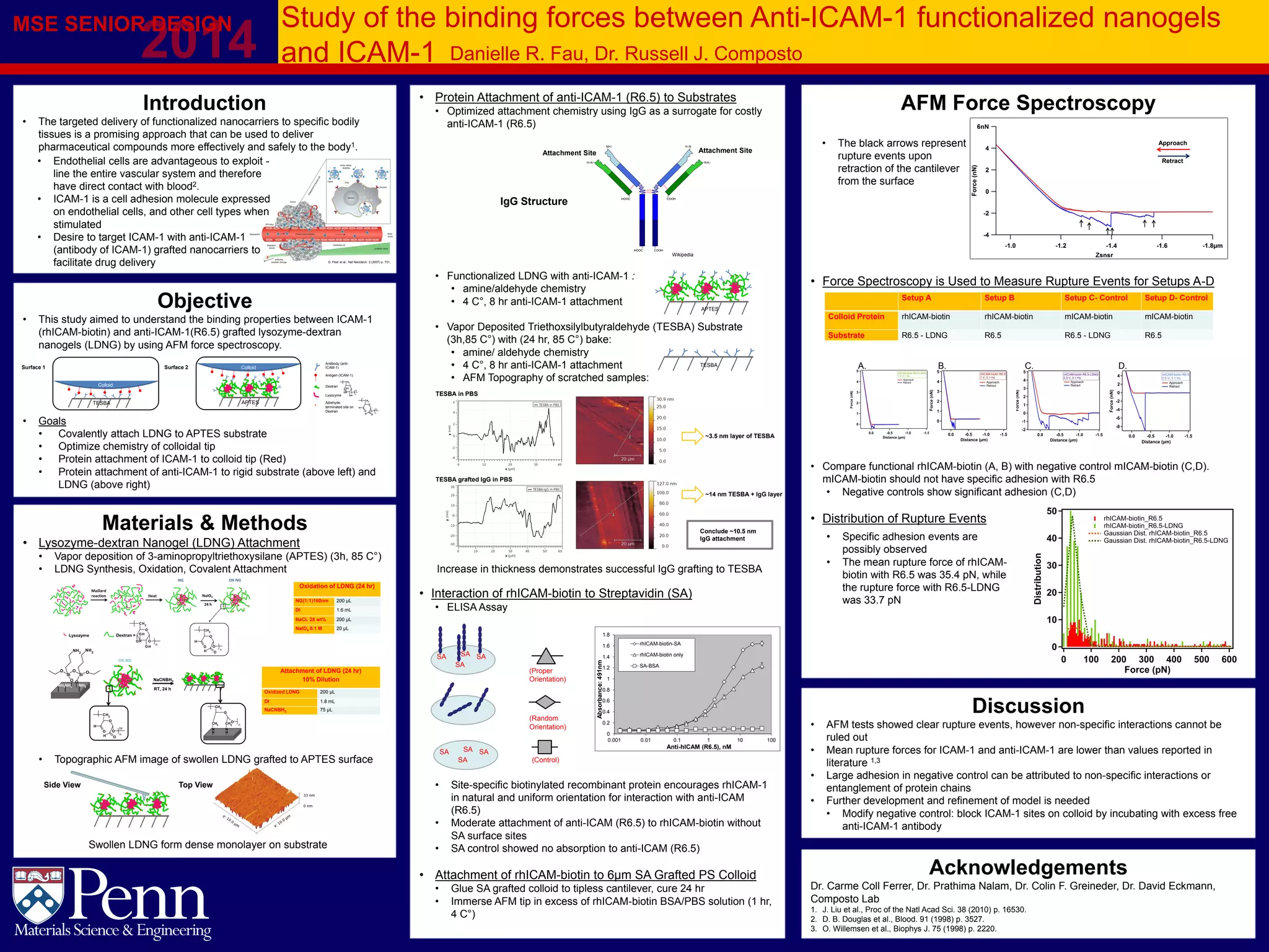
This study aimed to understand the binding forces between ICAM-1 and anti-ICAM-1 grafted nanogels using AFM force spectroscopy. Anti-ICAM-1 was attached to lysozyme-dextran nanogels (LDNG) and substrates. Recombinant human ICAM-1 tagged with biotin (rhICAM-biotin) was attached to AFM colloidal tips. Force spectroscopy measurements of specific rhICAM-biotin and anti-ICAM-1 interactions showed rupture events, though non-specific binding could not be ruled out. Further experiments are needed to optimize the model and reduce non-specific interactions.


![[Sigma Xi] Bioresponsive quantum dot lattices for applications in biosensing ...](https://cdn.slidesharecdn.com/ss_thumbnails/sigmaxibioresponsivequantumdotlatticesforapplicationsinbiosensingandconditionally-activatedrnainterf-190406055229-thumbnail.jpg?width=640&height=640&fit=bounds)


