Download as PPSX, PPTX
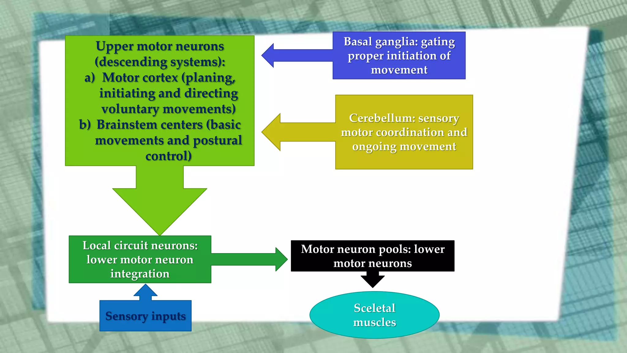
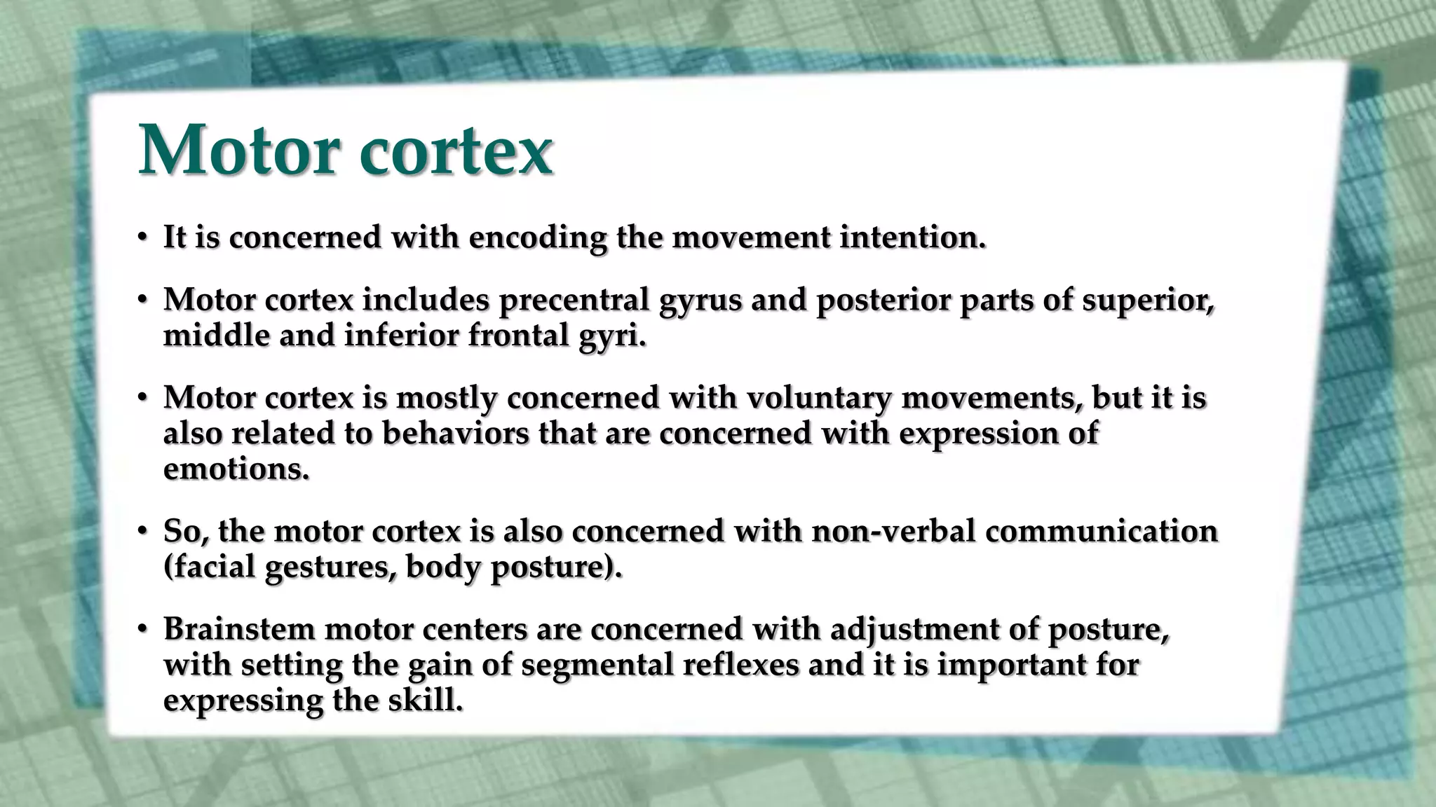
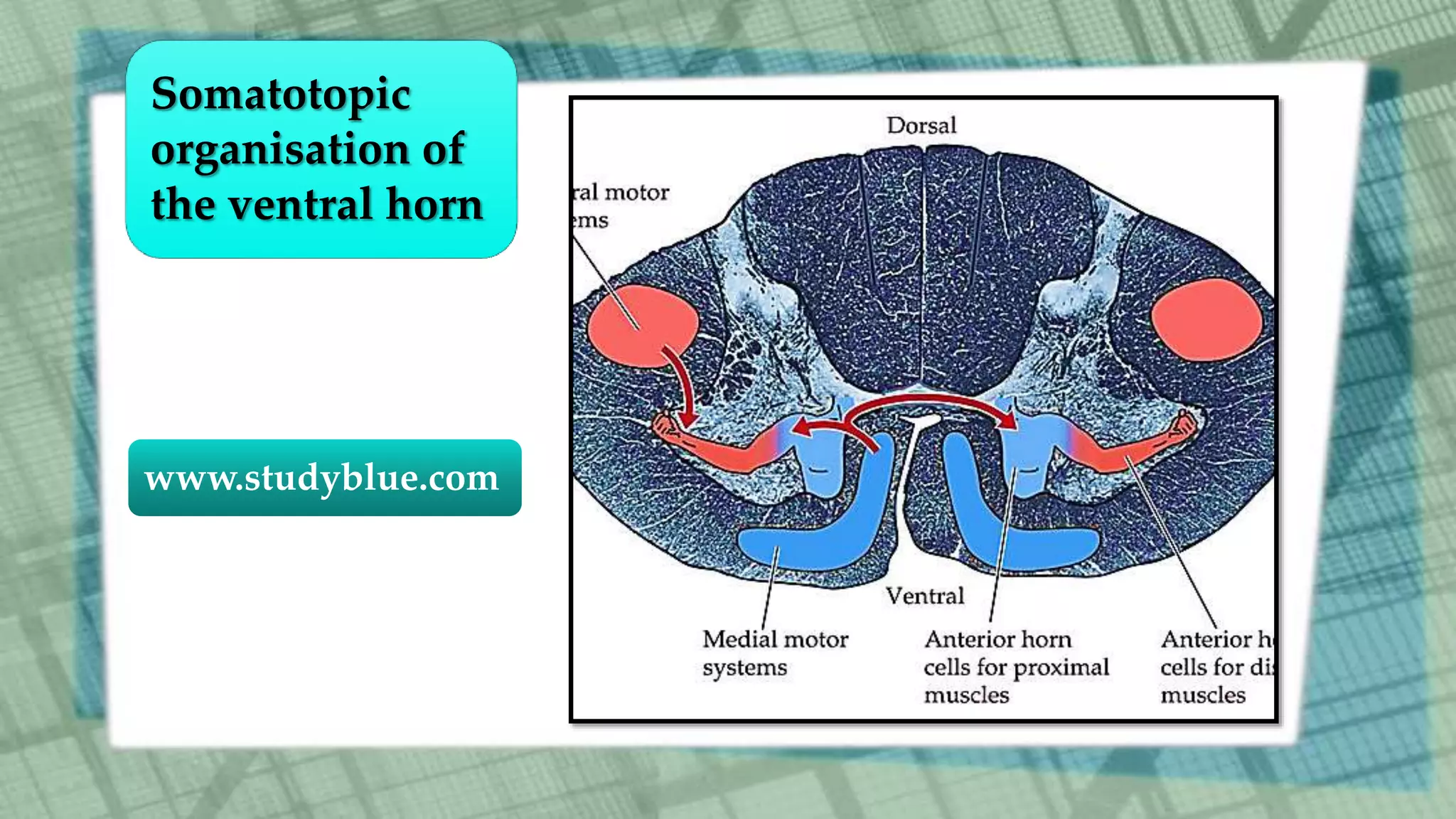
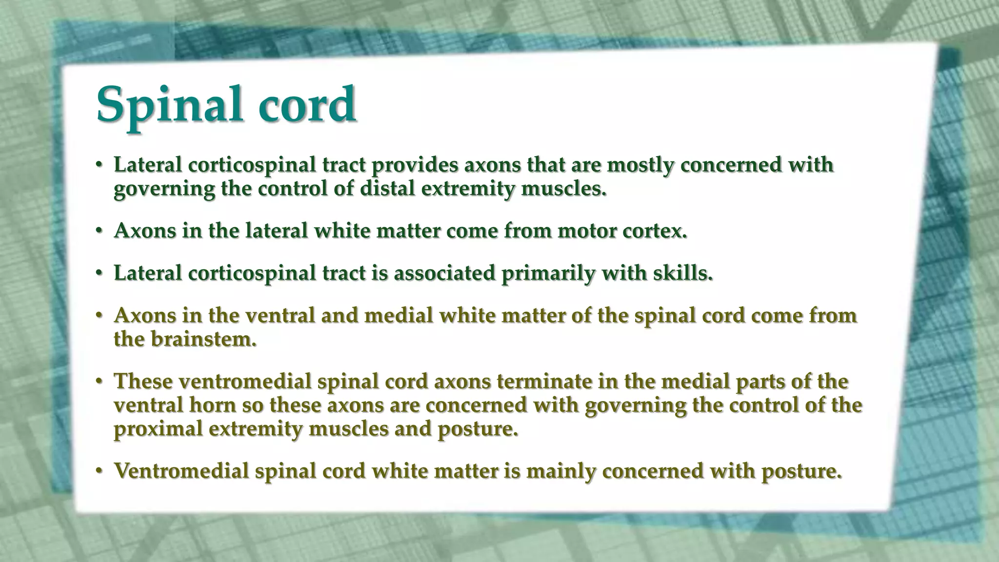
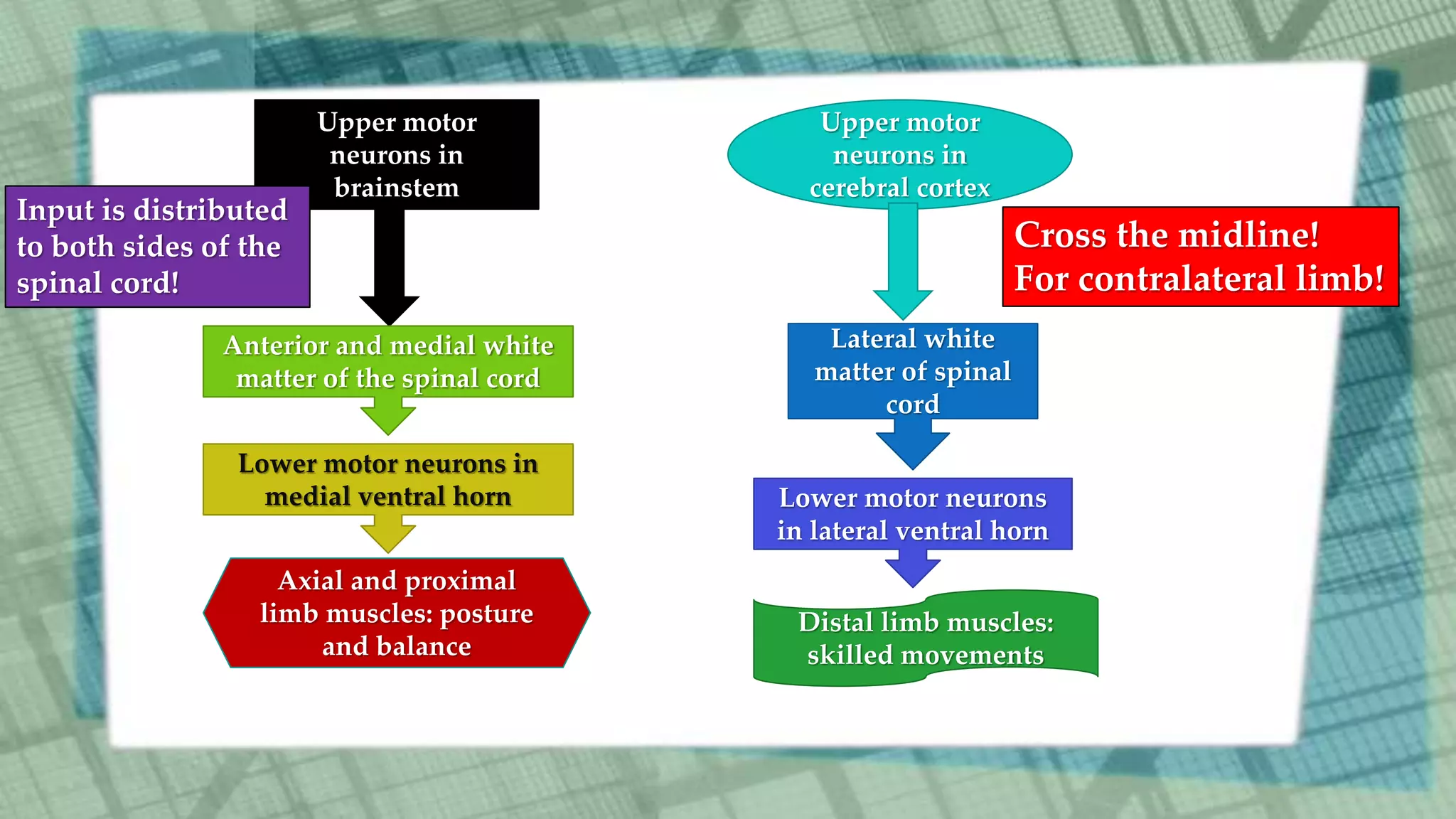
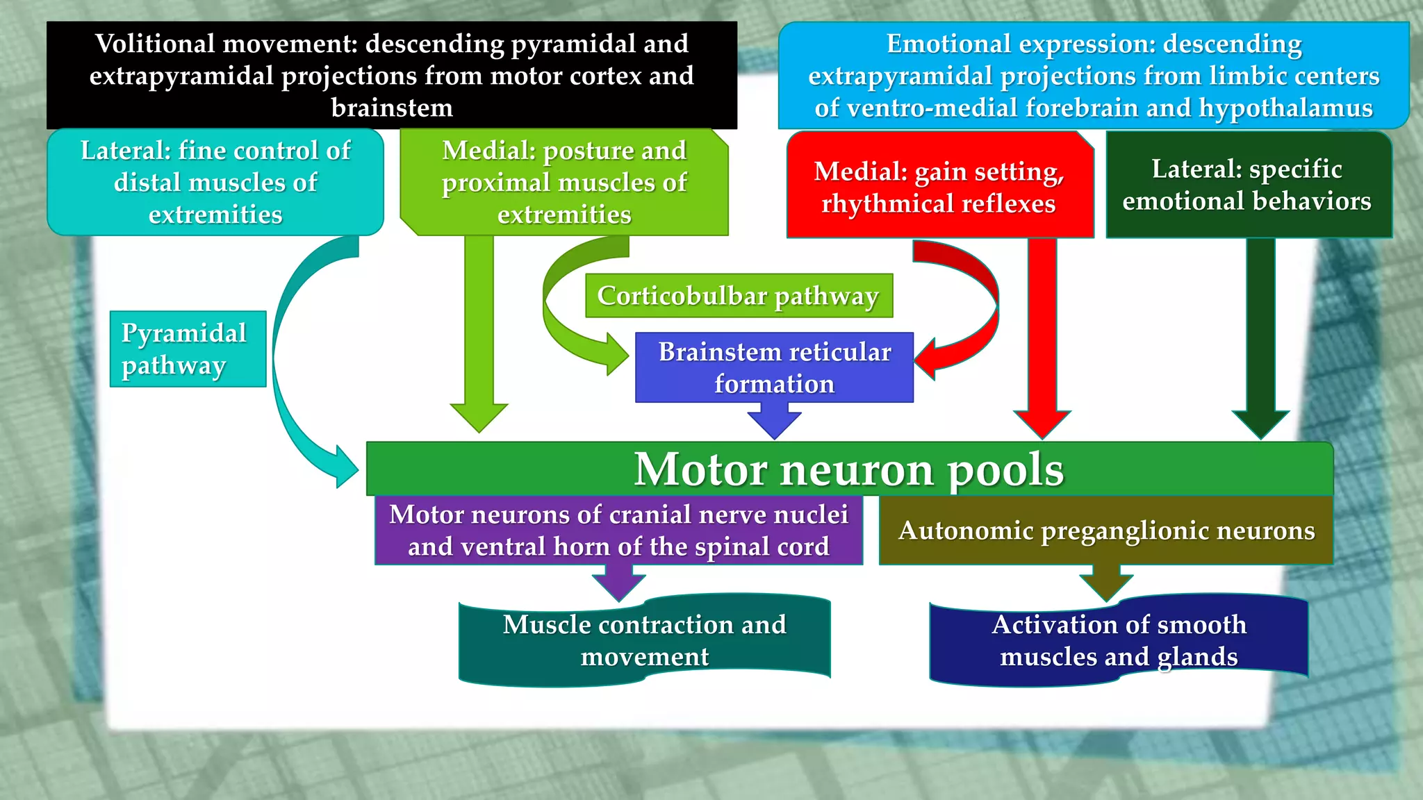
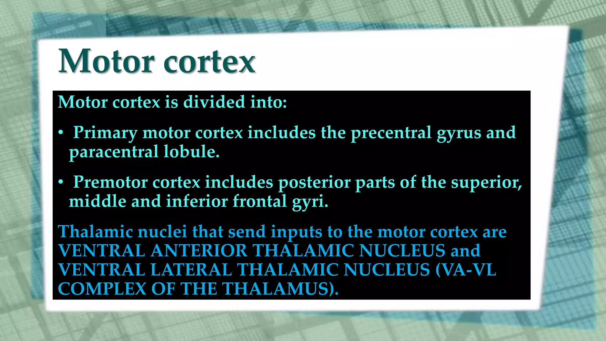
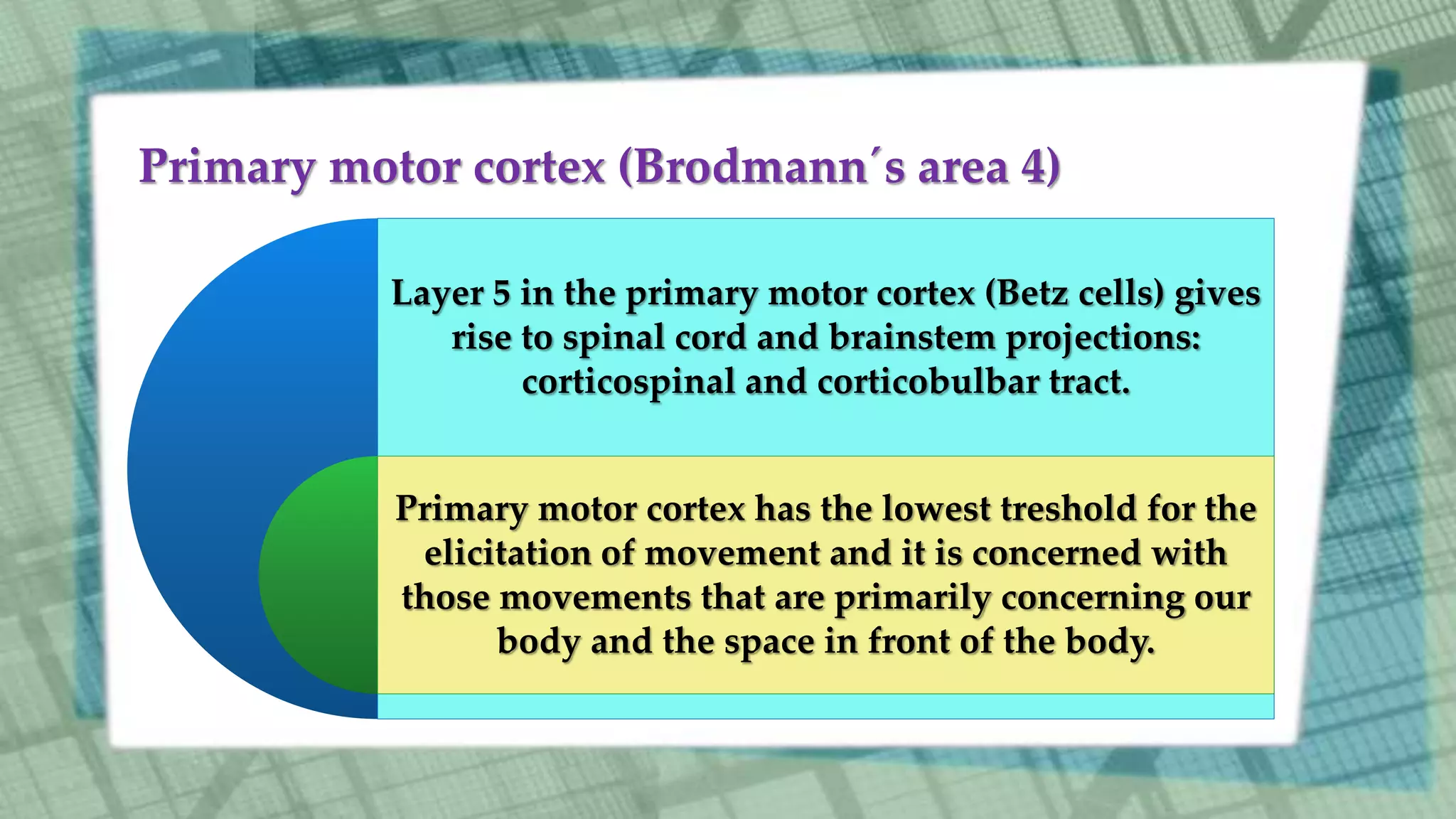
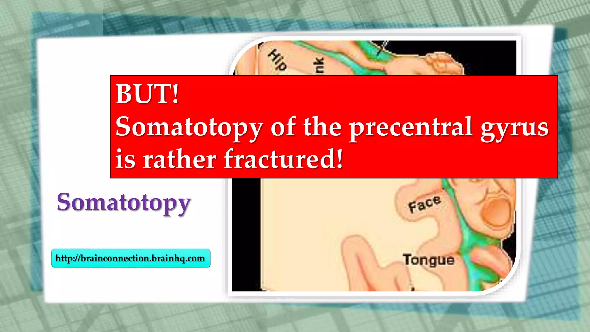
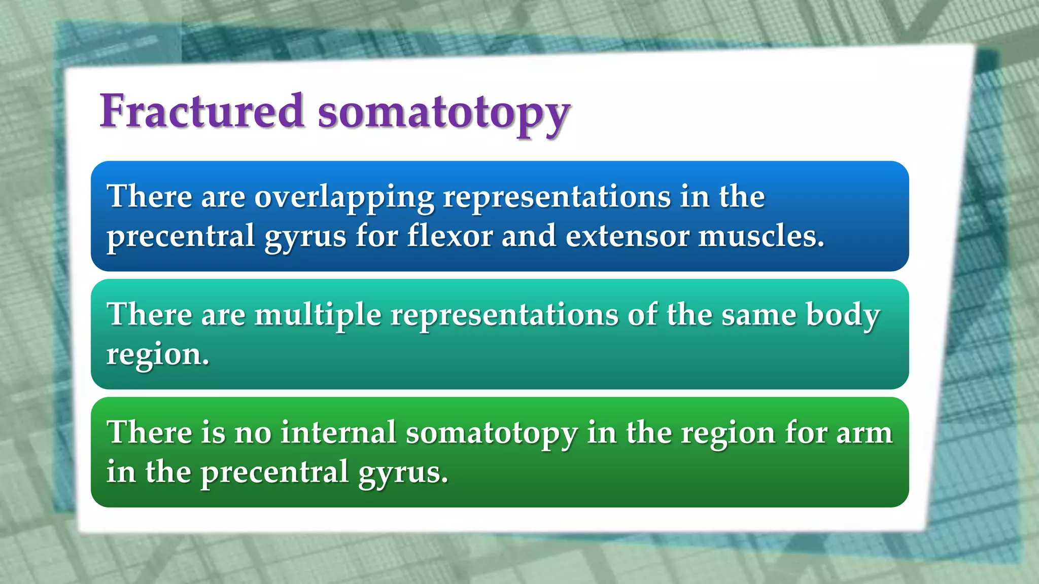
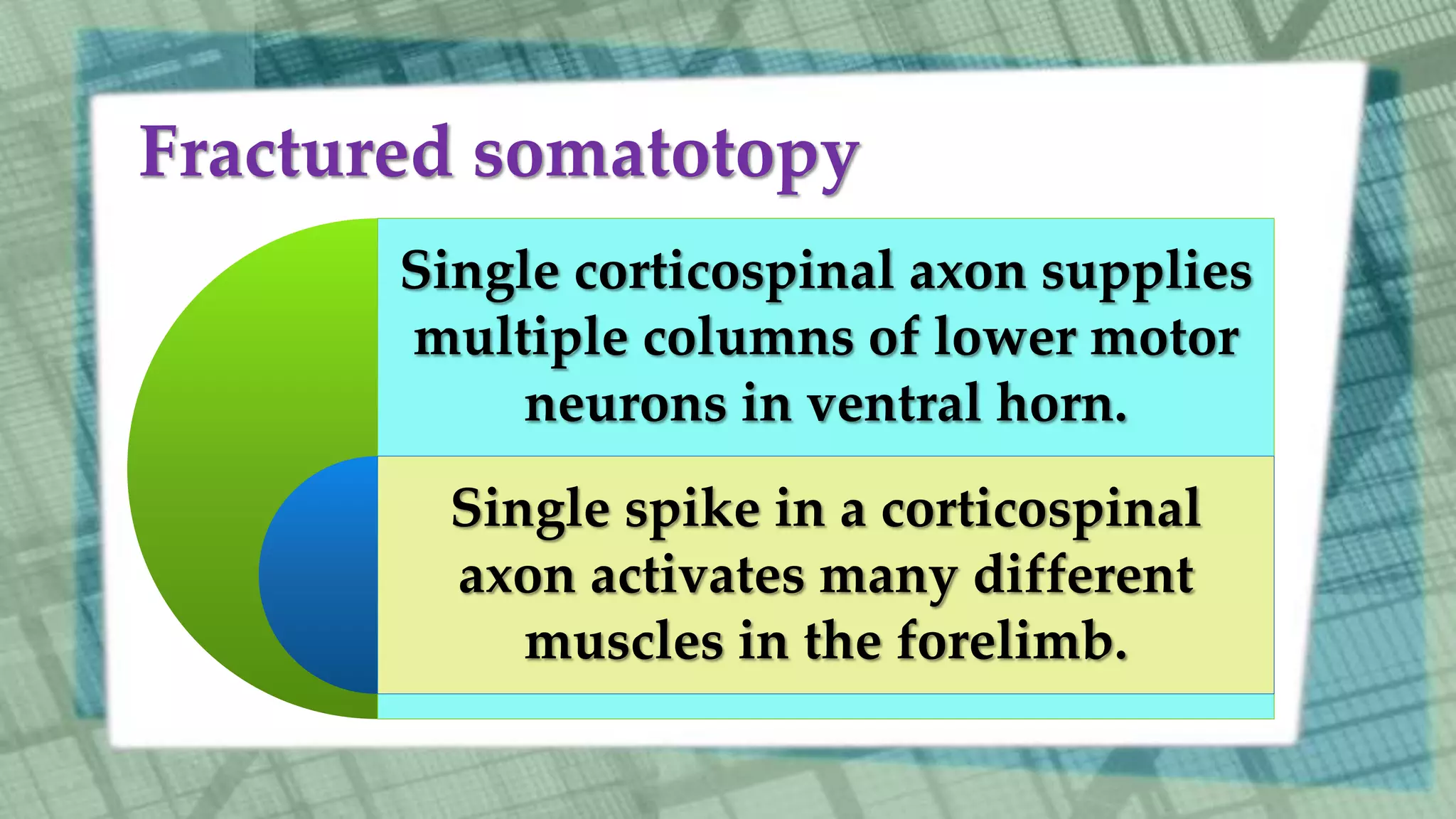


The primary motor cortex is responsible for planning, initiating, and directing voluntary movements. It contains the precentral gyrus and posterior parts of the superior, middle, and inferior frontal gyri. The primary motor cortex encodes movement intentions and is involved in both voluntary movements and the expression of emotions through facial gestures and body posture. Brainstem motor centers help control posture and reflexes. The lateral corticospinal tract governs distal limb muscles for skilled movements while the ventromedial tract governs proximal muscles and posture. The motor cortex has a fractured somatotopic organization and single neurons can activate multiple muscles.


