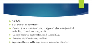
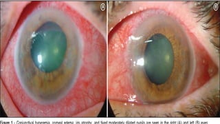
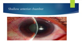
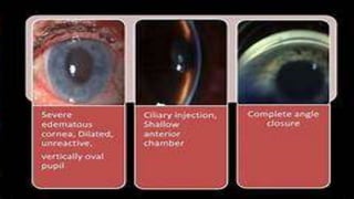
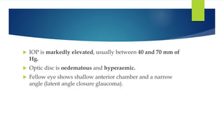
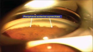
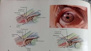
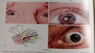
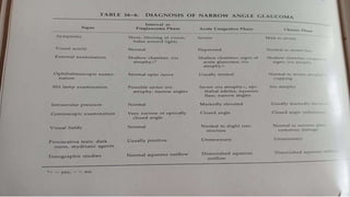
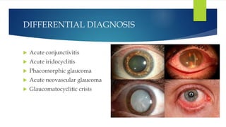
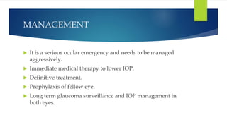
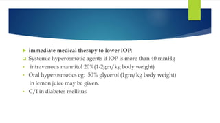
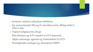
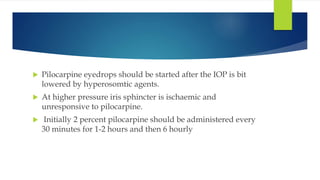
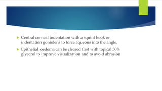
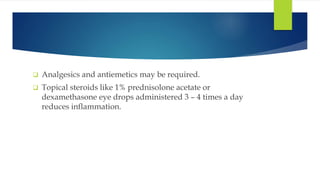
ACUTE CONGESTIVE
GLAUCOMA
BY
DR,B,UGANDHAR REDDY MS
PROFESSOR
REH,KNL.
 An attack of acute primary angle closure glaucoma occurs
due to a sudden total angle closure leading to severe rise in
IOP.
 May occur due to pupillary block.
 This is sight threatening emergency.
CLINICAL FEATURES
 SYMPTOMS
 Pain: Typically acute attack is characterised by sudden onset
of very severe pain in the eye which radiates along the
branches of 5th nerve.
 Nausea, vomiting and prostrations are frequently
associated with pain.
 Severe unilateral headache
 Rapidly progressive impairment of vision, redness,
photophobia and lacrimation develop in all cases.
 Past history: About 5 percent patients give history of typical
previous intermittent attacks of subacute angle-closure
glaucoma.
 SIGNS
 Lids may be oedematous.
 Conjunctiva is chemosed, and congested, (both conjunctival
and ciliary vessels are congested).
 Cornea becomes oedematous and insensitive.
 Anterior chamber is very shallow.
 Aqueous flare or cells may be seen in anterior chamber.
Shallow anterior chamber
 Angle of anterior chamber is completely closed as seen on
gonioscopy (shaffer grade 0).
 Iris may be discoloured.
 Pupil is semi-dilated, vertically oval and fixed. It is non-
reactive to both light and accommodation
Angle structures from anterior to
posterior
 Schwalbe’s line
 Trabecular meshwork
 Scleral spur
 Ciliary body band
 Root of iris
 IOP is markedly elevated, usually between 40 and 70 mm of
Hg.
 Optic disc is oedematous and hyperaemic.
 Fellow eye shows shallow anterior chamber and a narrow
angle (latent angle closure glaucoma).
DIFFERENTIAL DIAGNOSIS
 Acute conjunctivitis
 Acute iridocyclitis
 Phacomorphic glaucoma
 Acute neovascular glaucoma
 Glaucomatocyclitic crisis
MANAGEMENT
 It is a serious ocular emergency and needs to be managed
aggressively.
 Immediate medical therapy to lower IOP.
 Definitive treatment.
 Prophylaxis of fellow eye.
 Long term glaucoma surveillance and IOP management in
both eyes.
 immediate medical therapy to lower IOP:
 Systemic hyperosmotic agents if IOP is more than 40 mmHg
 intravenous mannitol 20%(1-2gm/kg body weight)
 Oral hyperosmotics eg: 50% glycerol (1gm/kg body weight)
in lemon juice may be given.
 C/I in diabetes mellitus
 Systemic carbonic anhydrase inhibitors:
 Eg: acetazolamide 500 mg IV stat followed by 250mg tablet 3
times a day.
 Topical antiglaucoma drugs:
 Beta-blockers eg: 0.5% timolol or 0.5% betaxolol.
 Alpha adrenergic agonists eg: brimonidine 0.1-0.2%
 Prostaglandin analogue eg: latanoprost 0.005%
 Pilocarpine eyedrops should be started after the IOP is bit
lowered by hyperosomtic agents.
 At higher pressure iris sphincter is ischaemic and
unresponsive to pilocarpine.
 Initially 2 percent pilocarpine should be administered every
30 minutes for 1-2 hours and then 6 hourly
 Central corneal indentation with a squint hook or
indentation goniolens to force aqueous into the angle.
 Epithelial oedema can be cleared first with topical 50%
glycerol to improve visualization and to avoid abrasion
 Analgesics and antiemetics may be required.
 Topical steroids like 1% prednisolone acetate or
dexamethasone eye drops administered 3 – 4 times a day
reduces inflammation.
 Definitive therapy
 Laser peripheral iriotomy:
 goniscopy should be performed as soon as cornea becomes
clear.
 Laser PI should be performed if PAS are seen in <270 angle.
 LPI re-establishes communication between posterior and
anterior chamber so it bypasses pupillary block and
immediately relieves the crowding of the angle.
 Filtration surgery: It should be performed in cases where
IOP is not controlled with the best medical therapy
following an attack of acute congestive glaucoma and also
when peripheral anterior synechiae are formed in more than
270 degrees of the angle of the anterior chamber.
 Mechanism: Filtration surgery provides an alternative to the
angle for drainage of aqueous from anterior chamber into
subconjunctival space.
 Clear lens extraction by phacoemulsification with
intraocular lens implantation by has recent been
recommended by some workers.
 Prophylactic treatment in the normal fellow eye
 Prophylactic laser iridotomy (preferably) or surgical
peripheral iridectomy should be performed on the fellow
asymptomatic eye.
 It should be done as early as possible as chances of acute
attack are 50% in such eyes.
 Long term glaucoma surveillance and IOP management in
both eyes.
 It is must to ultimately prevent glaucomatous blindness.
 Eyes treated with PI may develop PACG at any time, so it
should be treated as when required.
 Filtration surgery may fail anytime during course and hence
need to be repeated with antimetabolites.
Sequelae of acute PAC
 Postsurgical acute PAC
 Spontaneous angle reopening
 Ciliary body shut down
 Vogt’s triad
 Postsurgical acute PAC:
 This refers to the clinical status of the eye after laser
peripheral iridotomy (PI) for an attack of acute PAC. It may
occur in two clinical settings :
 i. With normalized IOP after successful laser PI, the eye
usually quitens after some time with or without marks of an
acute attack.
 ii. With raised IOP after unsuccessful laser PI, there occurs a
state of chronic congestive glaucoma. It needs to be treated
by trabeculectomy operation
 Spontaneous angle opening:
 It may very rarely occur in some cases and the attack of
acute PACG may subside itself without treatment.
 Treatment of such cases is laser peripheral iridotomy.
 Ciliary body shut down:
 It refers to temporary cessation of aqueous humour
secretion due to ischaemic damage to the ciliary epithelium
after an attack of acute PACG.
 Clinical features in this stage are similar to acute congestive
glaucoma except that the IOP is low and pain is markedly
reduced
 Treatment includes:
 Topical steroid drops to reduce inflammation.
 Laser iridotomy should be performed when the cornea
becomes clear and IOP should be monitored.
 Trabeculectomy is required when IOP rises constantly.
 Vogt’s triad
 It may be seen in patients with any type of postcongesive
glaucoma and in treated cases of acute congestive glaucoma.
 It is characterized by:
 Glaucomflecken (anterior subcapsular lenticular opacity),
 Patches of iris atrophy, and
 Slightly dilated non-reacting pupil (due to sphincter
atrophy).
 A patient presents with sudden onset of severe unilateral
eye pain, headache associated with blurred vision, rainbow
colored haloes around bright light, nausea and vomiting.
Examination revealed a fixed midpoint pupil and a hazy,
cloudy cornea with marked conjunctival congestion. What
could be the diagnosis? Discuss the pathophysiology and
treatment of the above condition.
 Differential diagnosis of acute red eye. Describe clinical
features and management of acute congestive glaucoma.
ACUTE CONGESTIVE GLAUCOMA.pptx

ACUTE CONGESTIVE GLAUCOMA.pptx

  • 1.
  • 2.
     An attackof acute primary angle closure glaucoma occurs due to a sudden total angle closure leading to severe rise in IOP.  May occur due to pupillary block.  This is sight threatening emergency.
  • 3.
    CLINICAL FEATURES  SYMPTOMS Pain: Typically acute attack is characterised by sudden onset of very severe pain in the eye which radiates along the branches of 5th nerve.  Nausea, vomiting and prostrations are frequently associated with pain.  Severe unilateral headache
  • 4.
     Rapidly progressiveimpairment of vision, redness, photophobia and lacrimation develop in all cases.  Past history: About 5 percent patients give history of typical previous intermittent attacks of subacute angle-closure glaucoma.
  • 5.
     SIGNS  Lidsmay be oedematous.  Conjunctiva is chemosed, and congested, (both conjunctival and ciliary vessels are congested).  Cornea becomes oedematous and insensitive.  Anterior chamber is very shallow.  Aqueous flare or cells may be seen in anterior chamber.
  • 7.
  • 8.
     Angle ofanterior chamber is completely closed as seen on gonioscopy (shaffer grade 0).  Iris may be discoloured.  Pupil is semi-dilated, vertically oval and fixed. It is non- reactive to both light and accommodation
  • 10.
    Angle structures fromanterior to posterior  Schwalbe’s line  Trabecular meshwork  Scleral spur  Ciliary body band  Root of iris
  • 12.
     IOP ismarkedly elevated, usually between 40 and 70 mm of Hg.  Optic disc is oedematous and hyperaemic.  Fellow eye shows shallow anterior chamber and a narrow angle (latent angle closure glaucoma).
  • 18.
    DIFFERENTIAL DIAGNOSIS  Acuteconjunctivitis  Acute iridocyclitis  Phacomorphic glaucoma  Acute neovascular glaucoma  Glaucomatocyclitic crisis
  • 19.
    MANAGEMENT  It isa serious ocular emergency and needs to be managed aggressively.  Immediate medical therapy to lower IOP.  Definitive treatment.  Prophylaxis of fellow eye.  Long term glaucoma surveillance and IOP management in both eyes.
  • 20.
     immediate medicaltherapy to lower IOP:  Systemic hyperosmotic agents if IOP is more than 40 mmHg  intravenous mannitol 20%(1-2gm/kg body weight)  Oral hyperosmotics eg: 50% glycerol (1gm/kg body weight) in lemon juice may be given.  C/I in diabetes mellitus
  • 21.
     Systemic carbonicanhydrase inhibitors:  Eg: acetazolamide 500 mg IV stat followed by 250mg tablet 3 times a day.  Topical antiglaucoma drugs:  Beta-blockers eg: 0.5% timolol or 0.5% betaxolol.  Alpha adrenergic agonists eg: brimonidine 0.1-0.2%  Prostaglandin analogue eg: latanoprost 0.005%
  • 22.
     Pilocarpine eyedropsshould be started after the IOP is bit lowered by hyperosomtic agents.  At higher pressure iris sphincter is ischaemic and unresponsive to pilocarpine.  Initially 2 percent pilocarpine should be administered every 30 minutes for 1-2 hours and then 6 hourly
  • 23.
     Central cornealindentation with a squint hook or indentation goniolens to force aqueous into the angle.  Epithelial oedema can be cleared first with topical 50% glycerol to improve visualization and to avoid abrasion
  • 24.
     Analgesics andantiemetics may be required.  Topical steroids like 1% prednisolone acetate or dexamethasone eye drops administered 3 – 4 times a day reduces inflammation.
  • 25.
     Definitive therapy Laser peripheral iriotomy:  goniscopy should be performed as soon as cornea becomes clear.  Laser PI should be performed if PAS are seen in <270 angle.  LPI re-establishes communication between posterior and anterior chamber so it bypasses pupillary block and immediately relieves the crowding of the angle.
  • 26.
     Filtration surgery:It should be performed in cases where IOP is not controlled with the best medical therapy following an attack of acute congestive glaucoma and also when peripheral anterior synechiae are formed in more than 270 degrees of the angle of the anterior chamber.  Mechanism: Filtration surgery provides an alternative to the angle for drainage of aqueous from anterior chamber into subconjunctival space.
  • 27.
     Clear lensextraction by phacoemulsification with intraocular lens implantation by has recent been recommended by some workers.
  • 28.
     Prophylactic treatmentin the normal fellow eye  Prophylactic laser iridotomy (preferably) or surgical peripheral iridectomy should be performed on the fellow asymptomatic eye.  It should be done as early as possible as chances of acute attack are 50% in such eyes.
  • 29.
     Long termglaucoma surveillance and IOP management in both eyes.  It is must to ultimately prevent glaucomatous blindness.  Eyes treated with PI may develop PACG at any time, so it should be treated as when required.  Filtration surgery may fail anytime during course and hence need to be repeated with antimetabolites.
  • 30.
    Sequelae of acutePAC  Postsurgical acute PAC  Spontaneous angle reopening  Ciliary body shut down  Vogt’s triad
  • 31.
     Postsurgical acutePAC:  This refers to the clinical status of the eye after laser peripheral iridotomy (PI) for an attack of acute PAC. It may occur in two clinical settings :  i. With normalized IOP after successful laser PI, the eye usually quitens after some time with or without marks of an acute attack.  ii. With raised IOP after unsuccessful laser PI, there occurs a state of chronic congestive glaucoma. It needs to be treated by trabeculectomy operation
  • 32.
     Spontaneous angleopening:  It may very rarely occur in some cases and the attack of acute PACG may subside itself without treatment.  Treatment of such cases is laser peripheral iridotomy.
  • 33.
     Ciliary bodyshut down:  It refers to temporary cessation of aqueous humour secretion due to ischaemic damage to the ciliary epithelium after an attack of acute PACG.  Clinical features in this stage are similar to acute congestive glaucoma except that the IOP is low and pain is markedly reduced
  • 34.
     Treatment includes: Topical steroid drops to reduce inflammation.  Laser iridotomy should be performed when the cornea becomes clear and IOP should be monitored.  Trabeculectomy is required when IOP rises constantly.
  • 35.
     Vogt’s triad It may be seen in patients with any type of postcongesive glaucoma and in treated cases of acute congestive glaucoma.  It is characterized by:  Glaucomflecken (anterior subcapsular lenticular opacity),  Patches of iris atrophy, and  Slightly dilated non-reacting pupil (due to sphincter atrophy).
  • 37.
     A patientpresents with sudden onset of severe unilateral eye pain, headache associated with blurred vision, rainbow colored haloes around bright light, nausea and vomiting. Examination revealed a fixed midpoint pupil and a hazy, cloudy cornea with marked conjunctival congestion. What could be the diagnosis? Discuss the pathophysiology and treatment of the above condition.
  • 38.
     Differential diagnosisof acute red eye. Describe clinical features and management of acute congestive glaucoma.