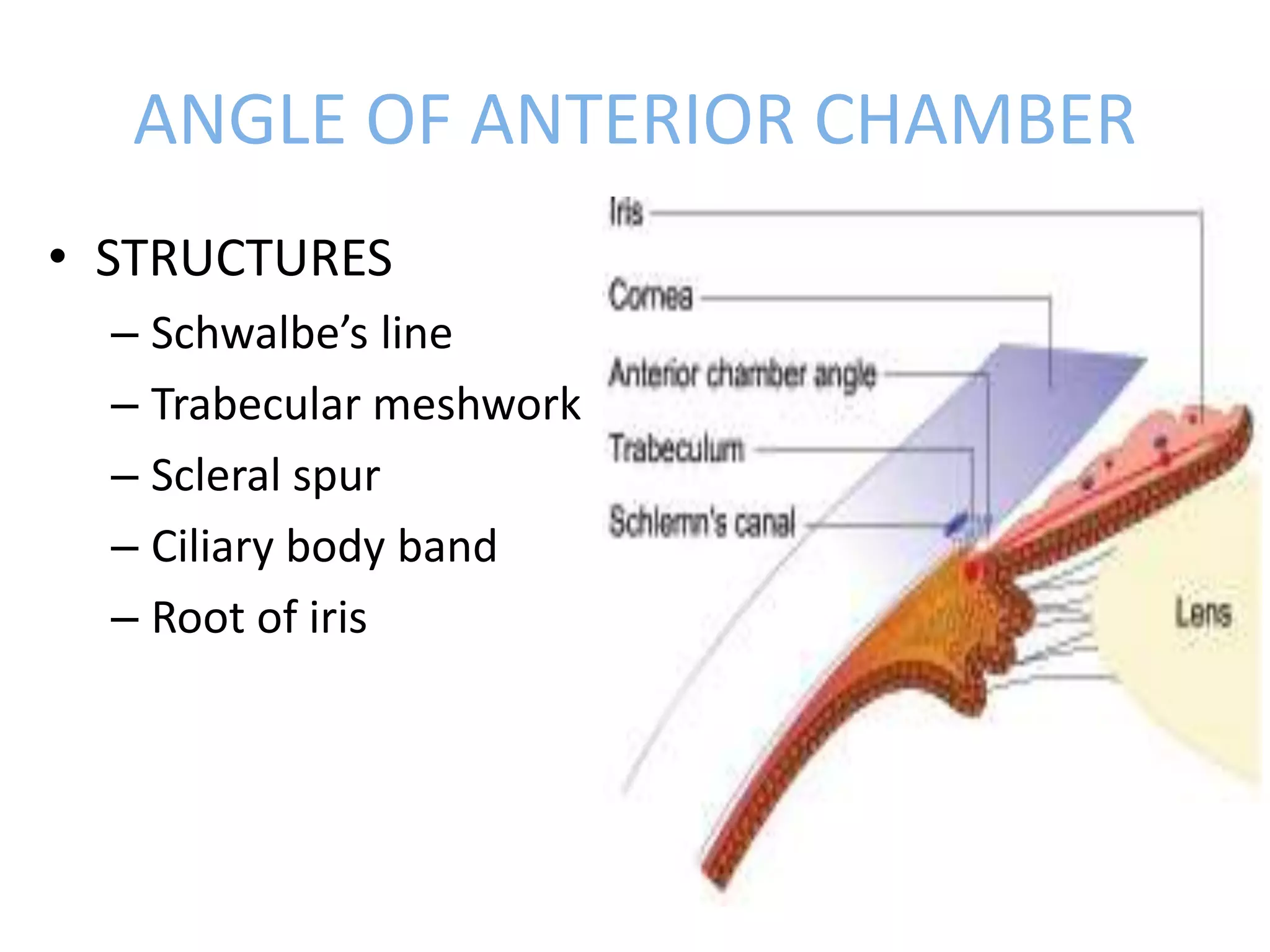
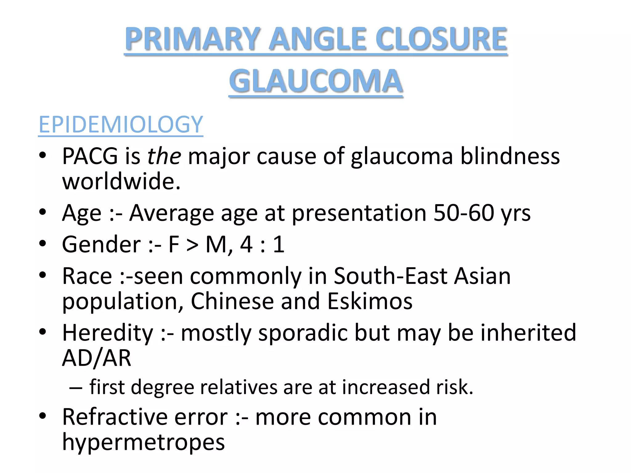
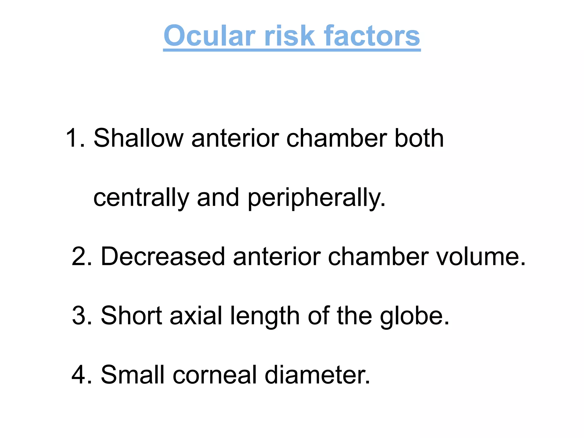
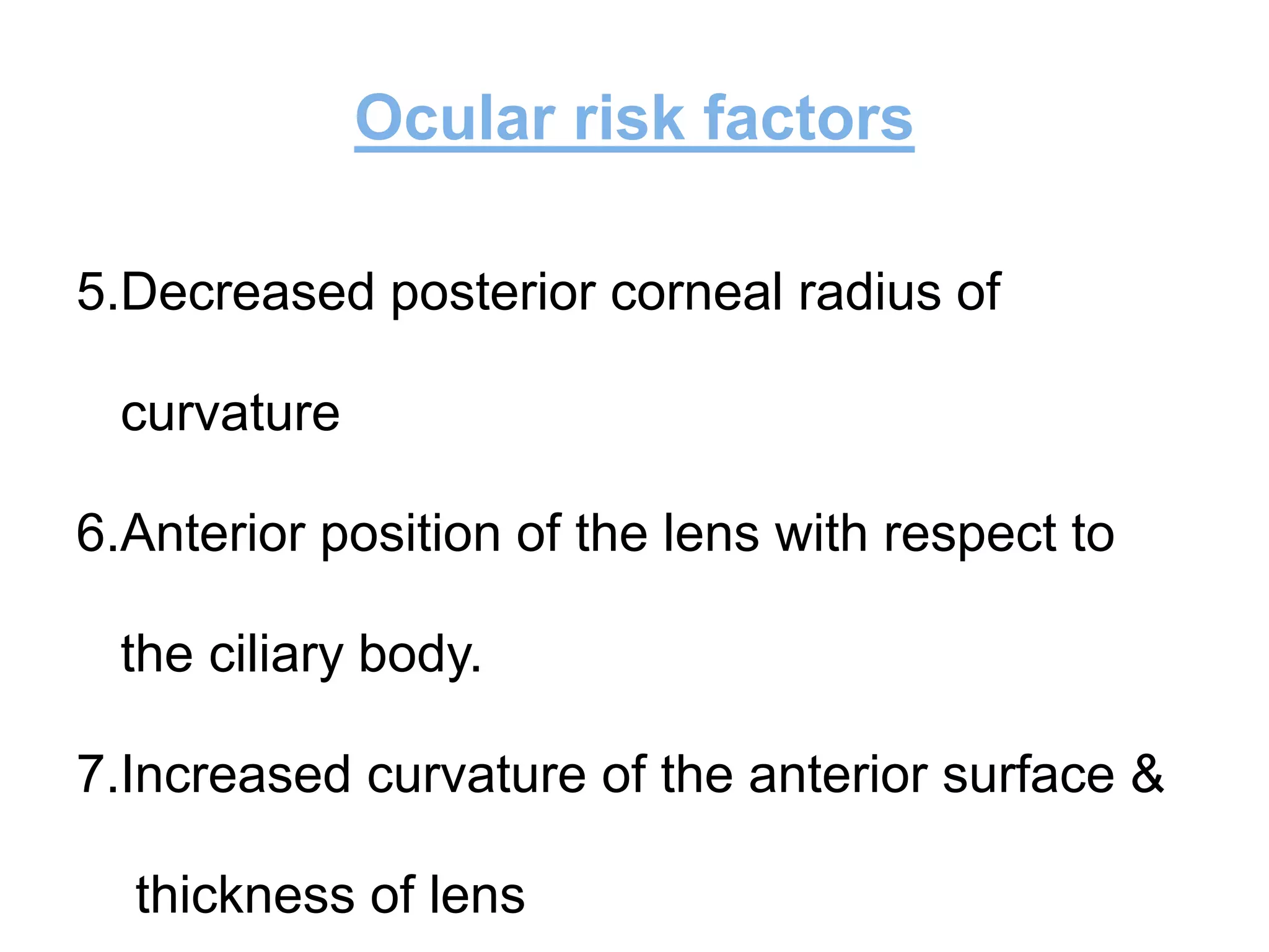
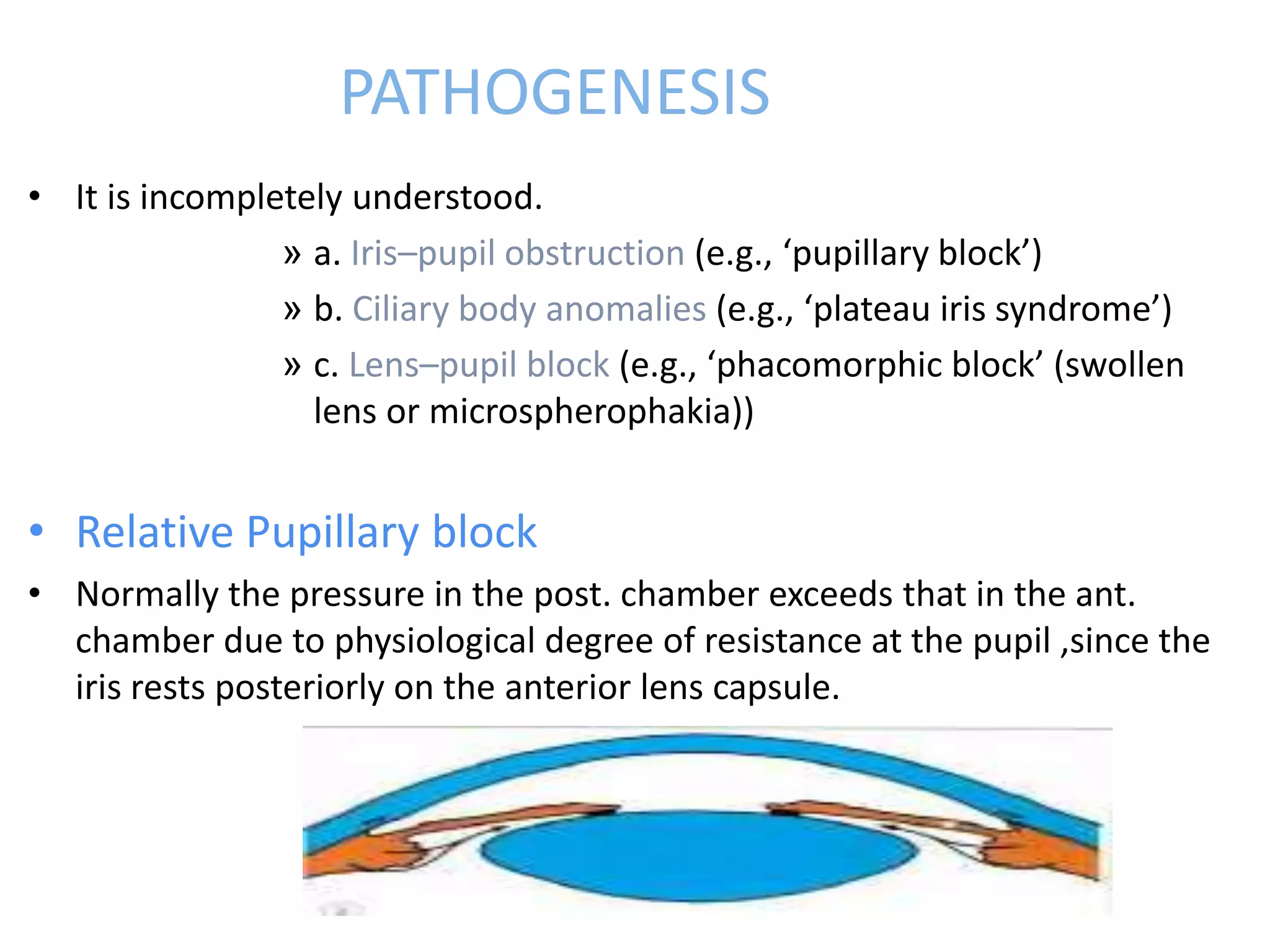
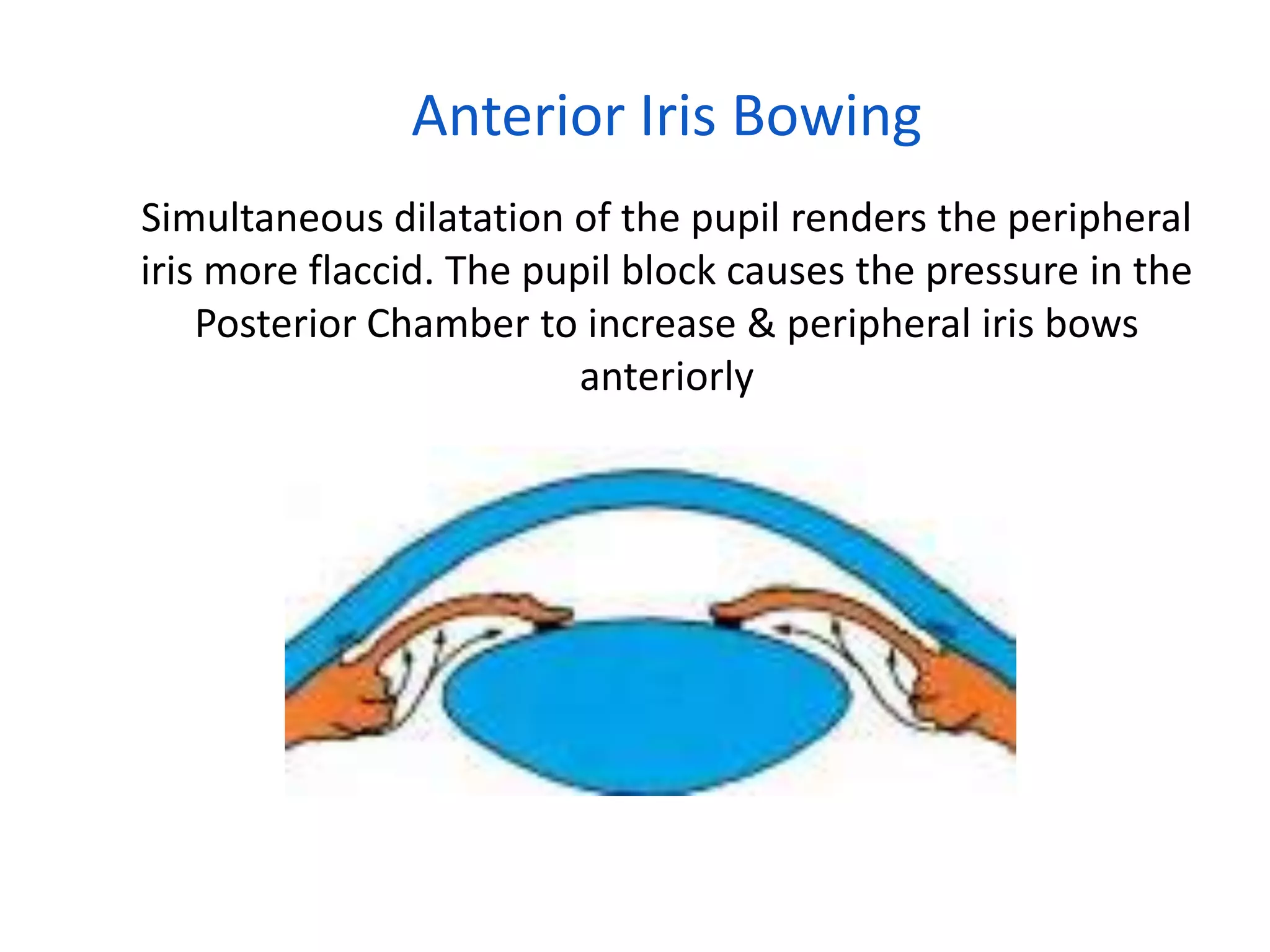
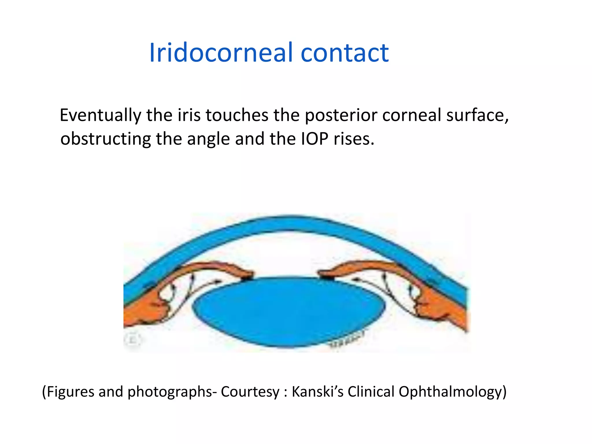
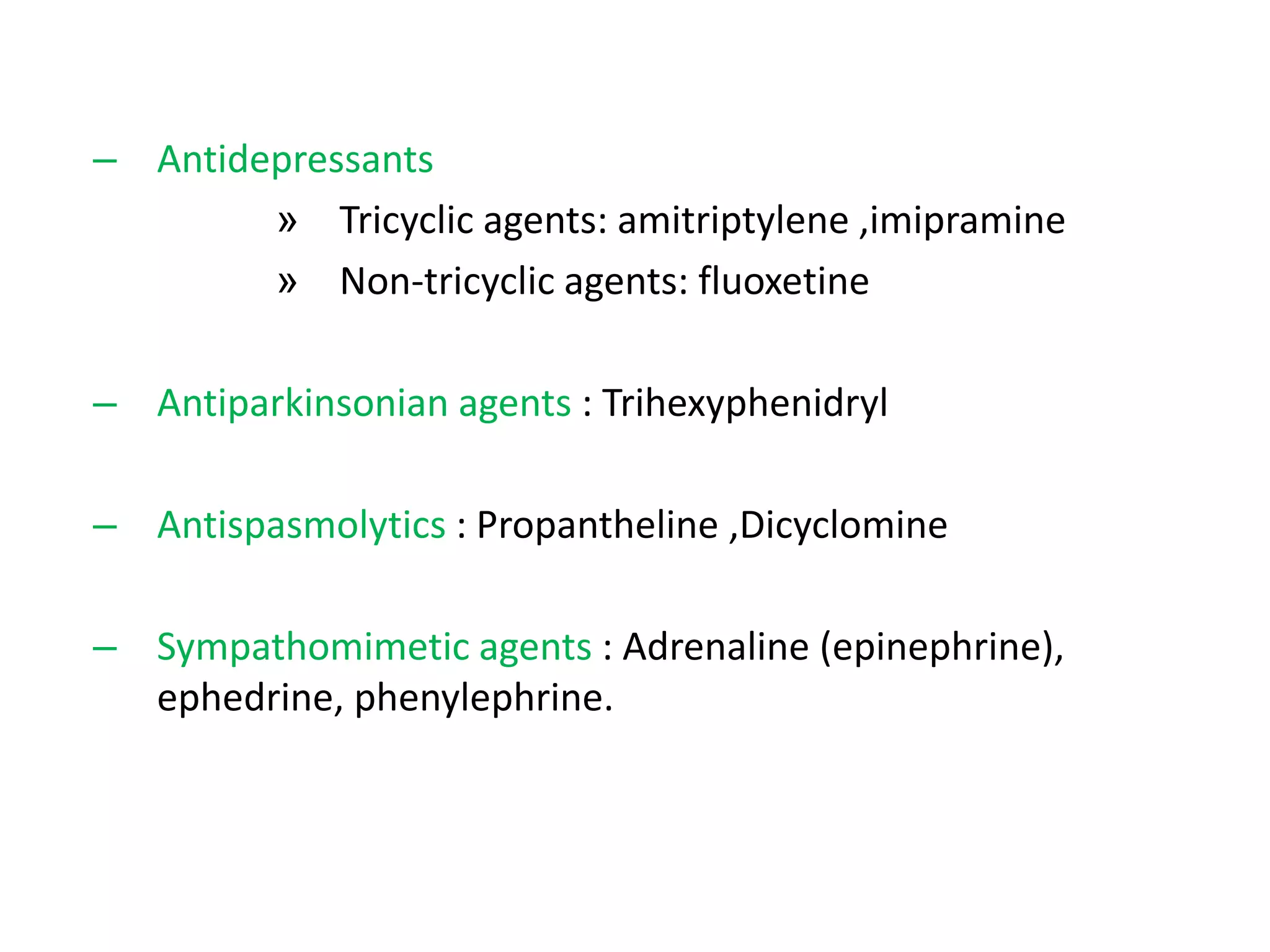
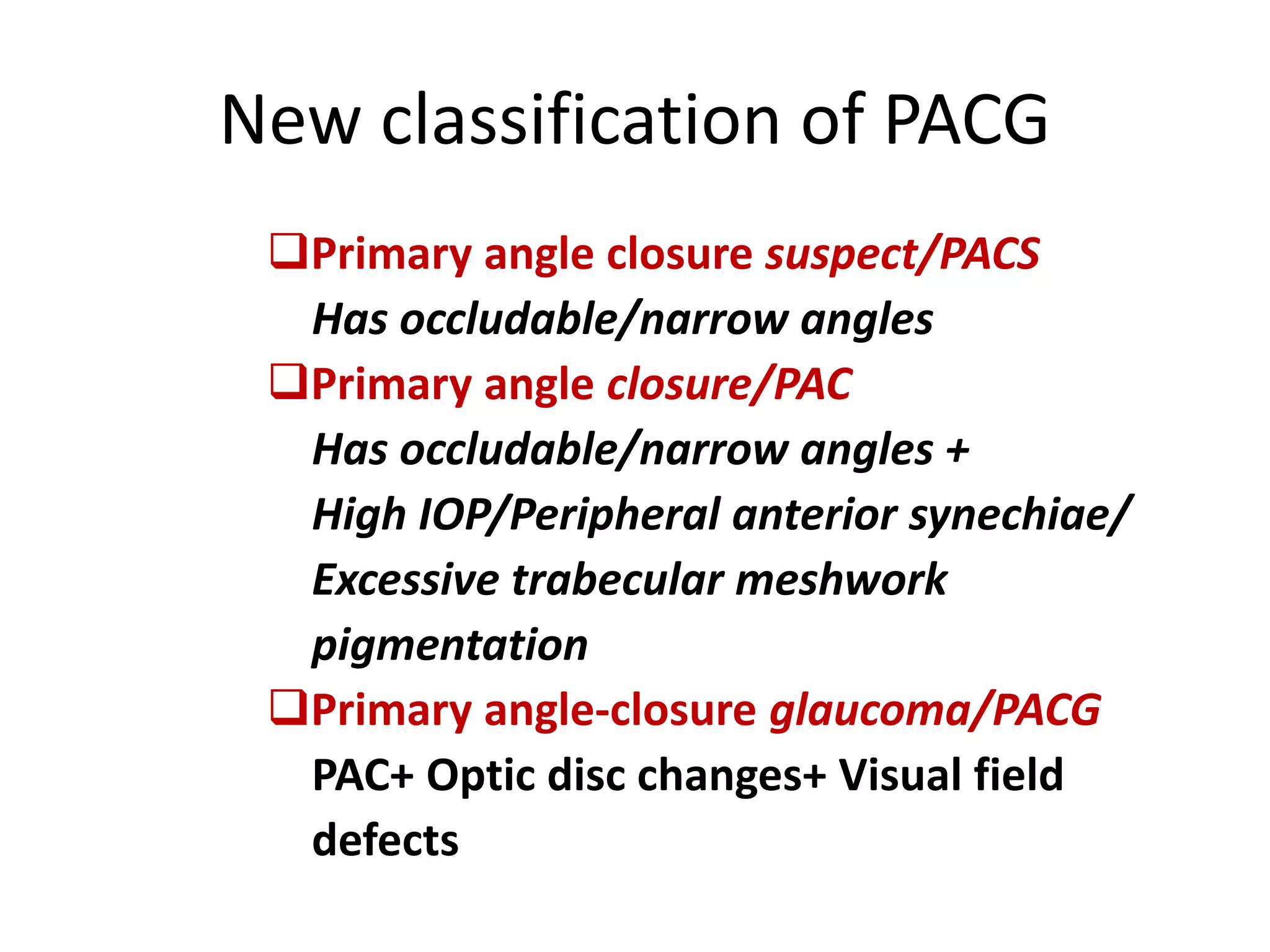
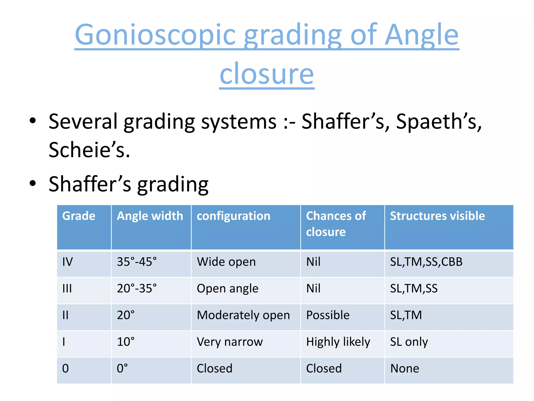
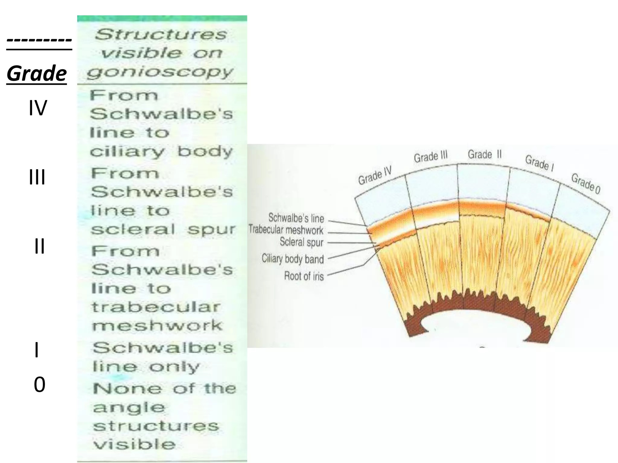
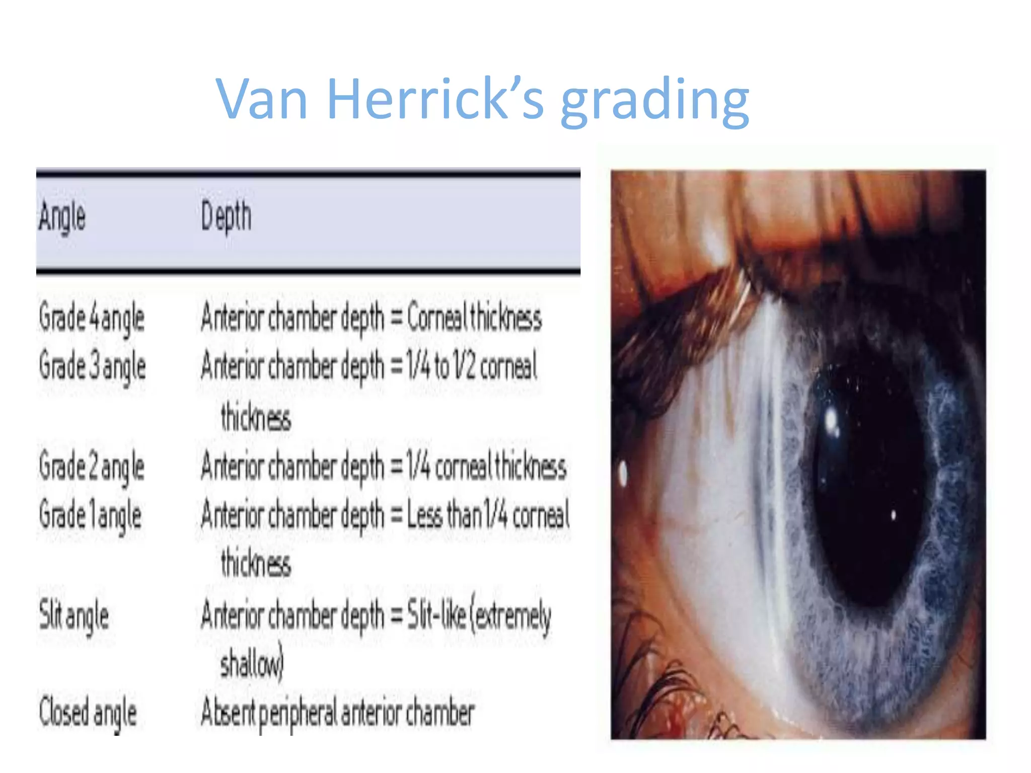
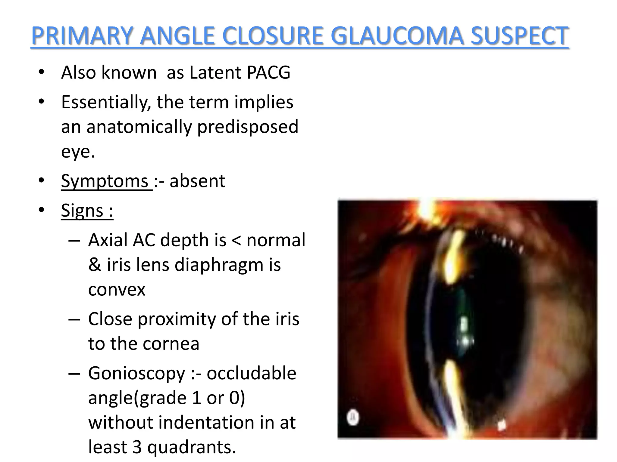
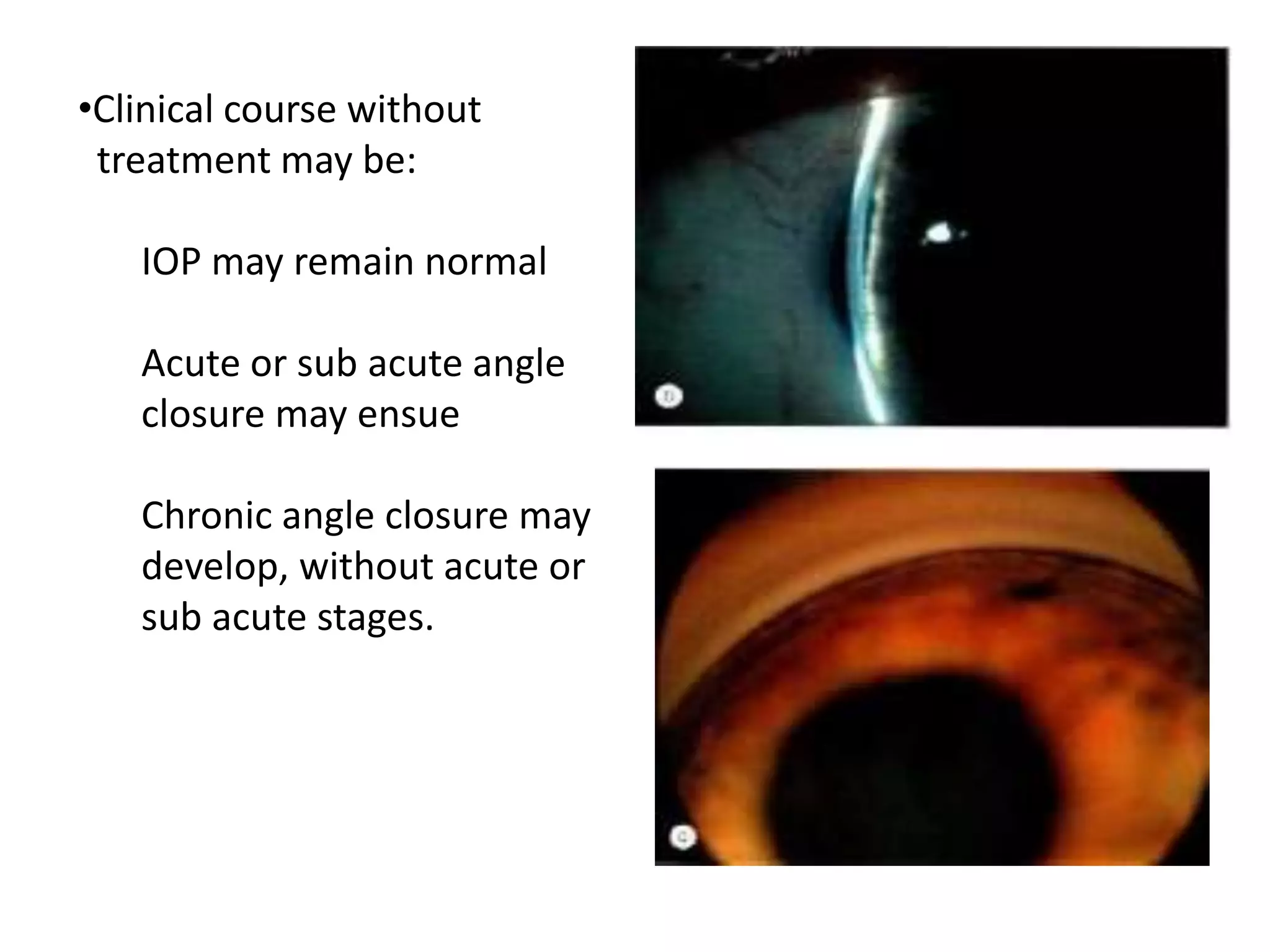
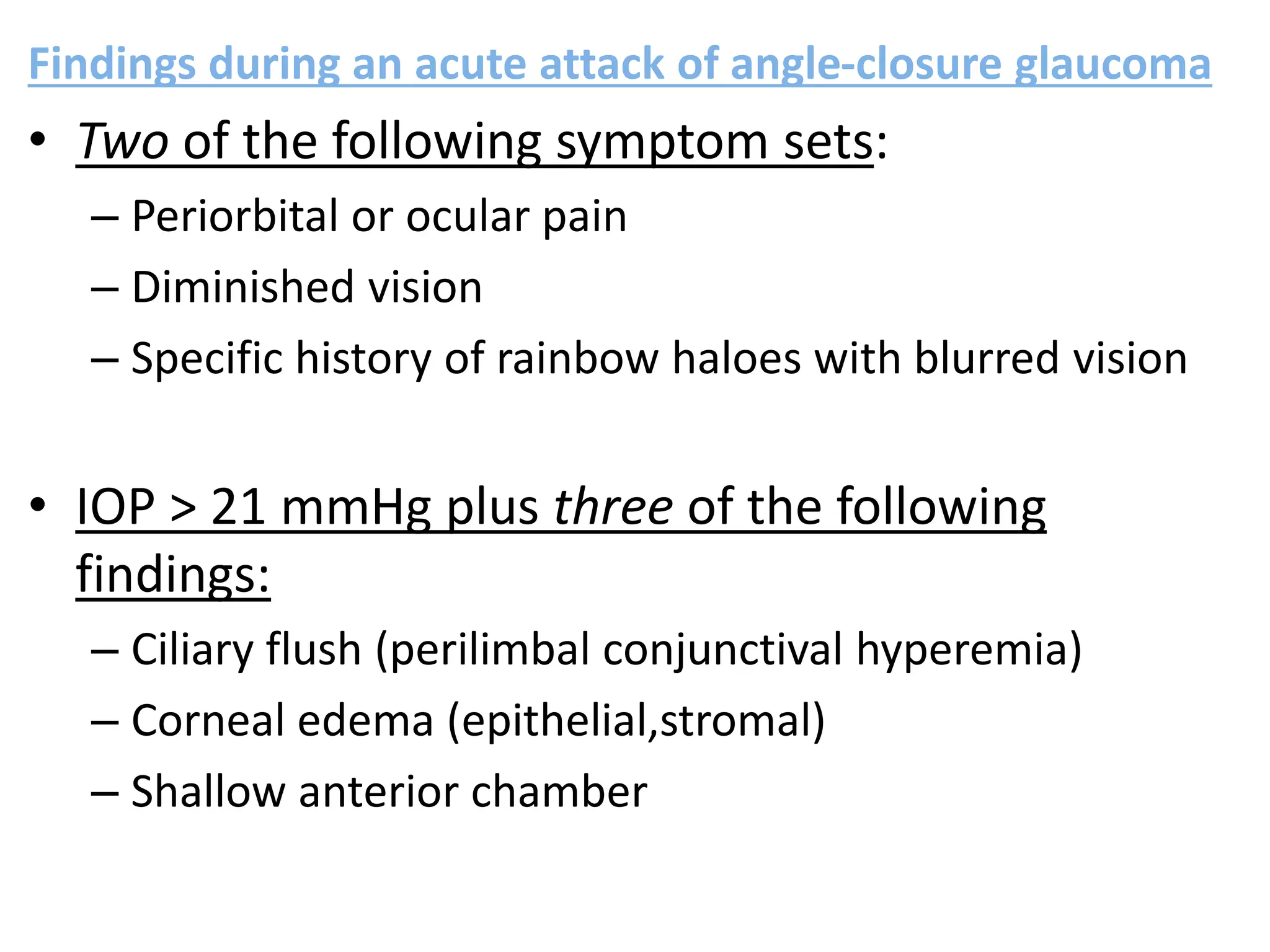
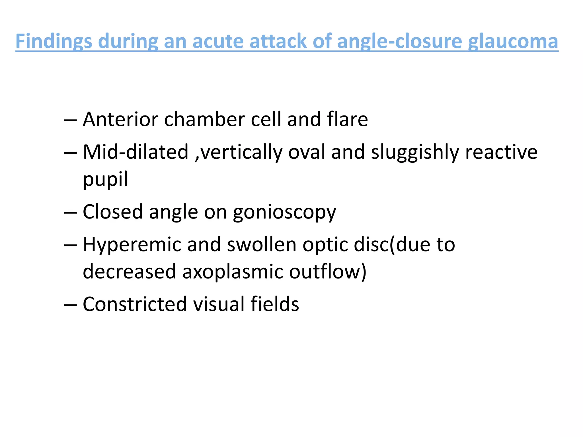
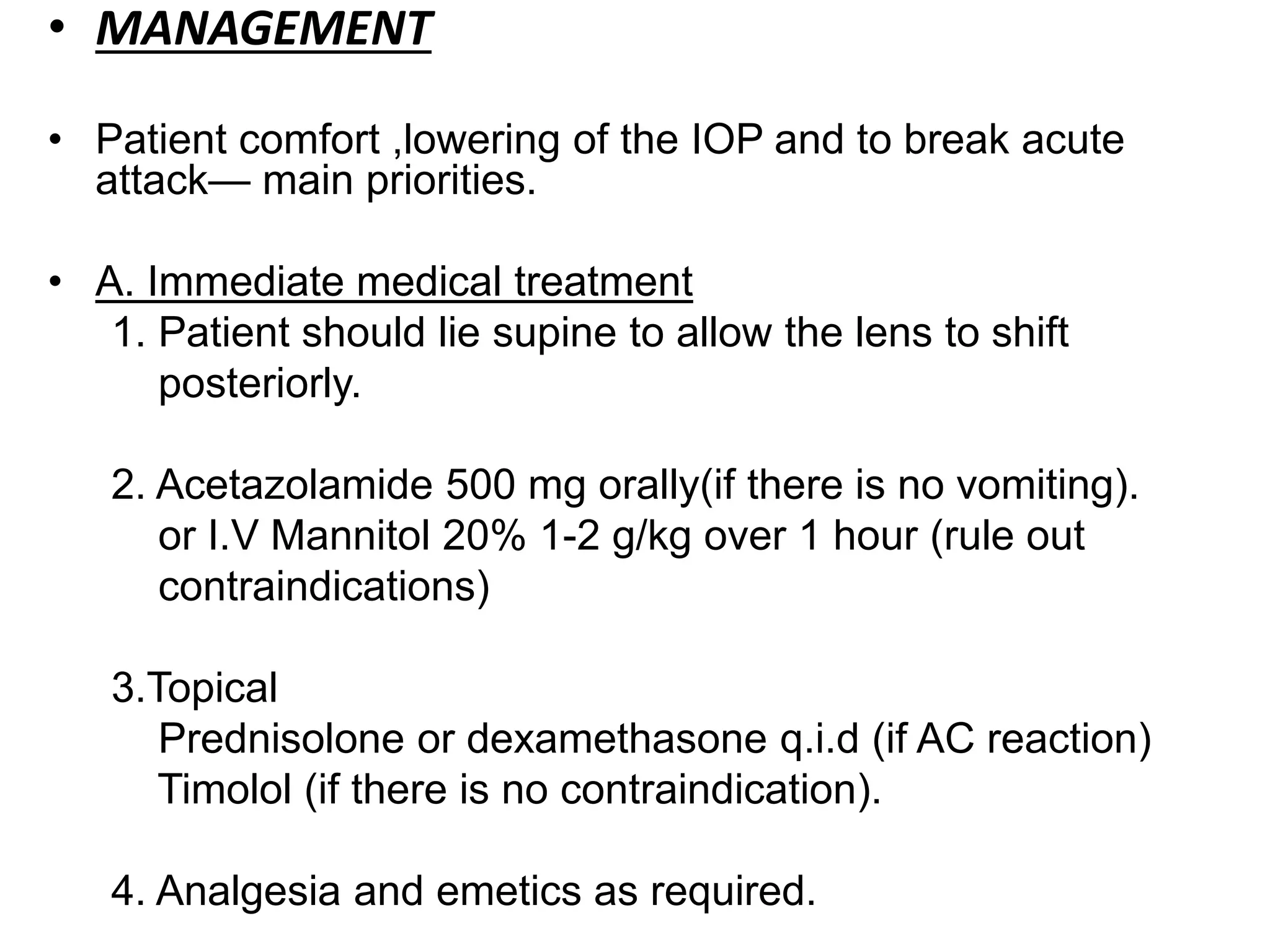
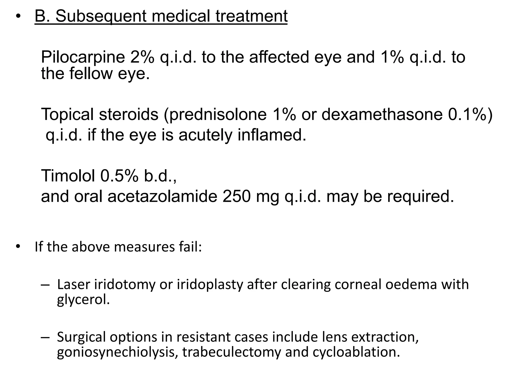
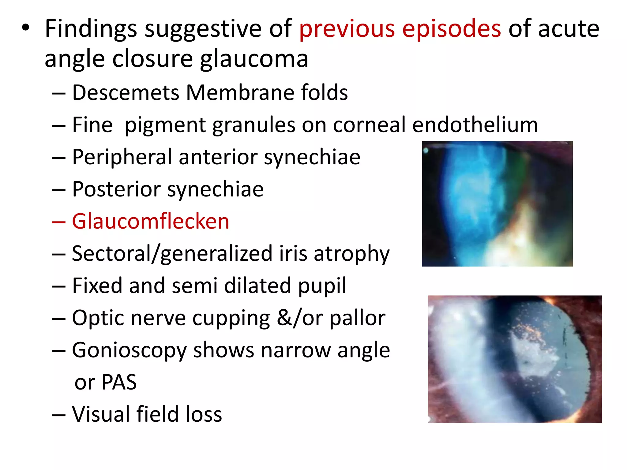
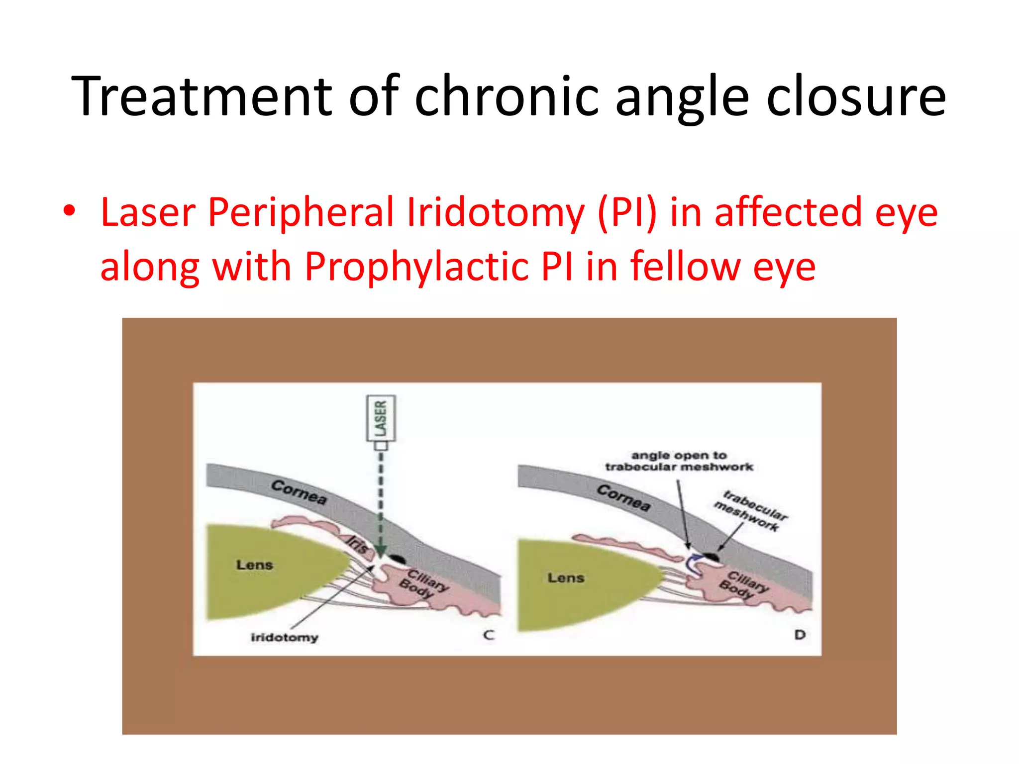
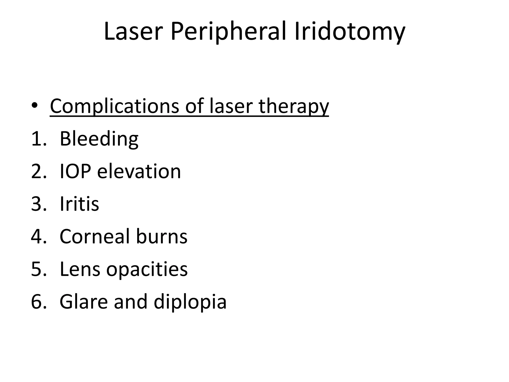
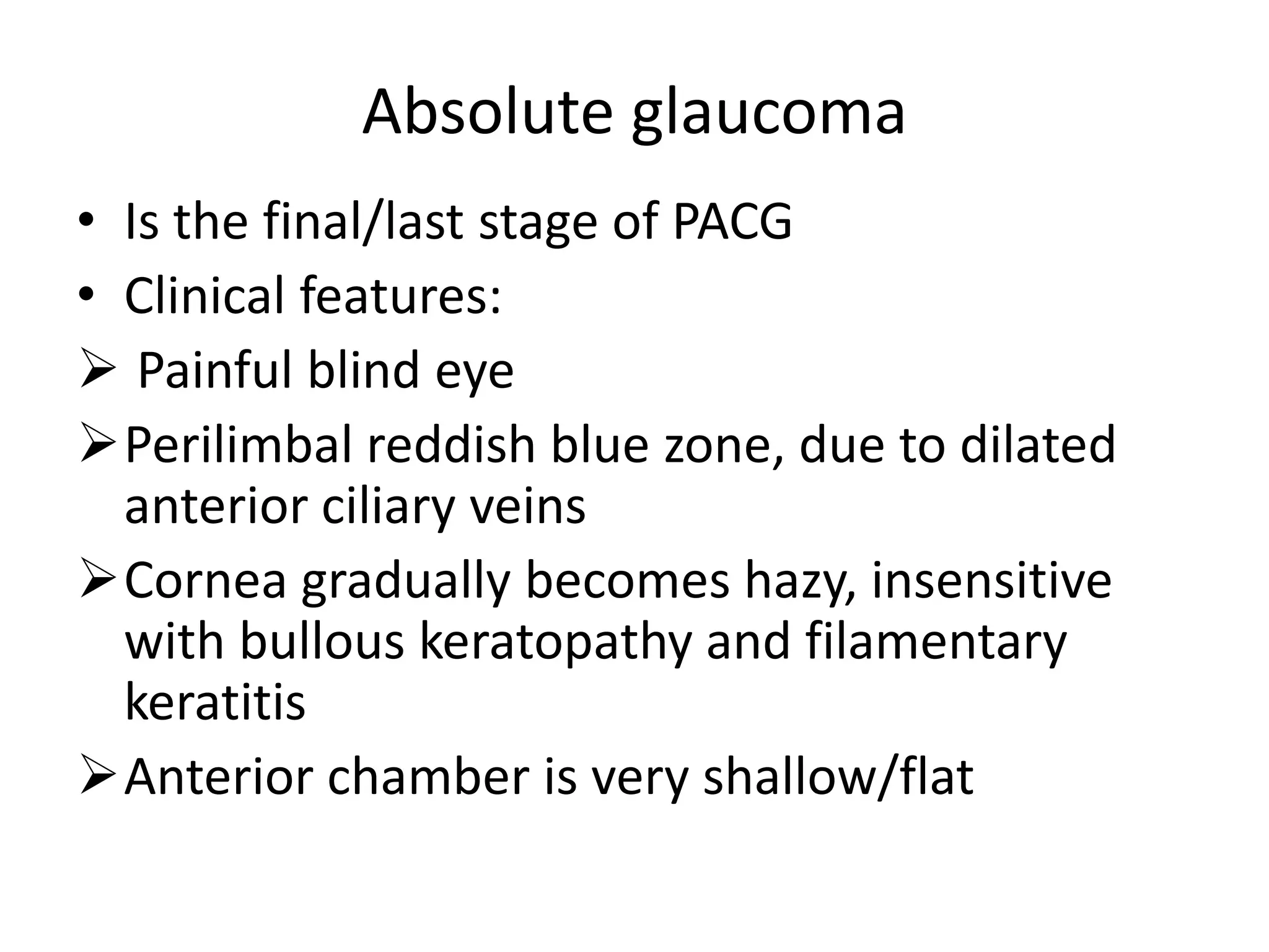
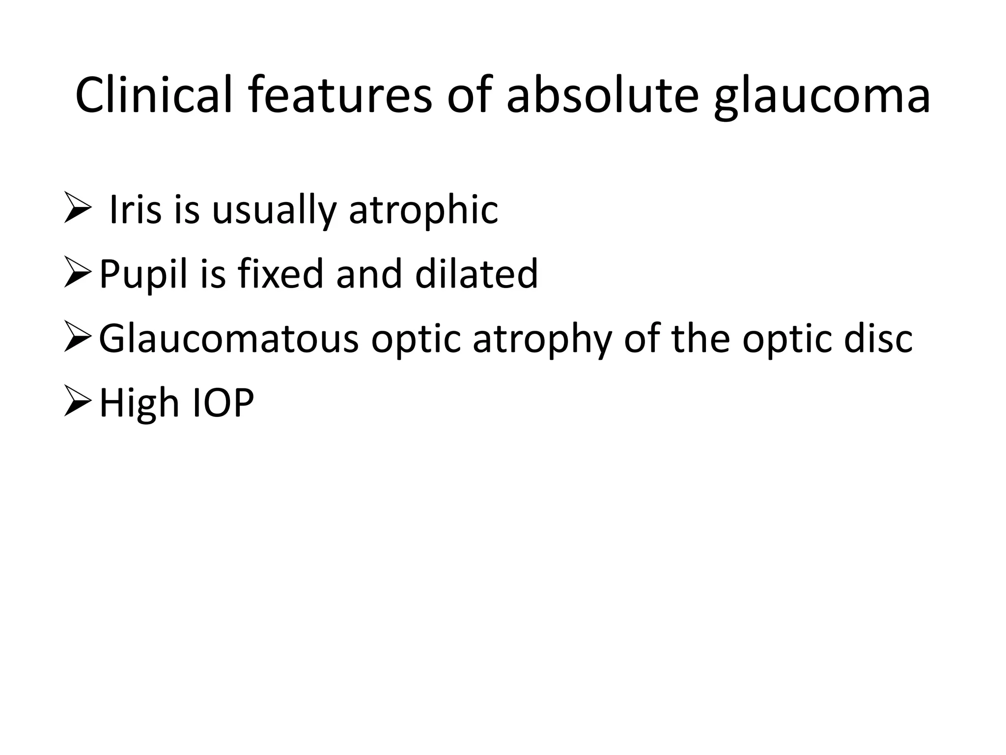
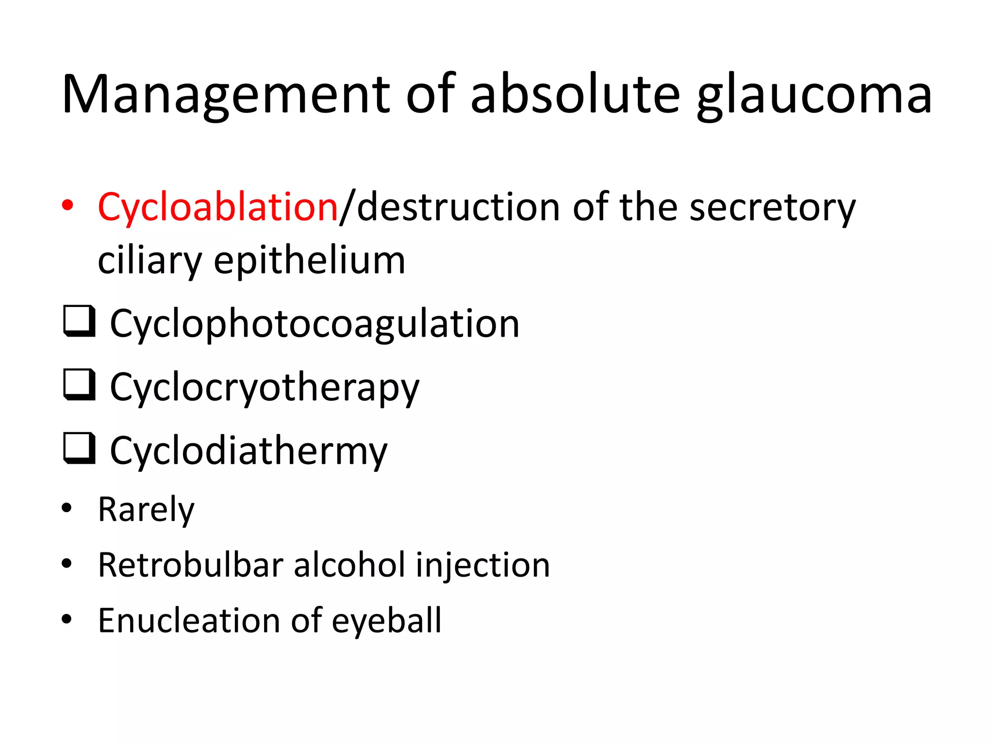
Dr.Ajai Agrawal
Associate Professor
Department of
Ophthalmology
AIIMS Rishikesh
Objectives
• At the end of this class the students shall be able
to :
• Define primary angle closure glaucoma.
• Understand the pathophysiology and the risk
factors.
• Be able to classify primary angle closure
glaucoma.
• Understand the fundamentals of managing
primary angle closure glaucoma
DEFINITION
• Primary angle closure glaucoma is a type of
primary glaucoma(with no obvious systemic or
ocular cause) characterized by
occludable/closed angles leading to
obstruction of aqueous outflow resulting in
rise of intra ocular pressure, optic nerve
damage and visual field defects.
ANGLE OF ANTERIOR CHAMBER
• STRUCTURES
– Schwalbe’s line
– Trabecular meshwork
– Scleral spur
– Ciliary body band
– Root of iris
DRAINAGE OF AQUEOUS HUMOR
PRIMARY ANGLE CLOSURE
GLAUCOMA
EPIDEMIOLOGY
• PACG is the major cause of glaucoma blindness
worldwide.
• Age :- Average age at presentation 50-60 yrs
• Gender :- F > M, 4 : 1
• Race :-seen commonly in South-East Asian
population, Chinese and Eskimos
• Heredity :- mostly sporadic but may be inherited
AD/AR
– first degree relatives are at increased risk.
• Refractive error :- more common in
hypermetropes
Ocular risk factors
1. Shallow anterior chamber both
centrally and peripherally.
2. Decreased anterior chamber volume.
3. Short axial length of the globe.
4. Small corneal diameter.
Ocular risk factors
5.Decreased posterior corneal radius of
curvature
6.Anterior position of the lens with respect to
the ciliary body.
7.Increased curvature of the anterior surface &
thickness of lens
PATHOGENESIS
• It is incompletely understood.
» a. Iris–pupil obstruction (e.g., ‘pupillary block’)
» b. Ciliary body anomalies (e.g., ‘plateau iris syndrome’)
» c. Lens–pupil block (e.g., ‘phacomorphic block’ (swollen
lens or microspherophakia))
• Relative Pupillary block
• Normally the pressure in the post. chamber exceeds that in the ant.
chamber due to physiological degree of resistance at the pupil ,since the
iris rests posteriorly on the anterior lens capsule.
Anterior Iris Bowing
Simultaneous dilatation of the pupil renders the peripheral
iris more flaccid. The pupil block causes the pressure in the
Posterior Chamber to increase & peripheral iris bows
anteriorly
Iridocorneal contact
Eventually the iris touches the posterior corneal surface,
obstructing the angle and the IOP rises.
(Figures and photographs- Courtesy : Kanski’s Clinical Ophthalmology)
Precipitating factors
1. Factors that produce mydriasis
• Dim illumination
• Emotional stress(due to increased sympathetic tone)
• Drugs
– Mydriatic agents :
• cyclopentolate, tropicamide, atropine, homatropine.
– Antipsychotic agents
» Phenothiazines: e.g., perphenazine ,fluphenazine
» Anticonvulsants e.g., Topiramate
– Antidepressants
» Tricyclic agents: amitriptylene ,imipramine
» Non-tricyclic agents: fluoxetine
– Antiparkinsonian agents : Trihexyphenidryl
– Antispasmolytics : Propantheline ,Dicyclomine
– Sympathomimetic agents : Adrenaline (epinephrine),
ephedrine, phenylephrine.
CLASSIFICATION
A. Primary angle-closure disease
• Irido-trabecular contact is the final common pathway of
angle closure disease, obstructing aqueous outflow
1. New classification
Primary angle closure suspect/PACS
Primary angle closure/PAC
Primary angle-closure glaucoma/PACG
2. Old classification
Angle closure suspect
Intermittent (sub acute) angle closure
Acute angle closure
Chronic angle closure
Absolute angle closure
New classification of PACG
Primary angle closure suspect/PACS
Has occludable/narrow angles
Primary angle closure/PAC
Has occludable/narrow angles +
High IOP/Peripheral anterior synechiae/
Excessive trabecular meshwork
pigmentation
Primary angle-closure glaucoma/PACG
PAC+ Optic disc changes+ Visual field
defects
Gonioscopic grading of Angle
closure
• Several grading systems :- Shaffer’s, Spaeth’s,
Scheie’s.
• Shaffer’s grading
Grade Angle width configuration Chances of
closure
Structures visible
IV 35°-45° Wide open Nil SL,TM,SS,CBB
III 20°-35° Open angle Nil SL,TM,SS
II 20° Moderately open Possible SL,TM
I 10° Very narrow Highly likely SL only
0 0° Closed Closed None
---------
Grade
IV
III
II
I
0
Van Herrick’s grading
Tests for Angle closure
• Eclipse test : uses flash light to make a rough
assessment of angle depth
• Provocative tests for PAC suspects
Prone- darkroom test: An increase in IOP of more
than 8mm Hg after one hour suggests PAC
Mydriatic provocative test: Not preferred now
Fincham’s Test: Also known as stenopaeic-slit test.
Glaucomatous halos remain intact , whereas
halos due to cataract are broken up into segments
PRIMARY ANGLE CLOSURE GLAUCOMA SUSPECT
• Also known as Latent PACG
• Essentially, the term implies
an anatomically predisposed
eye.
• Symptoms :- absent
• Signs :
– Axial AC depth is < normal
& iris lens diaphragm is
convex
– Close proximity of the iris
to the cornea
– Gonioscopy :- occludable
angle(grade 1 or 0)
without indentation in at
least 3 quadrants.
•Clinical course without
treatment may be:
IOP may remain normal
Acute or sub acute angle
closure may ensue
Chronic angle closure may
develop, without acute or
sub acute stages.
• Treatment
• Without treatment , risk of an acute pressure rise during the next
5 years is about 50 %.
• The need to treat is based on following criteria:-
– If one eye has had acute or subacute angle closure, then
fellow eye should undergo prophylactic peripheral laser
iridotomy (Laser PI)
– If both eyes have occludable angles, laser PI may be done
INTERMITTENT(SUBACUTE)PRIMARY ANGLE CLOSURE
GLAUCOMA
• A form of pupillary block glaucoma, which may not have
any recognizable symptoms.
• Occurs in a predisposed eye with an occludable angle in
association with intermittent pupillary block.
• Precipitating factors :- physiological mydriasis , or
physiological shallowing of AC when patient assumes a
prone or semi prone position ;emotional stress.
• Symptoms
– Characteristic h/o transient blurring of vision with haloes
around lights
– Ocular discomfort or frontal headache
– Attacks are recurrent and are usually broken after 1-2 hrs by
physiological miosis.
• Signs
– During an attack , eye is usually white
– In between attacks, eye looks normal although the angle is
narrow.
• Clinical course
– Without treatment is variable
• Some eyes develop an acute attack
• Others chronic angle closure
• Treatment:- Prophylactic laser PeripheraI Iridotomy(PI)
ACUTE PRIMARY ANGLE CLOSURE GLAUCOMA
• Sight threatening emergency
• Painful loss of vision due to sudden and total
closure of the angle.
• VA usually 6/60-Hand Movements.
• IOP is usually very high (40–70 mmHg).
Findings during an acute attack of angle-closure glaucoma
• Two of the following symptom sets:
– Periorbital or ocular pain
– Diminished vision
– Specific history of rainbow haloes with blurred vision
• IOP > 21 mmHg plus three of the following
findings:
– Ciliary flush (perilimbal conjunctival hyperemia)
– Corneal edema (epithelial,stromal)
– Shallow anterior chamber
Findings during an acute attack of angle-closure glaucoma
– Anterior chamber cell and flare
– Mid-dilated ,vertically oval and sluggishly reactive
pupil
– Closed angle on gonioscopy
– Hyperemic and swollen optic disc(due to
decreased axoplasmic outflow)
– Constricted visual fields
• MANAGEMENT
• Patient comfort ,lowering of the IOP and to break acute
attack— main priorities.
• A. Immediate medical treatment
1. Patient should lie supine to allow the lens to shift
posteriorly.
2. Acetazolamide 500 mg orally(if there is no vomiting).
or I.V Mannitol 20% 1-2 g/kg over 1 hour (rule out
contraindications)
3.Topical
Prednisolone or dexamethasone q.i.d (if AC reaction)
Timolol (if there is no contraindication).
4. Analgesia and emetics as required.
• B. Subsequent medical treatment
Pilocarpine 2% q.i.d. to the affected eye and 1% q.i.d. to
the fellow eye.
Topical steroids (prednisolone 1% or dexamethasone 0.1%)
q.i.d. if the eye is acutely inflamed.
Timolol 0.5% b.d.,
and oral acetazolamide 250 mg q.i.d. may be required.
• If the above measures fail:
– Laser iridotomy or iridoplasty after clearing corneal oedema with
glycerol.
– Surgical options in resistant cases include lens extraction,
goniosynechiolysis, trabeculectomy and cycloablation.
• Findings suggestive of previous episodes of acute
angle closure glaucoma
– Descemets Membrane folds
– Fine pigment granules on corneal endothelium
– Peripheral anterior synechiae
– Posterior synechiae
– Glaucomflecken
– Sectoral/generalized iris atrophy
– Fixed and semi dilated pupil
– Optic nerve cupping &/or pallor
– Gonioscopy shows narrow angle
or PAS
– Visual field loss
Chronic angle closure glaucoma
• Visual Acuity is normal unless damage is
advanced.
• Anterior chamber is shallower in pupillary
block than non-pupillary block.
• Optic nerve signs depend on severity of
damage.
• IOP elevation may be only intermittent.
• Gonioscopic abnormalities-Peripheral
Anterior Synechiae, narrow angle,
pigmentation of Schwalbe’s line.
Treatment of chronic angle closure
• Medical treatment is similar to that of POAG
• Prostaglandin/Prostamides
Latanoprost, Bimatoprost, Travoprost
• Beta blockers
Timolol maleate, Betaxolol
• Carbonic anhydrase inhibitors
Dorzolamide, Brinzolamide
• Sympathomimetics
Brimonidine, Apraclonidine
• Parasmpathomimetics
Pilocarpine
• Oral carbonic anhydrase inhibitors
Acetazolamide, Methazolamide
Treatment of chronic angle closure
• Laser Peripheral Iridotomy (PI) in affected eye
along with Prophylactic PI in fellow eye
Laser Peripheral Iridotomy
• Complications of laser therapy
1. Bleeding
2. IOP elevation
3. Iritis
4. Corneal burns
5. Lens opacities
6. Glare and diplopia
• Surgical treatment
Trabeculectomy (filtering surgery) is the
surgical procedure of choice
• Success:- 87- 100 % with multiple operations
• Complications:-
– Flat AC, hypotony
– Bleb related infections
– Cyclodialysis
• PATIENTS REQUIRE REGULAR AND LIFE LONG
FOLLOW UP
Absolute glaucoma
• Is the final/last stage of PACG
• Clinical features:
 Painful blind eye
Perilimbal reddish blue zone, due to dilated
anterior ciliary veins
Cornea gradually becomes hazy, insensitive
with bullous keratopathy and filamentary
keratitis
Anterior chamber is very shallow/flat
Clinical features of absolute glaucoma
 Iris is usually atrophic
Pupil is fixed and dilated
Glaucomatous optic atrophy of the optic disc
High IOP
Management of absolute glaucoma
• Cycloablation/destruction of the secretory
ciliary epithelium
 Cyclophotocoagulation
 Cyclocryotherapy
 Cyclodiathermy
• Rarely
• Retrobulbar alcohol injection
• Enucleation of eyeball
Complications
• Corneal ulceration
• Staphyloma formation (Ciliary/Equatorial)
• Atrophic bulbi (Shrunken eye)
Conclusion
• Primary angle closure glaucoma is a
potentially sight threatening condition,
characterized by occludable anterior chamber
angles.
• Obstruction of aqueous outflow results in rise
of intra ocular pressure, optic nerve damage
and visual field defects.
• Management may include medical, laser
and/or surgical modalities.
acinal.pptx

acinal.pptx

  • 1.
    Dr.Ajai Agrawal Associate Professor Departmentof Ophthalmology AIIMS Rishikesh
  • 2.
    Objectives • At theend of this class the students shall be able to : • Define primary angle closure glaucoma. • Understand the pathophysiology and the risk factors. • Be able to classify primary angle closure glaucoma. • Understand the fundamentals of managing primary angle closure glaucoma
  • 3.
    DEFINITION • Primary angleclosure glaucoma is a type of primary glaucoma(with no obvious systemic or ocular cause) characterized by occludable/closed angles leading to obstruction of aqueous outflow resulting in rise of intra ocular pressure, optic nerve damage and visual field defects.
  • 4.
    ANGLE OF ANTERIORCHAMBER • STRUCTURES – Schwalbe’s line – Trabecular meshwork – Scleral spur – Ciliary body band – Root of iris
  • 5.
  • 6.
    PRIMARY ANGLE CLOSURE GLAUCOMA EPIDEMIOLOGY •PACG is the major cause of glaucoma blindness worldwide. • Age :- Average age at presentation 50-60 yrs • Gender :- F > M, 4 : 1 • Race :-seen commonly in South-East Asian population, Chinese and Eskimos • Heredity :- mostly sporadic but may be inherited AD/AR – first degree relatives are at increased risk. • Refractive error :- more common in hypermetropes
  • 7.
    Ocular risk factors 1.Shallow anterior chamber both centrally and peripherally. 2. Decreased anterior chamber volume. 3. Short axial length of the globe. 4. Small corneal diameter.
  • 8.
    Ocular risk factors 5.Decreasedposterior corneal radius of curvature 6.Anterior position of the lens with respect to the ciliary body. 7.Increased curvature of the anterior surface & thickness of lens
  • 9.
    PATHOGENESIS • It isincompletely understood. » a. Iris–pupil obstruction (e.g., ‘pupillary block’) » b. Ciliary body anomalies (e.g., ‘plateau iris syndrome’) » c. Lens–pupil block (e.g., ‘phacomorphic block’ (swollen lens or microspherophakia)) • Relative Pupillary block • Normally the pressure in the post. chamber exceeds that in the ant. chamber due to physiological degree of resistance at the pupil ,since the iris rests posteriorly on the anterior lens capsule.
  • 10.
    Anterior Iris Bowing Simultaneousdilatation of the pupil renders the peripheral iris more flaccid. The pupil block causes the pressure in the Posterior Chamber to increase & peripheral iris bows anteriorly
  • 11.
    Iridocorneal contact Eventually theiris touches the posterior corneal surface, obstructing the angle and the IOP rises. (Figures and photographs- Courtesy : Kanski’s Clinical Ophthalmology)
  • 12.
    Precipitating factors 1. Factorsthat produce mydriasis • Dim illumination • Emotional stress(due to increased sympathetic tone) • Drugs – Mydriatic agents : • cyclopentolate, tropicamide, atropine, homatropine. – Antipsychotic agents » Phenothiazines: e.g., perphenazine ,fluphenazine » Anticonvulsants e.g., Topiramate
  • 13.
    – Antidepressants » Tricyclicagents: amitriptylene ,imipramine » Non-tricyclic agents: fluoxetine – Antiparkinsonian agents : Trihexyphenidryl – Antispasmolytics : Propantheline ,Dicyclomine – Sympathomimetic agents : Adrenaline (epinephrine), ephedrine, phenylephrine.
  • 14.
    CLASSIFICATION A. Primary angle-closuredisease • Irido-trabecular contact is the final common pathway of angle closure disease, obstructing aqueous outflow 1. New classification Primary angle closure suspect/PACS Primary angle closure/PAC Primary angle-closure glaucoma/PACG 2. Old classification Angle closure suspect Intermittent (sub acute) angle closure Acute angle closure Chronic angle closure Absolute angle closure
  • 15.
    New classification ofPACG Primary angle closure suspect/PACS Has occludable/narrow angles Primary angle closure/PAC Has occludable/narrow angles + High IOP/Peripheral anterior synechiae/ Excessive trabecular meshwork pigmentation Primary angle-closure glaucoma/PACG PAC+ Optic disc changes+ Visual field defects
  • 16.
    Gonioscopic grading ofAngle closure • Several grading systems :- Shaffer’s, Spaeth’s, Scheie’s. • Shaffer’s grading Grade Angle width configuration Chances of closure Structures visible IV 35°-45° Wide open Nil SL,TM,SS,CBB III 20°-35° Open angle Nil SL,TM,SS II 20° Moderately open Possible SL,TM I 10° Very narrow Highly likely SL only 0 0° Closed Closed None
  • 17.
  • 18.
  • 19.
    Tests for Angleclosure • Eclipse test : uses flash light to make a rough assessment of angle depth • Provocative tests for PAC suspects Prone- darkroom test: An increase in IOP of more than 8mm Hg after one hour suggests PAC Mydriatic provocative test: Not preferred now Fincham’s Test: Also known as stenopaeic-slit test. Glaucomatous halos remain intact , whereas halos due to cataract are broken up into segments
  • 20.
    PRIMARY ANGLE CLOSUREGLAUCOMA SUSPECT • Also known as Latent PACG • Essentially, the term implies an anatomically predisposed eye. • Symptoms :- absent • Signs : – Axial AC depth is < normal & iris lens diaphragm is convex – Close proximity of the iris to the cornea – Gonioscopy :- occludable angle(grade 1 or 0) without indentation in at least 3 quadrants.
  • 21.
    •Clinical course without treatmentmay be: IOP may remain normal Acute or sub acute angle closure may ensue Chronic angle closure may develop, without acute or sub acute stages.
  • 22.
    • Treatment • Withouttreatment , risk of an acute pressure rise during the next 5 years is about 50 %. • The need to treat is based on following criteria:- – If one eye has had acute or subacute angle closure, then fellow eye should undergo prophylactic peripheral laser iridotomy (Laser PI) – If both eyes have occludable angles, laser PI may be done
  • 23.
    INTERMITTENT(SUBACUTE)PRIMARY ANGLE CLOSURE GLAUCOMA •A form of pupillary block glaucoma, which may not have any recognizable symptoms. • Occurs in a predisposed eye with an occludable angle in association with intermittent pupillary block. • Precipitating factors :- physiological mydriasis , or physiological shallowing of AC when patient assumes a prone or semi prone position ;emotional stress.
  • 24.
    • Symptoms – Characteristich/o transient blurring of vision with haloes around lights – Ocular discomfort or frontal headache – Attacks are recurrent and are usually broken after 1-2 hrs by physiological miosis. • Signs – During an attack , eye is usually white – In between attacks, eye looks normal although the angle is narrow. • Clinical course – Without treatment is variable • Some eyes develop an acute attack • Others chronic angle closure • Treatment:- Prophylactic laser PeripheraI Iridotomy(PI)
  • 25.
    ACUTE PRIMARY ANGLECLOSURE GLAUCOMA • Sight threatening emergency • Painful loss of vision due to sudden and total closure of the angle. • VA usually 6/60-Hand Movements. • IOP is usually very high (40–70 mmHg).
  • 26.
    Findings during anacute attack of angle-closure glaucoma • Two of the following symptom sets: – Periorbital or ocular pain – Diminished vision – Specific history of rainbow haloes with blurred vision • IOP > 21 mmHg plus three of the following findings: – Ciliary flush (perilimbal conjunctival hyperemia) – Corneal edema (epithelial,stromal) – Shallow anterior chamber
  • 27.
    Findings during anacute attack of angle-closure glaucoma – Anterior chamber cell and flare – Mid-dilated ,vertically oval and sluggishly reactive pupil – Closed angle on gonioscopy – Hyperemic and swollen optic disc(due to decreased axoplasmic outflow) – Constricted visual fields
  • 28.
    • MANAGEMENT • Patientcomfort ,lowering of the IOP and to break acute attack— main priorities. • A. Immediate medical treatment 1. Patient should lie supine to allow the lens to shift posteriorly. 2. Acetazolamide 500 mg orally(if there is no vomiting). or I.V Mannitol 20% 1-2 g/kg over 1 hour (rule out contraindications) 3.Topical Prednisolone or dexamethasone q.i.d (if AC reaction) Timolol (if there is no contraindication). 4. Analgesia and emetics as required.
  • 29.
    • B. Subsequentmedical treatment Pilocarpine 2% q.i.d. to the affected eye and 1% q.i.d. to the fellow eye. Topical steroids (prednisolone 1% or dexamethasone 0.1%) q.i.d. if the eye is acutely inflamed. Timolol 0.5% b.d., and oral acetazolamide 250 mg q.i.d. may be required. • If the above measures fail: – Laser iridotomy or iridoplasty after clearing corneal oedema with glycerol. – Surgical options in resistant cases include lens extraction, goniosynechiolysis, trabeculectomy and cycloablation.
  • 30.
    • Findings suggestiveof previous episodes of acute angle closure glaucoma – Descemets Membrane folds – Fine pigment granules on corneal endothelium – Peripheral anterior synechiae – Posterior synechiae – Glaucomflecken – Sectoral/generalized iris atrophy – Fixed and semi dilated pupil – Optic nerve cupping &/or pallor – Gonioscopy shows narrow angle or PAS – Visual field loss
  • 31.
    Chronic angle closureglaucoma • Visual Acuity is normal unless damage is advanced. • Anterior chamber is shallower in pupillary block than non-pupillary block. • Optic nerve signs depend on severity of damage. • IOP elevation may be only intermittent. • Gonioscopic abnormalities-Peripheral Anterior Synechiae, narrow angle, pigmentation of Schwalbe’s line.
  • 32.
    Treatment of chronicangle closure • Medical treatment is similar to that of POAG • Prostaglandin/Prostamides Latanoprost, Bimatoprost, Travoprost • Beta blockers Timolol maleate, Betaxolol • Carbonic anhydrase inhibitors Dorzolamide, Brinzolamide • Sympathomimetics Brimonidine, Apraclonidine • Parasmpathomimetics Pilocarpine • Oral carbonic anhydrase inhibitors Acetazolamide, Methazolamide
  • 33.
    Treatment of chronicangle closure • Laser Peripheral Iridotomy (PI) in affected eye along with Prophylactic PI in fellow eye
  • 34.
    Laser Peripheral Iridotomy •Complications of laser therapy 1. Bleeding 2. IOP elevation 3. Iritis 4. Corneal burns 5. Lens opacities 6. Glare and diplopia
  • 35.
    • Surgical treatment Trabeculectomy(filtering surgery) is the surgical procedure of choice • Success:- 87- 100 % with multiple operations • Complications:- – Flat AC, hypotony – Bleb related infections – Cyclodialysis • PATIENTS REQUIRE REGULAR AND LIFE LONG FOLLOW UP
  • 36.
    Absolute glaucoma • Isthe final/last stage of PACG • Clinical features:  Painful blind eye Perilimbal reddish blue zone, due to dilated anterior ciliary veins Cornea gradually becomes hazy, insensitive with bullous keratopathy and filamentary keratitis Anterior chamber is very shallow/flat
  • 37.
    Clinical features ofabsolute glaucoma  Iris is usually atrophic Pupil is fixed and dilated Glaucomatous optic atrophy of the optic disc High IOP
  • 38.
    Management of absoluteglaucoma • Cycloablation/destruction of the secretory ciliary epithelium  Cyclophotocoagulation  Cyclocryotherapy  Cyclodiathermy • Rarely • Retrobulbar alcohol injection • Enucleation of eyeball
  • 39.
    Complications • Corneal ulceration •Staphyloma formation (Ciliary/Equatorial) • Atrophic bulbi (Shrunken eye)
  • 40.
    Conclusion • Primary angleclosure glaucoma is a potentially sight threatening condition, characterized by occludable anterior chamber angles. • Obstruction of aqueous outflow results in rise of intra ocular pressure, optic nerve damage and visual field defects. • Management may include medical, laser and/or surgical modalities.

Editor's Notes

  • #7 1st-10 Sibling 4