SlideShare a Scribd company logo
Dr/ ABD ALLAH NAZEER. MD.
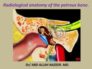
Radiological anatomy of the petrous bone.
Normal Anatomy of the Middle Ear
The temporal bone houses and is surrounded by many vital structures.
The temporal bone is actually composed of four bones, consisting of the
squamous, petrous, tympanic, and mastoid segments.
The bony framework of the temporal bone contains multiple air spaces.
The most complex of these spaces is the middle ear cavity, or tympanum.
The middle ear space is shaped somewhat in the form of a red blood cell
stood on end. This space is narrow in a medial-lateral direction and more
elongated anteroposteriorly and superoinferiorly.
Laterally, the tympanic membrane, annulus, and the handle of the malleus
make up the wall of the tympanum. The inner ear forms the medial wall of
the middle ear. The largest anatomic structure of the medial wall is the
promontory of the cochlea.
The roof of the middle ear space is formed by the tegmen tympani. This
structure separates the middle ear space from the middle cranial fossa.
The tegmen slopes inferiorly as it courses laterally along the temporal
bone; remember this point during mastoidectomy to avoid violating the
tegmen with the drill. In addition, the tegmen is located higher than the
superior border of the tympanic membrane, forming a space, the
epitympanum. The epitympanum houses the head of the malleus and the
body and short process of the incus.
The floor of the middle ear is primarily composed of the bone covering the
jugular bulb. The bone over the bulb may be dehiscent, rendering it more
susceptible to injury. The floor of the middle ear cavity is located further
inferiorly than the lowest extent of the tympanic membrane, creating a
space, the hypotympanum.
Anteriorly and inferiorly, the carotid artery limits the tympanum. More
superiorly, the eustachian tube, tensor tympani, and cochleariform
process can be observed along the anterior margin of the middle ear.
The mastoid air cells lie immediately posterior to the middle ear space.
The entrance to the mastoid air cells is the aditus ad antrum. The
pyramidal eminence (giving rise to the stapedius tendon) and the incudal
fossa can also be observed immediately posterior to the middle ear space.
The middle ear space contains several spaces of clinical significance. The
sinus tympani is located between the labyrinthine wall and the pyramidal
eminence. This area is a common site for recurrence of cholesteatoma.
The facial recess is found between the tympanic annulus and pyramidal
eminence. This recess provides improved access to the middle ear space
during a tympanomastoidectomy.
Normal Anatomy of the Inner Ear
The inner ear is housed in the bony labyrinth, which is well
demonstrated on CT scans. The cochlea lies anteriorly. The cochlea is
a conical structure, with its apex pointed anteriorly, inferiorly, and
laterally; its base rests near the internal auditory canal and extends
outward for 2.5-2.75 turns. Immediately anterior to the cochlea is
the carotid artery. The round window is located in the scala tympani
of the basal turn of the cochlea. The round window niche houses the
round window. This window is the termination of the scala tympani
of the cochlea. The niche protects the round window from direct
exposure to sound waves in the event of a tympanic membrane
perforation.
The vestibule lies posterior to the cochlea, abutting the internal
auditory canal medially. The stapes footplate transmits vibrations to
the vestibule at the oval window. The 3 semicircular canals emanate
from the vestibule. The lateral canal lies 30° from horizontal. The 3
canals lie at right angles to each other.
The endolymphatic sac is posteromedial to the semicircular
canals on the posterior margin of the petrous bone. The
seventh and eighth cranial nerves (CN VII and CN VIII) course
through the internal auditory canal. CN VIII enters the
structures of the inner ear to innervate them and CN VII passes
laterally and anteriorly to the geniculate ganglion, then
posteriorly along the medial wall of the tympanum before
heading inferiorly to the stylomastoid foramen.
A retrospective CT-scan study by Saxby et al found the rate of
semicircular canal dehiscence in pediatric patients to be
significantly lower than rates found in previous studies. The
study involved 334 children (649 temporal bones), with
temporal bone imaging revealing superior canal dehiscence in
3.3% of patients (1.7% of temporal bones) and posterior canal
dehiscence in 2.1% of patients (1.2% of temporal bones).
Radiographic positions of mastoids
The mastoid process is a part of the temporal bone which is also comprised of tympanic,
petrous and squamous parts. Accordingly, examination of the mastoid can be possible
using the following projections:
Law view: The X-ray beam is directed at a 15 degree oblique plain cephalocaudally while
the skull's sagittal plane is parallel to the X-ray film.
Law view: The X-ray beam is directed at a 15 degree oblique plain
cephalocaudally while the skull's sagittal plane is parallel to the X-ray film.
Stenver's view: It is also called an Axio-anterior oblique posterior view. The X-ray
beam is directed at a 14 degree angle caudally and the head faces the film with
slight flexion and rotation at an angle of 45 degrees to the opposite side.
Transorbital view: (posterioanterior and anteroposterior): The X-ray
beam is directed either postero anteriorly or anteroposteriorly along
the orbito-meatal line at a 90 degree angle to the film.
Town's view: It is an anteroposterior view with a 30 degree fronto-occipital axial tilt.
It helps in visualization of the temporal bones of, arcuate eminence, mastoid antrum,
superior semicircular canal, internal auditory meatus, cochlea and external auditory
meatus. It is also valuable in the diagnosis of apical petrositis and acoustic neuroma.
Axial anatomy from inferior to superior
At the most inferior level we see the facial nerve passing inferiorly to
finally reach the stylomastoid foramen (not shown in this image).
The carotid artery is shown within the carotid canal.
Also at this level is the top of the jugular bulb.
Coronal anatomy
The petrous bone is positioned in an oblique orientation from
posterolateral to anteromedial.
As a result most structures will be sectioned obliquely on coronal
images.
The following coronal images go from anterior to posterior.
First we will see the tympanic membrane with the ossicles, followed
by the cochlea, antrum and semicircular canals.
Finally the most posterior image will show the point where the facial
nerve exits the temporal bone at the stylomastoid foramen.
Glenoid fossa
Scissura eardrum
Fascial canal
Mastoid air cells.
Occipital condyle
Jugular foramen.
Foramen magnum
Carotid foramen.
Tensor tympani
Fascial canal Jugular foramen.
Tensor tympani
Eustachian tube
Corda tympani canal
Fascial canal
Jugular foramen.
Tensor tympani
Tensor tympani
Jugular foramen.
Fascial canal
Tympanic membrane
Corda tympani canal
Tendon for Tensor tympani
Jugular foramen.Osseous fascial canal
External auditory canal
Petrous Apex.
Osseous fascial canal
Inferior handle of malleus
Jugular
foramina.
Jugular Bulb.
Tendon for Tensor tympani
Apical turn of the cochlea
Basal turn of the cochlea
Handle of malleus
Osseous fascial canal
Handle of malleus
Long process of Incus
Osseous fascial canal
3rd portion
Tendon for Tensor tympani
Cochlear process
Basal turn of cochlea
Aqueduct of cochlea
Promontory
Head of malleus
Wall for middle ear
Body of Incus
Osseous fascial
canal (3rd portion)
Tendon for Tensor tympani
Cochlear process
Round window
Pyramidal eminence
Pyramidal eminence
VestibuleOsseous fascial
canal (3rd portion)
Ovale Window
Osseous fascial
canal (2nd portion)
Osseous fascial
canal (1st portion)
Mastoid antrum
Extreme upper
jugular foramen.
Internal acoustic meatus
Mastoid antrum
Aditus ad antrum
Aditus ad antrum
Lateral semicircular
canal
Mastoid antrum
Mastoid antrum
Mastoid foramen
Upper jugular
foramen.
Vestibular aqueduct
Mastoid foramens
Mastoid
canal
Superior semicircular canal.
Posterior semicircular canal.
jugular foramen.
Mastoid antrum
Ear pinna
Foramina for
internal jugular vein
internal jugular
vein foramen
Mastoid antrum
Mastoid antrum
Ear pinna
internal jugular
vein foramen
internal jugular
vein foramen
Posterior
semicircular canal.
Aditus ad antrum
Bony canal for
Corda tympanum
3rd portion of osseous fascial canal.
internal jugular
vein foramen
Pyramidal eminence
Lateral semicircular canal.
3rd portion of osseous fascial canal.
Ear pinna
internal jugular
vein foramen
Pyramidal eminence
Jugular -facial
foramen
Lateral semicircular
canal.
Superior semicircular
canal.
internal jugular
vein foramen
Pyramidal eminence
3rd portion of osseous fascial canal.
Lateral semicircular
canal.
Superior semicircular
canal.
internal jugular
vein foramen
Superior semicircular
canal.
internal jugular
vein foramen
Cochlear aqueduct
Round window.
Septum
epitympanum
jugulare
epitympanum
Round window.
Lateral semicircular
canal.
Tegmen tympani.
Lateral semicircular
canal.
2nd portion of
osseous fascial canal.
Ovale window.
Ear drum
Vestibule
Malleous
Epitympanum
Epitympanum
Tegmen tympani
Incus
Drum spur
External auditory
meatus
Tympanic membrane
Superior semicircular
canal.
Internal acoustic meatus
Promontory
Epitympanum
Tegmen tympani
Drum spur
Tympanic membrane
Handle of malleous
Petrous apex.
1st portion of
osseous fascial canal
Basal turn of cochlea
Tympanic
membrane
Drum spur
Tegmen tympani
malleous
1st portion of
osseous fascial canal
Internal acoustic meatus
1st portion of
osseous fascial canal
Cochlea
2nd portion of
osseous fascial canal
Malleous
Head
Neck
Handle
Tendon for Tensor tympani Septum epitympanum jugulare
Cochlea
Carotid
canal
Cochlear
process
Geniculate
ganglia.
Myotendinous junction
of tensor tympani
Carotid
canal
Apex of
Cochlea
Petrous apex
Geniculate
ganglia.
Muscle belly of
tensor tympani
Aditus antrum
Petrous
apex
Carotid
canal
Muscle belly of
tensor tympani
Petrous
apex
Carotid
canal
Aditus antrum
Head of malleous
Body of incus
Spiral laminar bone
with cochlear apex
Cochlea
(basal turn)
Pyramidal eminence
Promontory
Sinus tympani.
3rd portion of osseous
fascial canal
Cavernous
Tympanicum
The internal auditory canal:
- Has three parts: the internal acoustic meatus (medial opening), the
canal (an average of 8 mm) and the fundus, of irregular shape
(modulates the passage of the VII and VIII cranial nerves).
- Nervous contents: the facial nerve (the largest in size) and the
cochleo-vestibular nerve that divides into the cochlear nerve and the
vestibular nerve which further divides itself into the superior
(innervates the utricle and the ampulla of the superior and lateral
SCC), and the inferior branches (innervates the saccule and the
ampulla of the posterior SCC).
The singular nerve (or the posterior ampullary nerve) has its proper
canal, the singular canal, in the postero-inferior quadrant of the
fundus that can be often be observed with 3T imaging.
- Vascular content: arterial by the labyrinthine artery and venous
with three drainage pathways (internal auditory vein, vein of
cochlear aqueduct and vein of vestibular aqueduct).
The inner ear MR anatomy.
Axial section through the inner auditory canal (IAC) and the
labyrinthe with visualization of the cochlear and inferior vestibular
nerves. The utricular macula is also well depicted.
Anterior coronal section through the IAC. Outline of the facial nerve in
its complete cisternal course, the cochlear nerve is only partially viewed.
Posterior coronal section through the IAC. Vestibular nerve
division and vestibular ganglion (of Scarpa) are visualized.
Appearance variant of the vestibular nerve with inferior vestibular division into
the saccular nerve (that innerves the saccule) and the posterior ampullary nerve (for
the ampulla of the posterior semicircular canal). The singular canal is also frequently
observed, not shown in this figure. Coronal and sagittal sections through the IAC.
Sagittal seriate sections of the IAC from medial (left), showing the
pontocerebellar cistern, to lateral (right), showing the fundus and inner ear structures.
Cochlear nerve at the fundus of the IAC and its passage via the modiolus
to the cochlea in an oblique sagittal section. This finding is of clinical
importance when studying the inner ear malformations.
Heavily T2 coronal section respective to the IAC. Vestibular and cochlear
structures are seen, note the utricular macula and spiral lamina.
Sagittal section respective to the IAC through the inner ear in a 3D
Heavily T2 sequence. This section is also orthogonal to the macula of
the utricle and unfolds partially the cochlea.
Oblique coronal section through the anterior labyrinth
and fundus of the IAC, 3D Heavily T2 sequence.
FLAIR sequence in the axial plane four hours after Gd intravenous
injection, the saccule and part of the utricle are visualized.
Axial FLAIR Gd sequence through the utricle, the saccule is partially visualized.
Heavily T2 in the plane of the lateral semicircular canal (oblique
axial). The ampulla and its ampullary crest (low signal) are seen.
FLAIR Gd sequence section in the lateral SCC plane, passing through the utricle.
Section in the plane of the superior semicircular canal (plane of
Pöschl, sagittal to the petrous bone), with heavily T2 sequence.
Section in the same plane of the superior semicircular canal with FLAIR Gd sequence.
T2 sequence in the plane of the posterior SCC (plane of Stenver,
coronal to the petrous bone). Notice the common part of the
superior and posterior semicircular canals, i.e. the common crus.
FLAIR and Heavily T2 sequences, sections in the coronal plane. Notice the
position of the utricular macula (T2 sequence) relative to the utricle (FLAIR).
Thank You.

More Related Content

What's hot

Presentation1.pptx, radiological anatomy of the naso, oro and hypopharynx.
Presentation1.pptx, radiological anatomy of the naso, oro and hypopharynx.Presentation1.pptx, radiological anatomy of the naso, oro and hypopharynx.
Presentation1.pptx, radiological anatomy of the naso, oro and hypopharynx.Abdellah Nazeer
 
Orbit imaging anatomy
Orbit imaging anatomyOrbit imaging anatomy
Orbit imaging anatomy
Anish Choudhary
 
Larynx anatomy ct and mri
Larynx anatomy ct and mriLarynx anatomy ct and mri
Larynx anatomy ct and mri
Anish Choudhary
 
IMAGING OF TEMPORAL BONE
IMAGING OF TEMPORAL BONEIMAGING OF TEMPORAL BONE
IMAGING OF TEMPORAL BONE
Sameer Peer
 
Radiological anatomy of the brain
Radiological anatomy of the brainRadiological anatomy of the brain
Radiological anatomy of the brain
Mohamed M.A. Zaitoun
 
Radiological Imaging in Head and Neck and relevant anatomy
Radiological Imaging in Head and Neck and relevant anatomyRadiological Imaging in Head and Neck and relevant anatomy
Radiological Imaging in Head and Neck and relevant anatomy
Vibhay Pareek
 
ANATOMICAL VARIANTS OF CT PNS
ANATOMICAL VARIANTS OF CT PNSANATOMICAL VARIANTS OF CT PNS
ANATOMICAL VARIANTS OF CT PNS
susritha17
 
Imaging in Skull base
Imaging in Skull baseImaging in Skull base
Imaging in Skull base
Rakesh Ca
 
Larynx anatomy CT and MRI
Larynx anatomy CT and MRILarynx anatomy CT and MRI
Larynx anatomy CT and MRI
Dr. Mohit Goel
 
Presentation1.pptx, radiological anatomy of the neck.
Presentation1.pptx, radiological anatomy of the neck.Presentation1.pptx, radiological anatomy of the neck.
Presentation1.pptx, radiological anatomy of the neck.Abdellah Nazeer
 
Presentation1.pptx, ct high resolution anatomy of the petrous bone.
Presentation1.pptx, ct high resolution anatomy of the petrous bone.Presentation1.pptx, ct high resolution anatomy of the petrous bone.
Presentation1.pptx, ct high resolution anatomy of the petrous bone.
Abdellah Nazeer
 
Pre fess pns ct
Pre fess pns ctPre fess pns ct
Pre fess pns ct
Anish Choudhary
 
SKULL BASE IMAGING
SKULL BASE IMAGINGSKULL BASE IMAGING
CT anatomy of Neck Spaces RV
CT anatomy of Neck Spaces RVCT anatomy of Neck Spaces RV
CT anatomy of Neck Spaces RV
Roshan Valentine
 
Radiology day 3 mediastinal anatomy
Radiology day 3   mediastinal anatomyRadiology day 3   mediastinal anatomy
Radiology day 3 mediastinal anatomy
Vibhay Pareek
 
Radiological anatomy of_temporal_bone[1]
Radiological anatomy of_temporal_bone[1]Radiological anatomy of_temporal_bone[1]
Radiological anatomy of_temporal_bone[1]
suriyaprakash nagarajan
 
Ct of the larynx
Ct of the larynxCt of the larynx
Ct of the larynx
MuhammadSherwani
 
Ct anatomy of paranasal sinuses( PNS) pk.pdf ppt
Ct anatomy of paranasal sinuses( PNS) pk.pdf pptCt anatomy of paranasal sinuses( PNS) pk.pdf ppt
Ct anatomy of paranasal sinuses( PNS) pk.pdf ppt
Dr pradeep Kumar
 
Temporal bone Radiologic anatomy.. In depth
Temporal bone Radiologic anatomy.. In depthTemporal bone Radiologic anatomy.. In depth
Temporal bone Radiologic anatomy.. In depth
Ahmed Hamdy Hamdy
 
Internal Carotid Artery and Normal Variants
Internal Carotid Artery and Normal VariantsInternal Carotid Artery and Normal Variants
Internal Carotid Artery and Normal VariantsMATIAS FREITAS FH
 

What's hot (20)

Presentation1.pptx, radiological anatomy of the naso, oro and hypopharynx.
Presentation1.pptx, radiological anatomy of the naso, oro and hypopharynx.Presentation1.pptx, radiological anatomy of the naso, oro and hypopharynx.
Presentation1.pptx, radiological anatomy of the naso, oro and hypopharynx.
 
Orbit imaging anatomy
Orbit imaging anatomyOrbit imaging anatomy
Orbit imaging anatomy
 
Larynx anatomy ct and mri
Larynx anatomy ct and mriLarynx anatomy ct and mri
Larynx anatomy ct and mri
 
IMAGING OF TEMPORAL BONE
IMAGING OF TEMPORAL BONEIMAGING OF TEMPORAL BONE
IMAGING OF TEMPORAL BONE
 
Radiological anatomy of the brain
Radiological anatomy of the brainRadiological anatomy of the brain
Radiological anatomy of the brain
 
Radiological Imaging in Head and Neck and relevant anatomy
Radiological Imaging in Head and Neck and relevant anatomyRadiological Imaging in Head and Neck and relevant anatomy
Radiological Imaging in Head and Neck and relevant anatomy
 
ANATOMICAL VARIANTS OF CT PNS
ANATOMICAL VARIANTS OF CT PNSANATOMICAL VARIANTS OF CT PNS
ANATOMICAL VARIANTS OF CT PNS
 
Imaging in Skull base
Imaging in Skull baseImaging in Skull base
Imaging in Skull base
 
Larynx anatomy CT and MRI
Larynx anatomy CT and MRILarynx anatomy CT and MRI
Larynx anatomy CT and MRI
 
Presentation1.pptx, radiological anatomy of the neck.
Presentation1.pptx, radiological anatomy of the neck.Presentation1.pptx, radiological anatomy of the neck.
Presentation1.pptx, radiological anatomy of the neck.
 
Presentation1.pptx, ct high resolution anatomy of the petrous bone.
Presentation1.pptx, ct high resolution anatomy of the petrous bone.Presentation1.pptx, ct high resolution anatomy of the petrous bone.
Presentation1.pptx, ct high resolution anatomy of the petrous bone.
 
Pre fess pns ct
Pre fess pns ctPre fess pns ct
Pre fess pns ct
 
SKULL BASE IMAGING
SKULL BASE IMAGINGSKULL BASE IMAGING
SKULL BASE IMAGING
 
CT anatomy of Neck Spaces RV
CT anatomy of Neck Spaces RVCT anatomy of Neck Spaces RV
CT anatomy of Neck Spaces RV
 
Radiology day 3 mediastinal anatomy
Radiology day 3   mediastinal anatomyRadiology day 3   mediastinal anatomy
Radiology day 3 mediastinal anatomy
 
Radiological anatomy of_temporal_bone[1]
Radiological anatomy of_temporal_bone[1]Radiological anatomy of_temporal_bone[1]
Radiological anatomy of_temporal_bone[1]
 
Ct of the larynx
Ct of the larynxCt of the larynx
Ct of the larynx
 
Ct anatomy of paranasal sinuses( PNS) pk.pdf ppt
Ct anatomy of paranasal sinuses( PNS) pk.pdf pptCt anatomy of paranasal sinuses( PNS) pk.pdf ppt
Ct anatomy of paranasal sinuses( PNS) pk.pdf ppt
 
Temporal bone Radiologic anatomy.. In depth
Temporal bone Radiologic anatomy.. In depthTemporal bone Radiologic anatomy.. In depth
Temporal bone Radiologic anatomy.. In depth
 
Internal Carotid Artery and Normal Variants
Internal Carotid Artery and Normal VariantsInternal Carotid Artery and Normal Variants
Internal Carotid Artery and Normal Variants
 

Viewers also liked

Ct scan temporal bone Dr Zeeshan Ahmad
Ct scan temporal bone Dr Zeeshan AhmadCt scan temporal bone Dr Zeeshan Ahmad
Ct scan temporal bone Dr Zeeshan Ahmad
Dr Zeeshan Ahmad
 
52 DAVID SUTTON PICTURES THE PETROUS TEMPORAL BONE
52 DAVID SUTTON PICTURES THE PETROUS TEMPORAL BONE52 DAVID SUTTON PICTURES THE PETROUS TEMPORAL BONE
52 DAVID SUTTON PICTURES THE PETROUS TEMPORAL BONE
Dr. Muhammad Bin Zulfiqar
 
Ct temporal bone
Ct temporal bone Ct temporal bone
Ct temporal bone
Yasha Gupta
 
Anatomy of temporal bone and it’s surgical importance
Anatomy of temporal bone and it’s surgical importanceAnatomy of temporal bone and it’s surgical importance
Anatomy of temporal bone and it’s surgical importance
sritama1988
 
Led Zeppelin's Newest Album - The Complete BBC Sessions
Led Zeppelin's Newest Album - The Complete BBC SessionsLed Zeppelin's Newest Album - The Complete BBC Sessions
Led Zeppelin's Newest Album - The Complete BBC Sessions
Gerald Thermos
 
The Four Parts of the Temporal Bone
The Four Parts of the Temporal BoneThe Four Parts of the Temporal Bone
The Four Parts of the Temporal Bone
Gul Moonis
 
Skull,orbit temporal bones,facial bones,paranasal sinuses,mandible and opg
Skull,orbit temporal bones,facial bones,paranasal sinuses,mandible and opgSkull,orbit temporal bones,facial bones,paranasal sinuses,mandible and opg
Skull,orbit temporal bones,facial bones,paranasal sinuses,mandible and opg
Lalit Chandrawanshi
 
34 Dr Ahmed Esawy imaging oral board of breast imaging part II (magnetic reso...
34 Dr Ahmed Esawy imaging oral board of breast imaging part II (magnetic reso...34 Dr Ahmed Esawy imaging oral board of breast imaging part II (magnetic reso...
34 Dr Ahmed Esawy imaging oral board of breast imaging part II (magnetic reso...
AHMED ESAWY
 
Full story brain herniation imaging Dr Ahmed Esawy
Full story brain herniation imaging Dr Ahmed EsawyFull story brain herniation imaging Dr Ahmed Esawy
Full story brain herniation imaging Dr Ahmed Esawy
AHMED ESAWY
 
Presentation1.pptx, radiological imaging of intusussception.
Presentation1.pptx, radiological imaging of intusussception.Presentation1.pptx, radiological imaging of intusussception.
Presentation1.pptx, radiological imaging of intusussception.Abdellah Nazeer
 
Diabetes of stout people
Diabetes of stout peopleDiabetes of stout people
Diabetes of stout people
Usama Ragab
 
“Don’t touch” lesions new version Dr Ahmed Esawy
“Don’t touch” lesions new version Dr Ahmed Esawy“Don’t touch” lesions new version Dr Ahmed Esawy
“Don’t touch” lesions new version Dr Ahmed Esawy
AHMED ESAWY
 
66-Dr Ahmed Esawy imaging oral board of function MRI MRS DWI MRI PERFUSION
66-Dr Ahmed Esawy  imaging oral board of  function MRI MRS DWI MRI PERFUSION66-Dr Ahmed Esawy  imaging oral board of  function MRI MRS DWI MRI PERFUSION
66-Dr Ahmed Esawy imaging oral board of function MRI MRS DWI MRI PERFUSION
AHMED ESAWY
 
78-Dr Ahmed Esawy imaging oral board MRI shoulder jiont part III
78-Dr Ahmed Esawy  imaging oral board MRI shoulder jiont part III78-Dr Ahmed Esawy  imaging oral board MRI shoulder jiont part III
78-Dr Ahmed Esawy imaging oral board MRI shoulder jiont part III
AHMED ESAWY
 
Diagnostic Imaging of Pediatric Hip Lesions
Diagnostic Imaging of Pediatric Hip LesionsDiagnostic Imaging of Pediatric Hip Lesions
Diagnostic Imaging of Pediatric Hip Lesions
Mohamed M.A. Zaitoun
 
Temporal Bone Imaging
Temporal Bone ImagingTemporal Bone Imaging
Temporal Bone Imaging
Sasan Dabiri
 
Temporal Bone and Facial Nerve Anatomy
Temporal Bone and Facial Nerve AnatomyTemporal Bone and Facial Nerve Anatomy
Temporal Bone and Facial Nerve Anatomy
Anwaaar
 
MRI TMJ temporo mandibular jiont Dr Ahmed Esawy
MRI TMJ  temporo mandibular jiont Dr Ahmed EsawyMRI TMJ  temporo mandibular jiont Dr Ahmed Esawy
MRI TMJ temporo mandibular jiont Dr Ahmed Esawy
AHMED ESAWY
 

Viewers also liked (20)

Ct scan temporal bone Dr Zeeshan Ahmad
Ct scan temporal bone Dr Zeeshan AhmadCt scan temporal bone Dr Zeeshan Ahmad
Ct scan temporal bone Dr Zeeshan Ahmad
 
52 DAVID SUTTON PICTURES THE PETROUS TEMPORAL BONE
52 DAVID SUTTON PICTURES THE PETROUS TEMPORAL BONE52 DAVID SUTTON PICTURES THE PETROUS TEMPORAL BONE
52 DAVID SUTTON PICTURES THE PETROUS TEMPORAL BONE
 
Ct temporal bone
Ct temporal bone Ct temporal bone
Ct temporal bone
 
Ct petrous
Ct petrousCt petrous
Ct petrous
 
Anatomy of temporal bone and it’s surgical importance
Anatomy of temporal bone and it’s surgical importanceAnatomy of temporal bone and it’s surgical importance
Anatomy of temporal bone and it’s surgical importance
 
Led Zeppelin's Newest Album - The Complete BBC Sessions
Led Zeppelin's Newest Album - The Complete BBC SessionsLed Zeppelin's Newest Album - The Complete BBC Sessions
Led Zeppelin's Newest Album - The Complete BBC Sessions
 
The Four Parts of the Temporal Bone
The Four Parts of the Temporal BoneThe Four Parts of the Temporal Bone
The Four Parts of the Temporal Bone
 
Skull,orbit temporal bones,facial bones,paranasal sinuses,mandible and opg
Skull,orbit temporal bones,facial bones,paranasal sinuses,mandible and opgSkull,orbit temporal bones,facial bones,paranasal sinuses,mandible and opg
Skull,orbit temporal bones,facial bones,paranasal sinuses,mandible and opg
 
34 Dr Ahmed Esawy imaging oral board of breast imaging part II (magnetic reso...
34 Dr Ahmed Esawy imaging oral board of breast imaging part II (magnetic reso...34 Dr Ahmed Esawy imaging oral board of breast imaging part II (magnetic reso...
34 Dr Ahmed Esawy imaging oral board of breast imaging part II (magnetic reso...
 
Breast
BreastBreast
Breast
 
Full story brain herniation imaging Dr Ahmed Esawy
Full story brain herniation imaging Dr Ahmed EsawyFull story brain herniation imaging Dr Ahmed Esawy
Full story brain herniation imaging Dr Ahmed Esawy
 
Presentation1.pptx, radiological imaging of intusussception.
Presentation1.pptx, radiological imaging of intusussception.Presentation1.pptx, radiological imaging of intusussception.
Presentation1.pptx, radiological imaging of intusussception.
 
Diabetes of stout people
Diabetes of stout peopleDiabetes of stout people
Diabetes of stout people
 
“Don’t touch” lesions new version Dr Ahmed Esawy
“Don’t touch” lesions new version Dr Ahmed Esawy“Don’t touch” lesions new version Dr Ahmed Esawy
“Don’t touch” lesions new version Dr Ahmed Esawy
 
66-Dr Ahmed Esawy imaging oral board of function MRI MRS DWI MRI PERFUSION
66-Dr Ahmed Esawy  imaging oral board of  function MRI MRS DWI MRI PERFUSION66-Dr Ahmed Esawy  imaging oral board of  function MRI MRS DWI MRI PERFUSION
66-Dr Ahmed Esawy imaging oral board of function MRI MRS DWI MRI PERFUSION
 
78-Dr Ahmed Esawy imaging oral board MRI shoulder jiont part III
78-Dr Ahmed Esawy  imaging oral board MRI shoulder jiont part III78-Dr Ahmed Esawy  imaging oral board MRI shoulder jiont part III
78-Dr Ahmed Esawy imaging oral board MRI shoulder jiont part III
 
Diagnostic Imaging of Pediatric Hip Lesions
Diagnostic Imaging of Pediatric Hip LesionsDiagnostic Imaging of Pediatric Hip Lesions
Diagnostic Imaging of Pediatric Hip Lesions
 
Temporal Bone Imaging
Temporal Bone ImagingTemporal Bone Imaging
Temporal Bone Imaging
 
Temporal Bone and Facial Nerve Anatomy
Temporal Bone and Facial Nerve AnatomyTemporal Bone and Facial Nerve Anatomy
Temporal Bone and Facial Nerve Anatomy
 
MRI TMJ temporo mandibular jiont Dr Ahmed Esawy
MRI TMJ  temporo mandibular jiont Dr Ahmed EsawyMRI TMJ  temporo mandibular jiont Dr Ahmed Esawy
MRI TMJ temporo mandibular jiont Dr Ahmed Esawy
 

Similar to Presentation1.pptx, radiological anatomy of the petrous bone.

Anatomy of human ear
Anatomy of human earAnatomy of human ear
Anatomy of human ear
Dr Safika Zaman
 
High Resolution Computer TomographyTEMPORAL BONE Pathology.pptx
High Resolution Computer TomographyTEMPORAL BONE Pathology.pptxHigh Resolution Computer TomographyTEMPORAL BONE Pathology.pptx
High Resolution Computer TomographyTEMPORAL BONE Pathology.pptx
Nagasai Pelala
 
EXTERNAL ACOUSTIC MEATUS, MIDDLE EAR
EXTERNAL ACOUSTIC MEATUS, MIDDLE EAREXTERNAL ACOUSTIC MEATUS, MIDDLE EAR
EXTERNAL ACOUSTIC MEATUS, MIDDLE EAR
DHABHAI
 
Ear ppt
Ear pptEar ppt
Ear ppt
mani1708
 
Anatomy of middle ear
Anatomy of middle earAnatomy of middle ear
Anatomy of middle ear
Dr Soumya Singh
 
Middle ear by arifa
Middle      ear by arifaMiddle      ear by arifa
Middle ear by arifaarifahussain
 
Anatomy of ear
Anatomy of ear Anatomy of ear
Anatomy of ear
amit jha
 
Skull inside and some separate bones
Skull   inside and some separate bonesSkull   inside and some separate bones
Skull inside and some separate bones
Akram Jaffar
 
Lateral skull base anatomy and applied science by Dr, bomkar bam
Lateral skull base anatomy and applied science by Dr, bomkar bamLateral skull base anatomy and applied science by Dr, bomkar bam
Lateral skull base anatomy and applied science by Dr, bomkar bam
Bomkar Bam
 
Middle ear anatomy
Middle ear anatomyMiddle ear anatomy
Middle ear anatomy
Amro1988
 
Head & Neck Anatomy the EYEBALL lec 7.pdf
Head & Neck Anatomy the  EYEBALL lec 7.pdfHead & Neck Anatomy the  EYEBALL lec 7.pdf
Head & Neck Anatomy the EYEBALL lec 7.pdf
ssuser386649
 
Embryology and anatomy of external and middle ear
Embryology and anatomy of external and middle earEmbryology and anatomy of external and middle ear
Embryology and anatomy of external and middle ear
Ayesha Ather
 
USMLE NEUROANATOMY 019 Ear hearing balance ear anatomy .pdf
USMLE   NEUROANATOMY 019 Ear hearing balance ear anatomy .pdfUSMLE   NEUROANATOMY 019 Ear hearing balance ear anatomy .pdf
USMLE NEUROANATOMY 019 Ear hearing balance ear anatomy .pdf
AHMED ASHOUR
 
Ear functions
Ear functionsEar functions
Ear functions
RathiNivedhana
 
Presentation #13
Presentation #13Presentation #13
Presentation #13
Daley College
 
anatomy of middle ear.pptx
anatomy of middle ear.pptxanatomy of middle ear.pptx
anatomy of middle ear.pptx
Appu Aparna
 
ANATOMY OF EXTERNAL AUDITORY CANAL
ANATOMY OF EXTERNAL AUDITORY CANALANATOMY OF EXTERNAL AUDITORY CANAL
ANATOMY OF EXTERNAL AUDITORY CANAL
Kanu Saha
 
middle ear anatomy
middle ear anatomymiddle ear anatomy
middle ear anatomy
Shafiq38
 
Imaging in ent
Imaging in entImaging in ent
Imaging in ent
trisha srivastava
 
Vestibular and auditory apparatus-Dr.B.B.Gosai
Vestibular and auditory apparatus-Dr.B.B.GosaiVestibular and auditory apparatus-Dr.B.B.Gosai
Vestibular and auditory apparatus-Dr.B.B.Gosai
Dr.B.B. Gosai
 

Similar to Presentation1.pptx, radiological anatomy of the petrous bone. (20)

Anatomy of human ear
Anatomy of human earAnatomy of human ear
Anatomy of human ear
 
High Resolution Computer TomographyTEMPORAL BONE Pathology.pptx
High Resolution Computer TomographyTEMPORAL BONE Pathology.pptxHigh Resolution Computer TomographyTEMPORAL BONE Pathology.pptx
High Resolution Computer TomographyTEMPORAL BONE Pathology.pptx
 
EXTERNAL ACOUSTIC MEATUS, MIDDLE EAR
EXTERNAL ACOUSTIC MEATUS, MIDDLE EAREXTERNAL ACOUSTIC MEATUS, MIDDLE EAR
EXTERNAL ACOUSTIC MEATUS, MIDDLE EAR
 
Ear ppt
Ear pptEar ppt
Ear ppt
 
Anatomy of middle ear
Anatomy of middle earAnatomy of middle ear
Anatomy of middle ear
 
Middle ear by arifa
Middle      ear by arifaMiddle      ear by arifa
Middle ear by arifa
 
Anatomy of ear
Anatomy of ear Anatomy of ear
Anatomy of ear
 
Skull inside and some separate bones
Skull   inside and some separate bonesSkull   inside and some separate bones
Skull inside and some separate bones
 
Lateral skull base anatomy and applied science by Dr, bomkar bam
Lateral skull base anatomy and applied science by Dr, bomkar bamLateral skull base anatomy and applied science by Dr, bomkar bam
Lateral skull base anatomy and applied science by Dr, bomkar bam
 
Middle ear anatomy
Middle ear anatomyMiddle ear anatomy
Middle ear anatomy
 
Head & Neck Anatomy the EYEBALL lec 7.pdf
Head & Neck Anatomy the  EYEBALL lec 7.pdfHead & Neck Anatomy the  EYEBALL lec 7.pdf
Head & Neck Anatomy the EYEBALL lec 7.pdf
 
Embryology and anatomy of external and middle ear
Embryology and anatomy of external and middle earEmbryology and anatomy of external and middle ear
Embryology and anatomy of external and middle ear
 
USMLE NEUROANATOMY 019 Ear hearing balance ear anatomy .pdf
USMLE   NEUROANATOMY 019 Ear hearing balance ear anatomy .pdfUSMLE   NEUROANATOMY 019 Ear hearing balance ear anatomy .pdf
USMLE NEUROANATOMY 019 Ear hearing balance ear anatomy .pdf
 
Ear functions
Ear functionsEar functions
Ear functions
 
Presentation #13
Presentation #13Presentation #13
Presentation #13
 
anatomy of middle ear.pptx
anatomy of middle ear.pptxanatomy of middle ear.pptx
anatomy of middle ear.pptx
 
ANATOMY OF EXTERNAL AUDITORY CANAL
ANATOMY OF EXTERNAL AUDITORY CANALANATOMY OF EXTERNAL AUDITORY CANAL
ANATOMY OF EXTERNAL AUDITORY CANAL
 
middle ear anatomy
middle ear anatomymiddle ear anatomy
middle ear anatomy
 
Imaging in ent
Imaging in entImaging in ent
Imaging in ent
 
Vestibular and auditory apparatus-Dr.B.B.Gosai
Vestibular and auditory apparatus-Dr.B.B.GosaiVestibular and auditory apparatus-Dr.B.B.Gosai
Vestibular and auditory apparatus-Dr.B.B.Gosai
 

More from Abdellah Nazeer

Muculoskeletal Pediatic Imaging..pptx
Muculoskeletal Pediatic Imaging..pptxMuculoskeletal Pediatic Imaging..pptx
Muculoskeletal Pediatic Imaging..pptx
Abdellah Nazeer
 
Presentation1, Ultrasound of the bowel loops and the lymph nodes..pptx
Presentation1, Ultrasound of the bowel loops and the lymph nodes..pptxPresentation1, Ultrasound of the bowel loops and the lymph nodes..pptx
Presentation1, Ultrasound of the bowel loops and the lymph nodes..pptx
Abdellah Nazeer
 
Presentation1 Short cases MD..pptx
Presentation1 Short cases MD..pptxPresentation1 Short cases MD..pptx
Presentation1 Short cases MD..pptx
Abdellah Nazeer
 
Presentation1, MD MCQ Cases..pptx
Presentation1, MD MCQ Cases..pptxPresentation1, MD MCQ Cases..pptx
Presentation1, MD MCQ Cases..pptx
Abdellah Nazeer
 
Presentation1, Short Cases Quiz..pptx
Presentation1, Short Cases Quiz..pptxPresentation1, Short Cases Quiz..pptx
Presentation1, Short Cases Quiz..pptx
Abdellah Nazeer
 
Presentation1, radiological imaging of lateral hindfoot impingement.
Presentation1, radiological imaging of lateral hindfoot impingement.Presentation1, radiological imaging of lateral hindfoot impingement.
Presentation1, radiological imaging of lateral hindfoot impingement.
Abdellah Nazeer
 
Presentation2, radiological anatomy of the liver and spleen.
Presentation2, radiological anatomy of the liver and spleen.Presentation2, radiological anatomy of the liver and spleen.
Presentation2, radiological anatomy of the liver and spleen.
Abdellah Nazeer
 
Presentation1, artifacts and pitfalls of the wrist and elbow joints.
Presentation1, artifacts and pitfalls of the wrist and elbow joints.Presentation1, artifacts and pitfalls of the wrist and elbow joints.
Presentation1, artifacts and pitfalls of the wrist and elbow joints.
Abdellah Nazeer
 
Presentation1, artifact and pitfalls of the knee, hip and ankle joints.
Presentation1, artifact and pitfalls of the knee, hip and ankle joints.Presentation1, artifact and pitfalls of the knee, hip and ankle joints.
Presentation1, artifact and pitfalls of the knee, hip and ankle joints.
Abdellah Nazeer
 
Presentation1, radiological imaging of artifact and pitfalls in shoulder join...
Presentation1, radiological imaging of artifact and pitfalls in shoulder join...Presentation1, radiological imaging of artifact and pitfalls in shoulder join...
Presentation1, radiological imaging of artifact and pitfalls in shoulder join...
Abdellah Nazeer
 
Presentation1, radiological imaging of internal abdominal hernia.
Presentation1, radiological imaging of internal abdominal hernia.Presentation1, radiological imaging of internal abdominal hernia.
Presentation1, radiological imaging of internal abdominal hernia.
Abdellah Nazeer
 
Presentation11, radiological imaging of ovarian torsion.
Presentation11, radiological imaging of ovarian torsion.Presentation11, radiological imaging of ovarian torsion.
Presentation11, radiological imaging of ovarian torsion.
Abdellah Nazeer
 
Presentation1, musculoskeletal anatomy.
Presentation1, musculoskeletal anatomy.Presentation1, musculoskeletal anatomy.
Presentation1, musculoskeletal anatomy.
Abdellah Nazeer
 
Presentation1, new mri techniques in the diagnosis and monitoring of multiple...
Presentation1, new mri techniques in the diagnosis and monitoring of multiple...Presentation1, new mri techniques in the diagnosis and monitoring of multiple...
Presentation1, new mri techniques in the diagnosis and monitoring of multiple...
Abdellah Nazeer
 
Presentation1, radiological application of diffusion weighted mri in neck mas...
Presentation1, radiological application of diffusion weighted mri in neck mas...Presentation1, radiological application of diffusion weighted mri in neck mas...
Presentation1, radiological application of diffusion weighted mri in neck mas...
Abdellah Nazeer
 
Presentation1, radiological application of diffusion weighted images in breas...
Presentation1, radiological application of diffusion weighted images in breas...Presentation1, radiological application of diffusion weighted images in breas...
Presentation1, radiological application of diffusion weighted images in breas...
Abdellah Nazeer
 
Presentation1, radiological application of diffusion weighted images in abdom...
Presentation1, radiological application of diffusion weighted images in abdom...Presentation1, radiological application of diffusion weighted images in abdom...
Presentation1, radiological application of diffusion weighted images in abdom...
Abdellah Nazeer
 
Presentation1, radiological application of diffusion weighted imges in neuror...
Presentation1, radiological application of diffusion weighted imges in neuror...Presentation1, radiological application of diffusion weighted imges in neuror...
Presentation1, radiological application of diffusion weighted imges in neuror...
Abdellah Nazeer
 
Presentation1, mr physics.
Presentation1, mr physics.Presentation1, mr physics.
Presentation1, mr physics.
Abdellah Nazeer
 
Presentation1. ct physics.
Presentation1. ct physics.Presentation1. ct physics.
Presentation1. ct physics.
Abdellah Nazeer
 

More from Abdellah Nazeer (20)

Muculoskeletal Pediatic Imaging..pptx
Muculoskeletal Pediatic Imaging..pptxMuculoskeletal Pediatic Imaging..pptx
Muculoskeletal Pediatic Imaging..pptx
 
Presentation1, Ultrasound of the bowel loops and the lymph nodes..pptx
Presentation1, Ultrasound of the bowel loops and the lymph nodes..pptxPresentation1, Ultrasound of the bowel loops and the lymph nodes..pptx
Presentation1, Ultrasound of the bowel loops and the lymph nodes..pptx
 
Presentation1 Short cases MD..pptx
Presentation1 Short cases MD..pptxPresentation1 Short cases MD..pptx
Presentation1 Short cases MD..pptx
 
Presentation1, MD MCQ Cases..pptx
Presentation1, MD MCQ Cases..pptxPresentation1, MD MCQ Cases..pptx
Presentation1, MD MCQ Cases..pptx
 
Presentation1, Short Cases Quiz..pptx
Presentation1, Short Cases Quiz..pptxPresentation1, Short Cases Quiz..pptx
Presentation1, Short Cases Quiz..pptx
 
Presentation1, radiological imaging of lateral hindfoot impingement.
Presentation1, radiological imaging of lateral hindfoot impingement.Presentation1, radiological imaging of lateral hindfoot impingement.
Presentation1, radiological imaging of lateral hindfoot impingement.
 
Presentation2, radiological anatomy of the liver and spleen.
Presentation2, radiological anatomy of the liver and spleen.Presentation2, radiological anatomy of the liver and spleen.
Presentation2, radiological anatomy of the liver and spleen.
 
Presentation1, artifacts and pitfalls of the wrist and elbow joints.
Presentation1, artifacts and pitfalls of the wrist and elbow joints.Presentation1, artifacts and pitfalls of the wrist and elbow joints.
Presentation1, artifacts and pitfalls of the wrist and elbow joints.
 
Presentation1, artifact and pitfalls of the knee, hip and ankle joints.
Presentation1, artifact and pitfalls of the knee, hip and ankle joints.Presentation1, artifact and pitfalls of the knee, hip and ankle joints.
Presentation1, artifact and pitfalls of the knee, hip and ankle joints.
 
Presentation1, radiological imaging of artifact and pitfalls in shoulder join...
Presentation1, radiological imaging of artifact and pitfalls in shoulder join...Presentation1, radiological imaging of artifact and pitfalls in shoulder join...
Presentation1, radiological imaging of artifact and pitfalls in shoulder join...
 
Presentation1, radiological imaging of internal abdominal hernia.
Presentation1, radiological imaging of internal abdominal hernia.Presentation1, radiological imaging of internal abdominal hernia.
Presentation1, radiological imaging of internal abdominal hernia.
 
Presentation11, radiological imaging of ovarian torsion.
Presentation11, radiological imaging of ovarian torsion.Presentation11, radiological imaging of ovarian torsion.
Presentation11, radiological imaging of ovarian torsion.
 
Presentation1, musculoskeletal anatomy.
Presentation1, musculoskeletal anatomy.Presentation1, musculoskeletal anatomy.
Presentation1, musculoskeletal anatomy.
 
Presentation1, new mri techniques in the diagnosis and monitoring of multiple...
Presentation1, new mri techniques in the diagnosis and monitoring of multiple...Presentation1, new mri techniques in the diagnosis and monitoring of multiple...
Presentation1, new mri techniques in the diagnosis and monitoring of multiple...
 
Presentation1, radiological application of diffusion weighted mri in neck mas...
Presentation1, radiological application of diffusion weighted mri in neck mas...Presentation1, radiological application of diffusion weighted mri in neck mas...
Presentation1, radiological application of diffusion weighted mri in neck mas...
 
Presentation1, radiological application of diffusion weighted images in breas...
Presentation1, radiological application of diffusion weighted images in breas...Presentation1, radiological application of diffusion weighted images in breas...
Presentation1, radiological application of diffusion weighted images in breas...
 
Presentation1, radiological application of diffusion weighted images in abdom...
Presentation1, radiological application of diffusion weighted images in abdom...Presentation1, radiological application of diffusion weighted images in abdom...
Presentation1, radiological application of diffusion weighted images in abdom...
 
Presentation1, radiological application of diffusion weighted imges in neuror...
Presentation1, radiological application of diffusion weighted imges in neuror...Presentation1, radiological application of diffusion weighted imges in neuror...
Presentation1, radiological application of diffusion weighted imges in neuror...
 
Presentation1, mr physics.
Presentation1, mr physics.Presentation1, mr physics.
Presentation1, mr physics.
 
Presentation1. ct physics.
Presentation1. ct physics.Presentation1. ct physics.
Presentation1. ct physics.
 

Recently uploaded

basicmodesofventilation2022-220313203758.pdf
basicmodesofventilation2022-220313203758.pdfbasicmodesofventilation2022-220313203758.pdf
basicmodesofventilation2022-220313203758.pdf
aljamhori teaching hospital
 
Non-respiratory Functions of the Lungs.pdf
Non-respiratory Functions of the Lungs.pdfNon-respiratory Functions of the Lungs.pdf
Non-respiratory Functions of the Lungs.pdf
MedicoseAcademics
 
Antiulcer drugs Advance Pharmacology .pptx
Antiulcer drugs Advance Pharmacology .pptxAntiulcer drugs Advance Pharmacology .pptx
Antiulcer drugs Advance Pharmacology .pptx
Rohit chaurpagar
 
Ocular injury ppt Upendra pal optometrist upums saifai etawah
Ocular injury  ppt  Upendra pal  optometrist upums saifai etawahOcular injury  ppt  Upendra pal  optometrist upums saifai etawah
Ocular injury ppt Upendra pal optometrist upums saifai etawah
pal078100
 
Lung Cancer: Artificial Intelligence, Synergetics, Complex System Analysis, S...
Lung Cancer: Artificial Intelligence, Synergetics, Complex System Analysis, S...Lung Cancer: Artificial Intelligence, Synergetics, Complex System Analysis, S...
Lung Cancer: Artificial Intelligence, Synergetics, Complex System Analysis, S...
Oleg Kshivets
 
How to Give Better Lectures: Some Tips for Doctors
How to Give Better Lectures: Some Tips for DoctorsHow to Give Better Lectures: Some Tips for Doctors
How to Give Better Lectures: Some Tips for Doctors
LanceCatedral
 
KDIGO 2024 guidelines for diabetologists
KDIGO 2024 guidelines for diabetologistsKDIGO 2024 guidelines for diabetologists
KDIGO 2024 guidelines for diabetologists
د.محمود نجيب
 
MANAGEMENT OF ATRIOVENTRICULAR CONDUCTION BLOCK.pdf
MANAGEMENT OF ATRIOVENTRICULAR CONDUCTION BLOCK.pdfMANAGEMENT OF ATRIOVENTRICULAR CONDUCTION BLOCK.pdf
MANAGEMENT OF ATRIOVENTRICULAR CONDUCTION BLOCK.pdf
Jim Jacob Roy
 
Are There Any Natural Remedies To Treat Syphilis.pdf
Are There Any Natural Remedies To Treat Syphilis.pdfAre There Any Natural Remedies To Treat Syphilis.pdf
Are There Any Natural Remedies To Treat Syphilis.pdf
Little Cross Family Clinic
 
ANATOMY AND PHYSIOLOGY OF URINARY SYSTEM.pptx
ANATOMY AND PHYSIOLOGY OF URINARY SYSTEM.pptxANATOMY AND PHYSIOLOGY OF URINARY SYSTEM.pptx
ANATOMY AND PHYSIOLOGY OF URINARY SYSTEM.pptx
Swetaba Besh
 
Prix Galien International 2024 Forum Program
Prix Galien International 2024 Forum ProgramPrix Galien International 2024 Forum Program
Prix Galien International 2024 Forum Program
Levi Shapiro
 
Phone Us ❤85270-49040❤ #ℂall #gIRLS In Surat By Surat @ℂall @Girls Hotel With...
Phone Us ❤85270-49040❤ #ℂall #gIRLS In Surat By Surat @ℂall @Girls Hotel With...Phone Us ❤85270-49040❤ #ℂall #gIRLS In Surat By Surat @ℂall @Girls Hotel With...
Phone Us ❤85270-49040❤ #ℂall #gIRLS In Surat By Surat @ℂall @Girls Hotel With...
Savita Shen $i11
 
Pulmonary Thromboembolism - etilogy, types, medical- Surgical and nursing man...
Pulmonary Thromboembolism - etilogy, types, medical- Surgical and nursing man...Pulmonary Thromboembolism - etilogy, types, medical- Surgical and nursing man...
Pulmonary Thromboembolism - etilogy, types, medical- Surgical and nursing man...
VarunMahajani
 
HOT NEW PRODUCT! BIG SALES FAST SHIPPING NOW FROM CHINA!! EU KU DB BK substit...
HOT NEW PRODUCT! BIG SALES FAST SHIPPING NOW FROM CHINA!! EU KU DB BK substit...HOT NEW PRODUCT! BIG SALES FAST SHIPPING NOW FROM CHINA!! EU KU DB BK substit...
HOT NEW PRODUCT! BIG SALES FAST SHIPPING NOW FROM CHINA!! EU KU DB BK substit...
GL Anaacs
 
Ozempic: Preoperative Management of Patients on GLP-1 Receptor Agonists
Ozempic: Preoperative Management of Patients on GLP-1 Receptor Agonists  Ozempic: Preoperative Management of Patients on GLP-1 Receptor Agonists
Ozempic: Preoperative Management of Patients on GLP-1 Receptor Agonists
Saeid Safari
 
ARTIFICIAL INTELLIGENCE IN HEALTHCARE.pdf
ARTIFICIAL INTELLIGENCE IN  HEALTHCARE.pdfARTIFICIAL INTELLIGENCE IN  HEALTHCARE.pdf
ARTIFICIAL INTELLIGENCE IN HEALTHCARE.pdf
Anujkumaranit
 
Flu Vaccine Alert in Bangalore Karnataka
Flu Vaccine Alert in Bangalore KarnatakaFlu Vaccine Alert in Bangalore Karnataka
Flu Vaccine Alert in Bangalore Karnataka
addon Scans
 
Surgical Site Infections, pathophysiology, and prevention.pptx
Surgical Site Infections, pathophysiology, and prevention.pptxSurgical Site Infections, pathophysiology, and prevention.pptx
Surgical Site Infections, pathophysiology, and prevention.pptx
jval Landero
 
24 Upakrama.pptx class ppt useful in all
24 Upakrama.pptx class ppt useful in all24 Upakrama.pptx class ppt useful in all
24 Upakrama.pptx class ppt useful in all
DrSathishMS1
 
ARTHROLOGY PPT NCISM SYLLABUS AYURVEDA STUDENTS
ARTHROLOGY PPT NCISM SYLLABUS AYURVEDA STUDENTSARTHROLOGY PPT NCISM SYLLABUS AYURVEDA STUDENTS
ARTHROLOGY PPT NCISM SYLLABUS AYURVEDA STUDENTS
Dr. Vinay Pareek
 

Recently uploaded (20)

basicmodesofventilation2022-220313203758.pdf
basicmodesofventilation2022-220313203758.pdfbasicmodesofventilation2022-220313203758.pdf
basicmodesofventilation2022-220313203758.pdf
 
Non-respiratory Functions of the Lungs.pdf
Non-respiratory Functions of the Lungs.pdfNon-respiratory Functions of the Lungs.pdf
Non-respiratory Functions of the Lungs.pdf
 
Antiulcer drugs Advance Pharmacology .pptx
Antiulcer drugs Advance Pharmacology .pptxAntiulcer drugs Advance Pharmacology .pptx
Antiulcer drugs Advance Pharmacology .pptx
 
Ocular injury ppt Upendra pal optometrist upums saifai etawah
Ocular injury  ppt  Upendra pal  optometrist upums saifai etawahOcular injury  ppt  Upendra pal  optometrist upums saifai etawah
Ocular injury ppt Upendra pal optometrist upums saifai etawah
 
Lung Cancer: Artificial Intelligence, Synergetics, Complex System Analysis, S...
Lung Cancer: Artificial Intelligence, Synergetics, Complex System Analysis, S...Lung Cancer: Artificial Intelligence, Synergetics, Complex System Analysis, S...
Lung Cancer: Artificial Intelligence, Synergetics, Complex System Analysis, S...
 
How to Give Better Lectures: Some Tips for Doctors
How to Give Better Lectures: Some Tips for DoctorsHow to Give Better Lectures: Some Tips for Doctors
How to Give Better Lectures: Some Tips for Doctors
 
KDIGO 2024 guidelines for diabetologists
KDIGO 2024 guidelines for diabetologistsKDIGO 2024 guidelines for diabetologists
KDIGO 2024 guidelines for diabetologists
 
MANAGEMENT OF ATRIOVENTRICULAR CONDUCTION BLOCK.pdf
MANAGEMENT OF ATRIOVENTRICULAR CONDUCTION BLOCK.pdfMANAGEMENT OF ATRIOVENTRICULAR CONDUCTION BLOCK.pdf
MANAGEMENT OF ATRIOVENTRICULAR CONDUCTION BLOCK.pdf
 
Are There Any Natural Remedies To Treat Syphilis.pdf
Are There Any Natural Remedies To Treat Syphilis.pdfAre There Any Natural Remedies To Treat Syphilis.pdf
Are There Any Natural Remedies To Treat Syphilis.pdf
 
ANATOMY AND PHYSIOLOGY OF URINARY SYSTEM.pptx
ANATOMY AND PHYSIOLOGY OF URINARY SYSTEM.pptxANATOMY AND PHYSIOLOGY OF URINARY SYSTEM.pptx
ANATOMY AND PHYSIOLOGY OF URINARY SYSTEM.pptx
 
Prix Galien International 2024 Forum Program
Prix Galien International 2024 Forum ProgramPrix Galien International 2024 Forum Program
Prix Galien International 2024 Forum Program
 
Phone Us ❤85270-49040❤ #ℂall #gIRLS In Surat By Surat @ℂall @Girls Hotel With...
Phone Us ❤85270-49040❤ #ℂall #gIRLS In Surat By Surat @ℂall @Girls Hotel With...Phone Us ❤85270-49040❤ #ℂall #gIRLS In Surat By Surat @ℂall @Girls Hotel With...
Phone Us ❤85270-49040❤ #ℂall #gIRLS In Surat By Surat @ℂall @Girls Hotel With...
 
Pulmonary Thromboembolism - etilogy, types, medical- Surgical and nursing man...
Pulmonary Thromboembolism - etilogy, types, medical- Surgical and nursing man...Pulmonary Thromboembolism - etilogy, types, medical- Surgical and nursing man...
Pulmonary Thromboembolism - etilogy, types, medical- Surgical and nursing man...
 
HOT NEW PRODUCT! BIG SALES FAST SHIPPING NOW FROM CHINA!! EU KU DB BK substit...
HOT NEW PRODUCT! BIG SALES FAST SHIPPING NOW FROM CHINA!! EU KU DB BK substit...HOT NEW PRODUCT! BIG SALES FAST SHIPPING NOW FROM CHINA!! EU KU DB BK substit...
HOT NEW PRODUCT! BIG SALES FAST SHIPPING NOW FROM CHINA!! EU KU DB BK substit...
 
Ozempic: Preoperative Management of Patients on GLP-1 Receptor Agonists
Ozempic: Preoperative Management of Patients on GLP-1 Receptor Agonists  Ozempic: Preoperative Management of Patients on GLP-1 Receptor Agonists
Ozempic: Preoperative Management of Patients on GLP-1 Receptor Agonists
 
ARTIFICIAL INTELLIGENCE IN HEALTHCARE.pdf
ARTIFICIAL INTELLIGENCE IN  HEALTHCARE.pdfARTIFICIAL INTELLIGENCE IN  HEALTHCARE.pdf
ARTIFICIAL INTELLIGENCE IN HEALTHCARE.pdf
 
Flu Vaccine Alert in Bangalore Karnataka
Flu Vaccine Alert in Bangalore KarnatakaFlu Vaccine Alert in Bangalore Karnataka
Flu Vaccine Alert in Bangalore Karnataka
 
Surgical Site Infections, pathophysiology, and prevention.pptx
Surgical Site Infections, pathophysiology, and prevention.pptxSurgical Site Infections, pathophysiology, and prevention.pptx
Surgical Site Infections, pathophysiology, and prevention.pptx
 
24 Upakrama.pptx class ppt useful in all
24 Upakrama.pptx class ppt useful in all24 Upakrama.pptx class ppt useful in all
24 Upakrama.pptx class ppt useful in all
 
ARTHROLOGY PPT NCISM SYLLABUS AYURVEDA STUDENTS
ARTHROLOGY PPT NCISM SYLLABUS AYURVEDA STUDENTSARTHROLOGY PPT NCISM SYLLABUS AYURVEDA STUDENTS
ARTHROLOGY PPT NCISM SYLLABUS AYURVEDA STUDENTS
 

Presentation1.pptx, radiological anatomy of the petrous bone.

  • 1. Dr/ ABD ALLAH NAZEER. MD. Radiological anatomy of the petrous bone.
  • 2. Normal Anatomy of the Middle Ear The temporal bone houses and is surrounded by many vital structures. The temporal bone is actually composed of four bones, consisting of the squamous, petrous, tympanic, and mastoid segments. The bony framework of the temporal bone contains multiple air spaces. The most complex of these spaces is the middle ear cavity, or tympanum. The middle ear space is shaped somewhat in the form of a red blood cell stood on end. This space is narrow in a medial-lateral direction and more elongated anteroposteriorly and superoinferiorly. Laterally, the tympanic membrane, annulus, and the handle of the malleus make up the wall of the tympanum. The inner ear forms the medial wall of the middle ear. The largest anatomic structure of the medial wall is the promontory of the cochlea. The roof of the middle ear space is formed by the tegmen tympani. This structure separates the middle ear space from the middle cranial fossa. The tegmen slopes inferiorly as it courses laterally along the temporal bone; remember this point during mastoidectomy to avoid violating the tegmen with the drill. In addition, the tegmen is located higher than the superior border of the tympanic membrane, forming a space, the epitympanum. The epitympanum houses the head of the malleus and the body and short process of the incus.
  • 3. The floor of the middle ear is primarily composed of the bone covering the jugular bulb. The bone over the bulb may be dehiscent, rendering it more susceptible to injury. The floor of the middle ear cavity is located further inferiorly than the lowest extent of the tympanic membrane, creating a space, the hypotympanum. Anteriorly and inferiorly, the carotid artery limits the tympanum. More superiorly, the eustachian tube, tensor tympani, and cochleariform process can be observed along the anterior margin of the middle ear. The mastoid air cells lie immediately posterior to the middle ear space. The entrance to the mastoid air cells is the aditus ad antrum. The pyramidal eminence (giving rise to the stapedius tendon) and the incudal fossa can also be observed immediately posterior to the middle ear space. The middle ear space contains several spaces of clinical significance. The sinus tympani is located between the labyrinthine wall and the pyramidal eminence. This area is a common site for recurrence of cholesteatoma. The facial recess is found between the tympanic annulus and pyramidal eminence. This recess provides improved access to the middle ear space during a tympanomastoidectomy.
  • 4. Normal Anatomy of the Inner Ear The inner ear is housed in the bony labyrinth, which is well demonstrated on CT scans. The cochlea lies anteriorly. The cochlea is a conical structure, with its apex pointed anteriorly, inferiorly, and laterally; its base rests near the internal auditory canal and extends outward for 2.5-2.75 turns. Immediately anterior to the cochlea is the carotid artery. The round window is located in the scala tympani of the basal turn of the cochlea. The round window niche houses the round window. This window is the termination of the scala tympani of the cochlea. The niche protects the round window from direct exposure to sound waves in the event of a tympanic membrane perforation. The vestibule lies posterior to the cochlea, abutting the internal auditory canal medially. The stapes footplate transmits vibrations to the vestibule at the oval window. The 3 semicircular canals emanate from the vestibule. The lateral canal lies 30° from horizontal. The 3 canals lie at right angles to each other.
  • 5. The endolymphatic sac is posteromedial to the semicircular canals on the posterior margin of the petrous bone. The seventh and eighth cranial nerves (CN VII and CN VIII) course through the internal auditory canal. CN VIII enters the structures of the inner ear to innervate them and CN VII passes laterally and anteriorly to the geniculate ganglion, then posteriorly along the medial wall of the tympanum before heading inferiorly to the stylomastoid foramen. A retrospective CT-scan study by Saxby et al found the rate of semicircular canal dehiscence in pediatric patients to be significantly lower than rates found in previous studies. The study involved 334 children (649 temporal bones), with temporal bone imaging revealing superior canal dehiscence in 3.3% of patients (1.7% of temporal bones) and posterior canal dehiscence in 2.1% of patients (1.2% of temporal bones).
  • 6. Radiographic positions of mastoids The mastoid process is a part of the temporal bone which is also comprised of tympanic, petrous and squamous parts. Accordingly, examination of the mastoid can be possible using the following projections: Law view: The X-ray beam is directed at a 15 degree oblique plain cephalocaudally while the skull's sagittal plane is parallel to the X-ray film. Law view: The X-ray beam is directed at a 15 degree oblique plain cephalocaudally while the skull's sagittal plane is parallel to the X-ray film.
  • 7. Stenver's view: It is also called an Axio-anterior oblique posterior view. The X-ray beam is directed at a 14 degree angle caudally and the head faces the film with slight flexion and rotation at an angle of 45 degrees to the opposite side.
  • 8. Transorbital view: (posterioanterior and anteroposterior): The X-ray beam is directed either postero anteriorly or anteroposteriorly along the orbito-meatal line at a 90 degree angle to the film.
  • 9. Town's view: It is an anteroposterior view with a 30 degree fronto-occipital axial tilt. It helps in visualization of the temporal bones of, arcuate eminence, mastoid antrum, superior semicircular canal, internal auditory meatus, cochlea and external auditory meatus. It is also valuable in the diagnosis of apical petrositis and acoustic neuroma.
  • 10. Axial anatomy from inferior to superior At the most inferior level we see the facial nerve passing inferiorly to finally reach the stylomastoid foramen (not shown in this image). The carotid artery is shown within the carotid canal. Also at this level is the top of the jugular bulb. Coronal anatomy The petrous bone is positioned in an oblique orientation from posterolateral to anteromedial. As a result most structures will be sectioned obliquely on coronal images. The following coronal images go from anterior to posterior. First we will see the tympanic membrane with the ossicles, followed by the cochlea, antrum and semicircular canals. Finally the most posterior image will show the point where the facial nerve exits the temporal bone at the stylomastoid foramen.
  • 11. Glenoid fossa Scissura eardrum Fascial canal Mastoid air cells. Occipital condyle Jugular foramen. Foramen magnum Carotid foramen. Tensor tympani
  • 12. Fascial canal Jugular foramen. Tensor tympani Eustachian tube Corda tympani canal
  • 14. Tensor tympani Jugular foramen. Fascial canal Tympanic membrane Corda tympani canal
  • 15. Tendon for Tensor tympani Jugular foramen.Osseous fascial canal External auditory canal
  • 16. Petrous Apex. Osseous fascial canal Inferior handle of malleus Jugular foramina. Jugular Bulb. Tendon for Tensor tympani
  • 17. Tendon for Tensor tympani Apical turn of the cochlea Basal turn of the cochlea Handle of malleus Osseous fascial canal
  • 18. Handle of malleus Long process of Incus Osseous fascial canal 3rd portion Tendon for Tensor tympani Cochlear process Basal turn of cochlea Aqueduct of cochlea Promontory
  • 19. Head of malleus Wall for middle ear Body of Incus Osseous fascial canal (3rd portion) Tendon for Tensor tympani Cochlear process Round window Pyramidal eminence
  • 22. Osseous fascial canal (1st portion) Mastoid antrum Extreme upper jugular foramen.
  • 23. Internal acoustic meatus Mastoid antrum Aditus ad antrum
  • 24. Aditus ad antrum Lateral semicircular canal
  • 26. Mastoid antrum Mastoid foramen Upper jugular foramen. Vestibular aqueduct
  • 28. Mastoid canal Superior semicircular canal. Posterior semicircular canal. jugular foramen.
  • 29. Mastoid antrum Ear pinna Foramina for internal jugular vein
  • 31. Mastoid antrum Ear pinna internal jugular vein foramen
  • 33. Aditus ad antrum Bony canal for Corda tympanum 3rd portion of osseous fascial canal. internal jugular vein foramen Pyramidal eminence
  • 34. Lateral semicircular canal. 3rd portion of osseous fascial canal. Ear pinna internal jugular vein foramen Pyramidal eminence Jugular -facial foramen
  • 35. Lateral semicircular canal. Superior semicircular canal. internal jugular vein foramen Pyramidal eminence 3rd portion of osseous fascial canal.
  • 37. Superior semicircular canal. internal jugular vein foramen Cochlear aqueduct Round window. Septum epitympanum jugulare
  • 39. Lateral semicircular canal. 2nd portion of osseous fascial canal. Ovale window. Ear drum Vestibule Malleous Epitympanum
  • 40. Epitympanum Tegmen tympani Incus Drum spur External auditory meatus Tympanic membrane Superior semicircular canal. Internal acoustic meatus Promontory
  • 41. Epitympanum Tegmen tympani Drum spur Tympanic membrane Handle of malleous Petrous apex. 1st portion of osseous fascial canal Basal turn of cochlea
  • 42. Tympanic membrane Drum spur Tegmen tympani malleous 1st portion of osseous fascial canal Internal acoustic meatus
  • 43. 1st portion of osseous fascial canal Cochlea 2nd portion of osseous fascial canal Malleous Head Neck Handle Tendon for Tensor tympani Septum epitympanum jugulare
  • 45. Geniculate ganglia. Myotendinous junction of tensor tympani Carotid canal Apex of Cochlea Petrous apex
  • 46. Geniculate ganglia. Muscle belly of tensor tympani Aditus antrum Petrous apex Carotid canal
  • 47. Muscle belly of tensor tympani Petrous apex Carotid canal Aditus antrum
  • 49. Spiral laminar bone with cochlear apex Cochlea (basal turn)
  • 50. Pyramidal eminence Promontory Sinus tympani. 3rd portion of osseous fascial canal Cavernous Tympanicum
  • 51. The internal auditory canal: - Has three parts: the internal acoustic meatus (medial opening), the canal (an average of 8 mm) and the fundus, of irregular shape (modulates the passage of the VII and VIII cranial nerves). - Nervous contents: the facial nerve (the largest in size) and the cochleo-vestibular nerve that divides into the cochlear nerve and the vestibular nerve which further divides itself into the superior (innervates the utricle and the ampulla of the superior and lateral SCC), and the inferior branches (innervates the saccule and the ampulla of the posterior SCC). The singular nerve (or the posterior ampullary nerve) has its proper canal, the singular canal, in the postero-inferior quadrant of the fundus that can be often be observed with 3T imaging. - Vascular content: arterial by the labyrinthine artery and venous with three drainage pathways (internal auditory vein, vein of cochlear aqueduct and vein of vestibular aqueduct). The inner ear MR anatomy.
  • 52.
  • 53.
  • 54.
  • 55. Axial section through the inner auditory canal (IAC) and the labyrinthe with visualization of the cochlear and inferior vestibular nerves. The utricular macula is also well depicted.
  • 56. Anterior coronal section through the IAC. Outline of the facial nerve in its complete cisternal course, the cochlear nerve is only partially viewed.
  • 57. Posterior coronal section through the IAC. Vestibular nerve division and vestibular ganglion (of Scarpa) are visualized.
  • 58. Appearance variant of the vestibular nerve with inferior vestibular division into the saccular nerve (that innerves the saccule) and the posterior ampullary nerve (for the ampulla of the posterior semicircular canal). The singular canal is also frequently observed, not shown in this figure. Coronal and sagittal sections through the IAC.
  • 59. Sagittal seriate sections of the IAC from medial (left), showing the pontocerebellar cistern, to lateral (right), showing the fundus and inner ear structures.
  • 60. Cochlear nerve at the fundus of the IAC and its passage via the modiolus to the cochlea in an oblique sagittal section. This finding is of clinical importance when studying the inner ear malformations.
  • 61. Heavily T2 coronal section respective to the IAC. Vestibular and cochlear structures are seen, note the utricular macula and spiral lamina.
  • 62. Sagittal section respective to the IAC through the inner ear in a 3D Heavily T2 sequence. This section is also orthogonal to the macula of the utricle and unfolds partially the cochlea.
  • 63. Oblique coronal section through the anterior labyrinth and fundus of the IAC, 3D Heavily T2 sequence.
  • 64. FLAIR sequence in the axial plane four hours after Gd intravenous injection, the saccule and part of the utricle are visualized.
  • 65. Axial FLAIR Gd sequence through the utricle, the saccule is partially visualized.
  • 66. Heavily T2 in the plane of the lateral semicircular canal (oblique axial). The ampulla and its ampullary crest (low signal) are seen.
  • 67. FLAIR Gd sequence section in the lateral SCC plane, passing through the utricle.
  • 68. Section in the plane of the superior semicircular canal (plane of Pöschl, sagittal to the petrous bone), with heavily T2 sequence.
  • 69. Section in the same plane of the superior semicircular canal with FLAIR Gd sequence.
  • 70. T2 sequence in the plane of the posterior SCC (plane of Stenver, coronal to the petrous bone). Notice the common part of the superior and posterior semicircular canals, i.e. the common crus.
  • 71. FLAIR and Heavily T2 sequences, sections in the coronal plane. Notice the position of the utricular macula (T2 sequence) relative to the utricle (FLAIR).