SlideShare a Scribd company logo
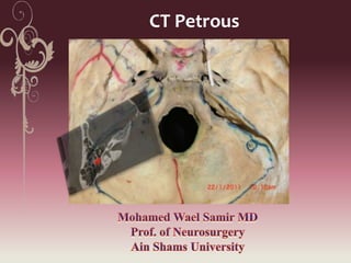
CT Petrous
Superior surface of petrous bone


        Trigeminal depression


                                Arcuate eminence
Medial surface of petrous bone
                          Subarcuate fossa & A


IAM
                                   Vestibular aqueduct

Cochlear canaliculus


        Jugular foramen
Arcuate eminence   Trigeminal impression
      (SSC)
Petro occipital
                suture
Arcuate eminence        Trigeminal
      (SSC)             impression


                                     PSC


                              IAM
Epitympanum


IAcanal


                                      LSC
                Mastoid
    vestibule   antrum          PSC
Middle ear cavity with Malleus & incus


         Labrynthine segment
            of facial nerve                       Tympanic segment of
                                                     facial nerve
          Cochlea


Meatal segment of
  facial nerve


     IAcanal


                                                  PSC
                    vestibule
Cochlea


Endolyphatic sac             Vestibular
                             aqueduct
External
                  Cochlea
                                           canal


                                         Middle ear

Jugular foramen
                  Cochlear canaliculus
Pharyngotympanic tube (auditory tube)


                                         External
                                          canal


                                        Middle ear


                        Carotid canal
                        Carotid canal
Pharyngotympanic tube
                                 (auditory tube)


Carotid canal


  Neural compartment


     Vascular compartment
                            Jugular foramen
 Pontine segment:
 Meatal segment:
 labyrinthine segment:
 tympanic or horizontal :
 mastoid or vertical segment:
 Extratemporal segment
Labrynthine segment
                           of facial nerve


                    Tympanic segment of
                       facial nerve


Meatal segment of
  facial nerve
Carotid canal (vertical segment)


                                   TMJ
Middle ear bony ossicles


                       External auditory canal


Cochlea   Middle ear
SCC
                Labrynthine
                  segment


 Meatal
segment


          Vestibule


                      LCC
Quiz
Quiz
Quiz
Quiz
        Carotid canal
TMJ
Quiz
Quiz
References
 Hugh D. Curtin, James D. Rabinov and Peter M. Som
(2003): Central Skull Base: Embryology Anatomy, and
Pathology in Central skull base in Head and Neck
Imaging. Peter M. Som & Hugh D. Curt (edit)
Mosby, Inc. St. Louis, Chapter 12 FOURTH EDITION.
Volume one 785- 863
Ct petrous

More Related Content

What's hot

Normal cerebral angiogram final
Normal cerebral angiogram finalNormal cerebral angiogram final
Normal cerebral angiogram final
nikhilzandu
 
How to read a brain ct scan moderate
How to read a brain ct scan moderateHow to read a brain ct scan moderate
How to read a brain ct scan moderate
Gauhar Azeem
 
Pineal region masses - radiology
Pineal region masses - radiologyPineal region masses - radiology
Pineal region masses - radiology
Dr. Mohit Goel
 
Csf flowmetry
Csf flowmetryCsf flowmetry
Csf flowmetry
Dalia El Said
 
Larynx anatomy CT and MRI
Larynx anatomy CT and MRILarynx anatomy CT and MRI
Larynx anatomy CT and MRI
Dr. Mohit Goel
 
Temporal bone Radiologic anatomy.. In depth
Temporal bone Radiologic anatomy.. In depthTemporal bone Radiologic anatomy.. In depth
Temporal bone Radiologic anatomy.. In depth
Ahmed Hamdy Hamdy
 
Presentation1.pptx, radiological anatomy of the neck.
Presentation1.pptx, radiological anatomy of the neck.Presentation1.pptx, radiological anatomy of the neck.
Presentation1.pptx, radiological anatomy of the neck.Abdellah Nazeer
 
Anatomy of hippocampus ( radiology )
Anatomy of hippocampus ( radiology )Anatomy of hippocampus ( radiology )
Anatomy of hippocampus ( radiology )Sajith Selvaganesan
 
Imaging sectional anatomy of brain part 1
Imaging sectional  anatomy  of  brain  part 1Imaging sectional  anatomy  of  brain  part 1
Imaging sectional anatomy of brain part 1drnaveent
 
Skull base tumors & perineural spread radiology ppt
Skull base tumors & perineural spread radiology pptSkull base tumors & perineural spread radiology ppt
Skull base tumors & perineural spread radiology ppt
Dr pradeep Kumar
 
Triple Rule Out Computed Tomography
Triple Rule Out Computed TomographyTriple Rule Out Computed Tomography
Triple Rule Out Computed Tomography
Daniel Steven
 
Presentation1.pptx, radiological imaging of the larngeal diseases.
Presentation1.pptx, radiological imaging of the larngeal diseases.Presentation1.pptx, radiological imaging of the larngeal diseases.
Presentation1.pptx, radiological imaging of the larngeal diseases.Abdellah Nazeer
 
Brain anatomy new
Brain anatomy newBrain anatomy new
Brain anatomy new
MD RADIODIAGNOSIS
 
SKULL BASE IMAGING
SKULL BASE IMAGINGSKULL BASE IMAGING
Mediastnum ppt
Mediastnum pptMediastnum ppt
Mediastnum pptjyomita
 
E.N.T 5th year, 1st lecture (Dr. Hiwa)
E.N.T 5th year, 1st lecture (Dr. Hiwa)E.N.T 5th year, 1st lecture (Dr. Hiwa)
E.N.T 5th year, 1st lecture (Dr. Hiwa)
College of Medicine, Sulaymaniyah
 
Internal Carotid Artery and Normal Variants
Internal Carotid Artery and Normal VariantsInternal Carotid Artery and Normal Variants
Internal Carotid Artery and Normal VariantsMATIAS FREITAS FH
 
Imaging in Skull base
Imaging in Skull baseImaging in Skull base
Imaging in Skull base
Rakesh Ca
 
Neurosonogram.. Dr.Padmesh
Neurosonogram.. Dr.PadmeshNeurosonogram.. Dr.Padmesh
Neurosonogram.. Dr.Padmesh
Dr Padmesh Vadakepat
 
Ccf(1)
Ccf(1)Ccf(1)

What's hot (20)

Normal cerebral angiogram final
Normal cerebral angiogram finalNormal cerebral angiogram final
Normal cerebral angiogram final
 
How to read a brain ct scan moderate
How to read a brain ct scan moderateHow to read a brain ct scan moderate
How to read a brain ct scan moderate
 
Pineal region masses - radiology
Pineal region masses - radiologyPineal region masses - radiology
Pineal region masses - radiology
 
Csf flowmetry
Csf flowmetryCsf flowmetry
Csf flowmetry
 
Larynx anatomy CT and MRI
Larynx anatomy CT and MRILarynx anatomy CT and MRI
Larynx anatomy CT and MRI
 
Temporal bone Radiologic anatomy.. In depth
Temporal bone Radiologic anatomy.. In depthTemporal bone Radiologic anatomy.. In depth
Temporal bone Radiologic anatomy.. In depth
 
Presentation1.pptx, radiological anatomy of the neck.
Presentation1.pptx, radiological anatomy of the neck.Presentation1.pptx, radiological anatomy of the neck.
Presentation1.pptx, radiological anatomy of the neck.
 
Anatomy of hippocampus ( radiology )
Anatomy of hippocampus ( radiology )Anatomy of hippocampus ( radiology )
Anatomy of hippocampus ( radiology )
 
Imaging sectional anatomy of brain part 1
Imaging sectional  anatomy  of  brain  part 1Imaging sectional  anatomy  of  brain  part 1
Imaging sectional anatomy of brain part 1
 
Skull base tumors & perineural spread radiology ppt
Skull base tumors & perineural spread radiology pptSkull base tumors & perineural spread radiology ppt
Skull base tumors & perineural spread radiology ppt
 
Triple Rule Out Computed Tomography
Triple Rule Out Computed TomographyTriple Rule Out Computed Tomography
Triple Rule Out Computed Tomography
 
Presentation1.pptx, radiological imaging of the larngeal diseases.
Presentation1.pptx, radiological imaging of the larngeal diseases.Presentation1.pptx, radiological imaging of the larngeal diseases.
Presentation1.pptx, radiological imaging of the larngeal diseases.
 
Brain anatomy new
Brain anatomy newBrain anatomy new
Brain anatomy new
 
SKULL BASE IMAGING
SKULL BASE IMAGINGSKULL BASE IMAGING
SKULL BASE IMAGING
 
Mediastnum ppt
Mediastnum pptMediastnum ppt
Mediastnum ppt
 
E.N.T 5th year, 1st lecture (Dr. Hiwa)
E.N.T 5th year, 1st lecture (Dr. Hiwa)E.N.T 5th year, 1st lecture (Dr. Hiwa)
E.N.T 5th year, 1st lecture (Dr. Hiwa)
 
Internal Carotid Artery and Normal Variants
Internal Carotid Artery and Normal VariantsInternal Carotid Artery and Normal Variants
Internal Carotid Artery and Normal Variants
 
Imaging in Skull base
Imaging in Skull baseImaging in Skull base
Imaging in Skull base
 
Neurosonogram.. Dr.Padmesh
Neurosonogram.. Dr.PadmeshNeurosonogram.. Dr.Padmesh
Neurosonogram.. Dr.Padmesh
 
Ccf(1)
Ccf(1)Ccf(1)
Ccf(1)
 

Viewers also liked

Pineal Body Cyst
Pineal Body CystPineal Body Cyst
Orbital ct and mr
Orbital ct and mrOrbital ct and mr
Orbital ct and mrkebaplik
 
Temporal bone
Temporal boneTemporal bone
Temporal bone
Adila Iqbal
 
Detecting Early Liver Fibrosis - A Nutshell for Primary Care
Detecting Early Liver Fibrosis - A Nutshell for Primary CareDetecting Early Liver Fibrosis - A Nutshell for Primary Care
Detecting Early Liver Fibrosis - A Nutshell for Primary Care
Jarrod Lee
 
Imaging modalities of the petrous bone
Imaging modalities of the petrous boneImaging modalities of the petrous bone
Imaging modalities of the petrous boneAbdellah Nazeer
 
Ct scan eye slide presentation
Ct scan eye slide presentationCt scan eye slide presentation
Ct scan eye slide presentation
REKHAKHARE
 
Presentation1.pptx, supratentorial brain tumour
Presentation1.pptx, supratentorial brain tumourPresentation1.pptx, supratentorial brain tumour
Presentation1.pptx, supratentorial brain tumourAbdellah Nazeer
 
Imaging in Ocular Pathologies
Imaging in Ocular PathologiesImaging in Ocular Pathologies
Imaging in Ocular Pathologies
Sahil Chaudhry
 
Imaging of Bile Duct - Columbia Asia Workshop
Imaging of Bile Duct - Columbia Asia WorkshopImaging of Bile Duct - Columbia Asia Workshop
Imaging of Bile Duct - Columbia Asia Workshopinjoosweb
 
Presentation2.pptx imaging of the biliary system
Presentation2.pptx  imaging of the biliary systemPresentation2.pptx  imaging of the biliary system
Presentation2.pptx imaging of the biliary systemAbdellah Nazeer
 
CT brain in emergencies ... for ER doctors
CT brain in emergencies ... for ER doctorsCT brain in emergencies ... for ER doctors
CT brain in emergencies ... for ER doctors
Mohamed Shaaban
 
Sono elastography - gut
Sono elastography - gutSono elastography - gut
Sono elastography - gut
Mohamed Shaaban
 
The pathway to uroradiology
The pathway to uroradiologyThe pathway to uroradiology
The pathway to uroradiology
Mohamed Shaaban
 
MRCP Classical Presentations
MRCP Classical PresentationsMRCP Classical Presentations
MRCP Classical Presentations
Sherif Elbadrawy
 
Liver stiffness measurement (fibroscan®)
Liver stiffness measurement (fibroscan®)Liver stiffness measurement (fibroscan®)
Liver stiffness measurement (fibroscan®)
Samir Haffar
 

Viewers also liked (20)

Mohamed Sobhi Hamza CV
Mohamed Sobhi Hamza CVMohamed Sobhi Hamza CV
Mohamed Sobhi Hamza CV
 
Fungal infections2012
Fungal infections2012Fungal infections2012
Fungal infections2012
 
Pineal Body Cyst
Pineal Body CystPineal Body Cyst
Pineal Body Cyst
 
Orbital ct and mr
Orbital ct and mrOrbital ct and mr
Orbital ct and mr
 
Ct brain by prof. Wael samir
Ct brain by prof. Wael samirCt brain by prof. Wael samir
Ct brain by prof. Wael samir
 
Temporal bone
Temporal boneTemporal bone
Temporal bone
 
Detecting Early Liver Fibrosis - A Nutshell for Primary Care
Detecting Early Liver Fibrosis - A Nutshell for Primary CareDetecting Early Liver Fibrosis - A Nutshell for Primary Care
Detecting Early Liver Fibrosis - A Nutshell for Primary Care
 
Imaging modalities of the petrous bone
Imaging modalities of the petrous boneImaging modalities of the petrous bone
Imaging modalities of the petrous bone
 
Ct orbit
Ct orbitCt orbit
Ct orbit
 
Ct scan eye slide presentation
Ct scan eye slide presentationCt scan eye slide presentation
Ct scan eye slide presentation
 
Primary CNS lymphoma
Primary CNS lymphomaPrimary CNS lymphoma
Primary CNS lymphoma
 
Presentation1.pptx, supratentorial brain tumour
Presentation1.pptx, supratentorial brain tumourPresentation1.pptx, supratentorial brain tumour
Presentation1.pptx, supratentorial brain tumour
 
Imaging in Ocular Pathologies
Imaging in Ocular PathologiesImaging in Ocular Pathologies
Imaging in Ocular Pathologies
 
Imaging of Bile Duct - Columbia Asia Workshop
Imaging of Bile Duct - Columbia Asia WorkshopImaging of Bile Duct - Columbia Asia Workshop
Imaging of Bile Duct - Columbia Asia Workshop
 
Presentation2.pptx imaging of the biliary system
Presentation2.pptx  imaging of the biliary systemPresentation2.pptx  imaging of the biliary system
Presentation2.pptx imaging of the biliary system
 
CT brain in emergencies ... for ER doctors
CT brain in emergencies ... for ER doctorsCT brain in emergencies ... for ER doctors
CT brain in emergencies ... for ER doctors
 
Sono elastography - gut
Sono elastography - gutSono elastography - gut
Sono elastography - gut
 
The pathway to uroradiology
The pathway to uroradiologyThe pathway to uroradiology
The pathway to uroradiology
 
MRCP Classical Presentations
MRCP Classical PresentationsMRCP Classical Presentations
MRCP Classical Presentations
 
Liver stiffness measurement (fibroscan®)
Liver stiffness measurement (fibroscan®)Liver stiffness measurement (fibroscan®)
Liver stiffness measurement (fibroscan®)
 

Similar to Ct petrous

Skull, dura & sinuses
Skull, dura & sinusesSkull, dura & sinuses
Skull, dura & sinuses
Dr. Noura El Tahawy
 
127166 gross-anatomy-guide
127166 gross-anatomy-guide127166 gross-anatomy-guide
127166 gross-anatomy-guideYoAmoNYC
 
127166 gross-anatomy-guide
127166 gross-anatomy-guide127166 gross-anatomy-guide
127166 gross-anatomy-guideabctutor
 
Facial anatomy
Facial anatomyFacial anatomy
Facial anatomywebzforu
 
Temporal bone anatomy and surgical significancepptx
Temporal bone anatomy and surgical significancepptxTemporal bone anatomy and surgical significancepptx
Temporal bone anatomy and surgical significancepptx
druttamnepal
 
Respiratory system notes std 3 2011
Respiratory system notes std 3 2011Respiratory system notes std 3 2011
Respiratory system notes std 3 2011ZygomaticMonkey
 
Rubzzzz's notes on Artery system
Rubzzzz's notes on Artery systemRubzzzz's notes on Artery system
Rubzzzz's notes on Artery systemDr. Rubz
 
Anatomy revision
Anatomy revisionAnatomy revision
Anatomy revisionbbyrne725
 
Pituitary gland anatomy
Pituitary gland anatomyPituitary gland anatomy
Pituitary gland anatomy
Dr. Noura El Tahawy
 
IMAGING OF TEMPORAL BONE
IMAGING OF TEMPORAL BONEIMAGING OF TEMPORAL BONE
IMAGING OF TEMPORAL BONE
Sameer Peer
 
Head and Neck
Head and NeckHead and Neck
Head and Neck
Selva Kumar
 
Zoology lab exam ii
Zoology lab exam iiZoology lab exam ii
Zoology lab exam iiKeriann King
 
Anatomy of ear
Anatomy of ear Anatomy of ear
Anatomy of ear
amit jha
 
Anatomy of skull by RW..
Anatomy of skull by RW..Anatomy of skull by RW..
Anatomy of skull by RW..Ramsha Wahid
 

Similar to Ct petrous (20)

Ct skull base
Ct skull baseCt skull base
Ct skull base
 
Skull, dura & sinuses
Skull, dura & sinusesSkull, dura & sinuses
Skull, dura & sinuses
 
127166 gross-anatomy-guide
127166 gross-anatomy-guide127166 gross-anatomy-guide
127166 gross-anatomy-guide
 
127166 gross-anatomy-guide
127166 gross-anatomy-guide127166 gross-anatomy-guide
127166 gross-anatomy-guide
 
Table 1 Foramina Of Skull
Table 1 Foramina Of SkullTable 1 Foramina Of Skull
Table 1 Foramina Of Skull
 
Table 2 Foramina Of Skull
Table 2 Foramina Of SkullTable 2 Foramina Of Skull
Table 2 Foramina Of Skull
 
Facial anatomy
Facial anatomyFacial anatomy
Facial anatomy
 
Temporal bone anatomy and surgical significancepptx
Temporal bone anatomy and surgical significancepptxTemporal bone anatomy and surgical significancepptx
Temporal bone anatomy and surgical significancepptx
 
Respiratory system notes std 3 2011
Respiratory system notes std 3 2011Respiratory system notes std 3 2011
Respiratory system notes std 3 2011
 
Rubzzzz's notes on Artery system
Rubzzzz's notes on Artery systemRubzzzz's notes on Artery system
Rubzzzz's notes on Artery system
 
Anatomy revision
Anatomy revisionAnatomy revision
Anatomy revision
 
Pituitary gland anatomy
Pituitary gland anatomyPituitary gland anatomy
Pituitary gland anatomy
 
IMAGING OF TEMPORAL BONE
IMAGING OF TEMPORAL BONEIMAGING OF TEMPORAL BONE
IMAGING OF TEMPORAL BONE
 
Norma basalis interna
Norma basalis internaNorma basalis interna
Norma basalis interna
 
Head and Neck
Head and NeckHead and Neck
Head and Neck
 
Zoology lab exam ii
Zoology lab exam iiZoology lab exam ii
Zoology lab exam ii
 
Ear models
Ear modelsEar models
Ear models
 
Anatomy of ear
Anatomy of ear Anatomy of ear
Anatomy of ear
 
Anatomy of skull by RW..
Anatomy of skull by RW..Anatomy of skull by RW..
Anatomy of skull by RW..
 
Bld supply
Bld supplyBld supply
Bld supply
 

Recently uploaded

Report Back from SGO 2024: What’s the Latest in Cervical Cancer?
Report Back from SGO 2024: What’s the Latest in Cervical Cancer?Report Back from SGO 2024: What’s the Latest in Cervical Cancer?
Report Back from SGO 2024: What’s the Latest in Cervical Cancer?
bkling
 
Tom Selleck Health: A Comprehensive Look at the Iconic Actor’s Wellness Journey
Tom Selleck Health: A Comprehensive Look at the Iconic Actor’s Wellness JourneyTom Selleck Health: A Comprehensive Look at the Iconic Actor’s Wellness Journey
Tom Selleck Health: A Comprehensive Look at the Iconic Actor’s Wellness Journey
greendigital
 
Superficial & Deep Fascia of the NECK.pptx
Superficial & Deep Fascia of the NECK.pptxSuperficial & Deep Fascia of the NECK.pptx
Superficial & Deep Fascia of the NECK.pptx
Dr. Rabia Inam Gandapore
 
Are There Any Natural Remedies To Treat Syphilis.pdf
Are There Any Natural Remedies To Treat Syphilis.pdfAre There Any Natural Remedies To Treat Syphilis.pdf
Are There Any Natural Remedies To Treat Syphilis.pdf
Little Cross Family Clinic
 
micro teaching on communication m.sc nursing.pdf
micro teaching on communication m.sc nursing.pdfmicro teaching on communication m.sc nursing.pdf
micro teaching on communication m.sc nursing.pdf
Anurag Sharma
 
New Drug Discovery and Development .....
New Drug Discovery and Development .....New Drug Discovery and Development .....
New Drug Discovery and Development .....
NEHA GUPTA
 
Pharynx and Clinical Correlations BY Dr.Rabia Inam Gandapore.pptx
Pharynx and Clinical Correlations BY Dr.Rabia Inam Gandapore.pptxPharynx and Clinical Correlations BY Dr.Rabia Inam Gandapore.pptx
Pharynx and Clinical Correlations BY Dr.Rabia Inam Gandapore.pptx
Dr. Rabia Inam Gandapore
 
Phone Us ❤85270-49040❤ #ℂall #gIRLS In Surat By Surat @ℂall @Girls Hotel With...
Phone Us ❤85270-49040❤ #ℂall #gIRLS In Surat By Surat @ℂall @Girls Hotel With...Phone Us ❤85270-49040❤ #ℂall #gIRLS In Surat By Surat @ℂall @Girls Hotel With...
Phone Us ❤85270-49040❤ #ℂall #gIRLS In Surat By Surat @ℂall @Girls Hotel With...
Savita Shen $i11
 
ARTHROLOGY PPT NCISM SYLLABUS AYURVEDA STUDENTS
ARTHROLOGY PPT NCISM SYLLABUS AYURVEDA STUDENTSARTHROLOGY PPT NCISM SYLLABUS AYURVEDA STUDENTS
ARTHROLOGY PPT NCISM SYLLABUS AYURVEDA STUDENTS
Dr. Vinay Pareek
 
Flu Vaccine Alert in Bangalore Karnataka
Flu Vaccine Alert in Bangalore KarnatakaFlu Vaccine Alert in Bangalore Karnataka
Flu Vaccine Alert in Bangalore Karnataka
addon Scans
 
Non-respiratory Functions of the Lungs.pdf
Non-respiratory Functions of the Lungs.pdfNon-respiratory Functions of the Lungs.pdf
Non-respiratory Functions of the Lungs.pdf
MedicoseAcademics
 
Evaluation of antidepressant activity of clitoris ternatea in animals
Evaluation of antidepressant activity of clitoris ternatea in animalsEvaluation of antidepressant activity of clitoris ternatea in animals
Evaluation of antidepressant activity of clitoris ternatea in animals
Shweta
 
Pictures of Superficial & Deep Fascia.ppt.pdf
Pictures of Superficial & Deep Fascia.ppt.pdfPictures of Superficial & Deep Fascia.ppt.pdf
Pictures of Superficial & Deep Fascia.ppt.pdf
Dr. Rabia Inam Gandapore
 
Physiology of Chemical Sensation of smell.pdf
Physiology of Chemical Sensation of smell.pdfPhysiology of Chemical Sensation of smell.pdf
Physiology of Chemical Sensation of smell.pdf
MedicoseAcademics
 
Thyroid Gland- Gross Anatomy by Dr. Rabia Inam Gandapore.pptx
Thyroid Gland- Gross Anatomy by Dr. Rabia Inam Gandapore.pptxThyroid Gland- Gross Anatomy by Dr. Rabia Inam Gandapore.pptx
Thyroid Gland- Gross Anatomy by Dr. Rabia Inam Gandapore.pptx
Dr. Rabia Inam Gandapore
 
Light House Retreats: Plant Medicine Retreat Europe
Light House Retreats: Plant Medicine Retreat EuropeLight House Retreats: Plant Medicine Retreat Europe
Light House Retreats: Plant Medicine Retreat Europe
Lighthouse Retreat
 
Ozempic: Preoperative Management of Patients on GLP-1 Receptor Agonists
Ozempic: Preoperative Management of Patients on GLP-1 Receptor Agonists  Ozempic: Preoperative Management of Patients on GLP-1 Receptor Agonists
Ozempic: Preoperative Management of Patients on GLP-1 Receptor Agonists
Saeid Safari
 
For Better Surat #ℂall #Girl Service ❤85270-49040❤ Surat #ℂall #Girls
For Better Surat #ℂall #Girl Service ❤85270-49040❤ Surat #ℂall #GirlsFor Better Surat #ℂall #Girl Service ❤85270-49040❤ Surat #ℂall #Girls
For Better Surat #ℂall #Girl Service ❤85270-49040❤ Surat #ℂall #Girls
Savita Shen $i11
 
Maxilla, Mandible & Hyoid Bone & Clinical Correlations by Dr. RIG.pptx
Maxilla, Mandible & Hyoid Bone & Clinical Correlations by Dr. RIG.pptxMaxilla, Mandible & Hyoid Bone & Clinical Correlations by Dr. RIG.pptx
Maxilla, Mandible & Hyoid Bone & Clinical Correlations by Dr. RIG.pptx
Dr. Rabia Inam Gandapore
 
Lung Cancer: Artificial Intelligence, Synergetics, Complex System Analysis, S...
Lung Cancer: Artificial Intelligence, Synergetics, Complex System Analysis, S...Lung Cancer: Artificial Intelligence, Synergetics, Complex System Analysis, S...
Lung Cancer: Artificial Intelligence, Synergetics, Complex System Analysis, S...
Oleg Kshivets
 

Recently uploaded (20)

Report Back from SGO 2024: What’s the Latest in Cervical Cancer?
Report Back from SGO 2024: What’s the Latest in Cervical Cancer?Report Back from SGO 2024: What’s the Latest in Cervical Cancer?
Report Back from SGO 2024: What’s the Latest in Cervical Cancer?
 
Tom Selleck Health: A Comprehensive Look at the Iconic Actor’s Wellness Journey
Tom Selleck Health: A Comprehensive Look at the Iconic Actor’s Wellness JourneyTom Selleck Health: A Comprehensive Look at the Iconic Actor’s Wellness Journey
Tom Selleck Health: A Comprehensive Look at the Iconic Actor’s Wellness Journey
 
Superficial & Deep Fascia of the NECK.pptx
Superficial & Deep Fascia of the NECK.pptxSuperficial & Deep Fascia of the NECK.pptx
Superficial & Deep Fascia of the NECK.pptx
 
Are There Any Natural Remedies To Treat Syphilis.pdf
Are There Any Natural Remedies To Treat Syphilis.pdfAre There Any Natural Remedies To Treat Syphilis.pdf
Are There Any Natural Remedies To Treat Syphilis.pdf
 
micro teaching on communication m.sc nursing.pdf
micro teaching on communication m.sc nursing.pdfmicro teaching on communication m.sc nursing.pdf
micro teaching on communication m.sc nursing.pdf
 
New Drug Discovery and Development .....
New Drug Discovery and Development .....New Drug Discovery and Development .....
New Drug Discovery and Development .....
 
Pharynx and Clinical Correlations BY Dr.Rabia Inam Gandapore.pptx
Pharynx and Clinical Correlations BY Dr.Rabia Inam Gandapore.pptxPharynx and Clinical Correlations BY Dr.Rabia Inam Gandapore.pptx
Pharynx and Clinical Correlations BY Dr.Rabia Inam Gandapore.pptx
 
Phone Us ❤85270-49040❤ #ℂall #gIRLS In Surat By Surat @ℂall @Girls Hotel With...
Phone Us ❤85270-49040❤ #ℂall #gIRLS In Surat By Surat @ℂall @Girls Hotel With...Phone Us ❤85270-49040❤ #ℂall #gIRLS In Surat By Surat @ℂall @Girls Hotel With...
Phone Us ❤85270-49040❤ #ℂall #gIRLS In Surat By Surat @ℂall @Girls Hotel With...
 
ARTHROLOGY PPT NCISM SYLLABUS AYURVEDA STUDENTS
ARTHROLOGY PPT NCISM SYLLABUS AYURVEDA STUDENTSARTHROLOGY PPT NCISM SYLLABUS AYURVEDA STUDENTS
ARTHROLOGY PPT NCISM SYLLABUS AYURVEDA STUDENTS
 
Flu Vaccine Alert in Bangalore Karnataka
Flu Vaccine Alert in Bangalore KarnatakaFlu Vaccine Alert in Bangalore Karnataka
Flu Vaccine Alert in Bangalore Karnataka
 
Non-respiratory Functions of the Lungs.pdf
Non-respiratory Functions of the Lungs.pdfNon-respiratory Functions of the Lungs.pdf
Non-respiratory Functions of the Lungs.pdf
 
Evaluation of antidepressant activity of clitoris ternatea in animals
Evaluation of antidepressant activity of clitoris ternatea in animalsEvaluation of antidepressant activity of clitoris ternatea in animals
Evaluation of antidepressant activity of clitoris ternatea in animals
 
Pictures of Superficial & Deep Fascia.ppt.pdf
Pictures of Superficial & Deep Fascia.ppt.pdfPictures of Superficial & Deep Fascia.ppt.pdf
Pictures of Superficial & Deep Fascia.ppt.pdf
 
Physiology of Chemical Sensation of smell.pdf
Physiology of Chemical Sensation of smell.pdfPhysiology of Chemical Sensation of smell.pdf
Physiology of Chemical Sensation of smell.pdf
 
Thyroid Gland- Gross Anatomy by Dr. Rabia Inam Gandapore.pptx
Thyroid Gland- Gross Anatomy by Dr. Rabia Inam Gandapore.pptxThyroid Gland- Gross Anatomy by Dr. Rabia Inam Gandapore.pptx
Thyroid Gland- Gross Anatomy by Dr. Rabia Inam Gandapore.pptx
 
Light House Retreats: Plant Medicine Retreat Europe
Light House Retreats: Plant Medicine Retreat EuropeLight House Retreats: Plant Medicine Retreat Europe
Light House Retreats: Plant Medicine Retreat Europe
 
Ozempic: Preoperative Management of Patients on GLP-1 Receptor Agonists
Ozempic: Preoperative Management of Patients on GLP-1 Receptor Agonists  Ozempic: Preoperative Management of Patients on GLP-1 Receptor Agonists
Ozempic: Preoperative Management of Patients on GLP-1 Receptor Agonists
 
For Better Surat #ℂall #Girl Service ❤85270-49040❤ Surat #ℂall #Girls
For Better Surat #ℂall #Girl Service ❤85270-49040❤ Surat #ℂall #GirlsFor Better Surat #ℂall #Girl Service ❤85270-49040❤ Surat #ℂall #Girls
For Better Surat #ℂall #Girl Service ❤85270-49040❤ Surat #ℂall #Girls
 
Maxilla, Mandible & Hyoid Bone & Clinical Correlations by Dr. RIG.pptx
Maxilla, Mandible & Hyoid Bone & Clinical Correlations by Dr. RIG.pptxMaxilla, Mandible & Hyoid Bone & Clinical Correlations by Dr. RIG.pptx
Maxilla, Mandible & Hyoid Bone & Clinical Correlations by Dr. RIG.pptx
 
Lung Cancer: Artificial Intelligence, Synergetics, Complex System Analysis, S...
Lung Cancer: Artificial Intelligence, Synergetics, Complex System Analysis, S...Lung Cancer: Artificial Intelligence, Synergetics, Complex System Analysis, S...
Lung Cancer: Artificial Intelligence, Synergetics, Complex System Analysis, S...
 

Ct petrous

Editor's Notes

  1. The superior surface of petrosal part of temporal bone: Middle fossa surface of petrous part of temporal bone called tegmen. It contains vestibular and cochlear labrynth. It roofs carotid canal, EA canal, facial canal, & tympanic cavity. It has following surface landmarks: 1) Trigeminal impression:: on the upper surface of the petrous bone where Meckel’s cave and the semilunar ganglion sit. 2) GSP groove:Lateral to this impression is a small groove on the anterior, deep surface of the petrous portion of the temporal bone. The groove opens posteriorly into a canal, the hiatus of the facial canal. Grooves for the greater and lesser petrosal nerves: found on the upper surface of the petrous bone. Lateral to itrigeminal impression there are two small grooves, a medial sulcus of the greater petrosal nerve (sulcus n. petrosimajoris) and a lateral sulcus of the lesser petrosal nerve (sulcus n. petrosiminoris). They lead to two openings of the same name, a medial the greater petrosal opening (hiatus canalis n. petrosimajoris) and a lateral the lesser petrosal nerve opening (hiatus canalis n. petrosiminoris). 3) Arcuate eminence (SSC): Above and lateral to the hiatus is a bony prominence, the arcuate eminence, overlying the superior semicircular canal. The arcuate eminence (eminentiaarcuata) is lateral to these openings; it forms due to prominence of the vigorously developing labyrinth, particularly the superior semicircular canal. The internal auditory canal can be identified below the floor of the middle fossa by drilling along a line approximately 60 degrees medial to the arcuate eminence, near the middle portion of the angle between the greater petrosal nerve and arcuate eminence4) Tegmen tympani: A thin lamina of bone, the tegmen tympani, roofs the area above the middle ear and auditory ossicles on the anterolateral side of the arcuate eminence (between the petrosquamous fissure and the arcuate eminence).5) The carotid canal extends upward and medially and provides passage to the internal carotid artery and carotid sympathetic nerves in their course to the cavernous sinus6) Groove for superior petrosal sinus: The superior-most portion of the petrous portion of the temporal bone is a thin ridge, which constitutes part of the posterior border of the middle cranial fossa. This ridge contains the groove for the superior petrosal sinus.
  2. Posterior fossa surface of petrous part of temporal bone blends with mastoid surface of temporal bone. It contains 1) IAM midway between apex and base of petrous. 2) Subarcuatefossafound posterior to EAM and is the site for penetration of subarcuate artery (branch of AICA) which supply SSC.3) vestibular aqueduct: Inferior lateral to EAC is opening for vestibular aqueduct that transmit enodlymphatic duct that communicate endolymphatic sac found between the dural layers and the endolymph of labrynth.4) Cochlear canaliculuswhere cochlear aqueduct (contain perilymph) opens. found at the antromedial edge of jugular foramen just superior and lateral to glossopharyngeal nerve where it enters the jugular foramen. During drilling of posterior lip of IAM care should be given to avoid injury of 1) Common crus of posterior and superior canals which found lateral to the entry of subarcuate artery. 2) Vestibule and Posterior semicircular canal: which are away from EAM by average 7mm 3) Vestibular aqueduct: the endolymphatic duct that marked by the endolymphatic ridge at the level of posterior canal. 4) Inferiorly: the high arched jugular bulb.
  3. The course of the facial nerve can be roughly divided into 5 segments, 1) The pontine segment, between the brainstem and porus, measures 23 to 24 mm in length. At this point the facial nerve is anterior to the cochleovestibular nerve. The special sensory and visceral efferent components of the facial nerve pass in a separate bundle adjacent to the main motor trunk as the nervusintermedius.2) The meatal segment, within the internal auditory canal, is 7 to 8 mm in length. The facial nerve passes superior to the falciform crest in the lateral aspect of the canal and is separated from the superior vestibular nerve by a vertical crest of bone (Bill's bar).3) The labyrinthine segment, between the meatal segment and geniculate ganglion, is 4 mm in length. The osseous canal surrounding the facial nerve is narrowest at the most proximal portion of the labyrinthine segment. This segment passes anterolaterally, paralleling the axis of the arcuate eminence of the superior semicircular canal, and passes superior and in proximity to the basal turn of the cochlea. The geniculate ganglion is triangular and averages 1.09 mm in length. 4) The tympanic or horizontal segment, between the geniculate ganglion and second genu, is 12 to 13 mm in length. The proximal edge of the geniculate ganglion is 5 mm anterosuperior to the posterior edge of the processuscochleariformis. The facial nerve passes superior to the oval window niche, a region where in approximately 55% of cases it is dehisc 5) The mastoid or vertical segment, between the second genu and stylomastoid foramen, measures 15 to 20 mm in length. At the second genu, the semicircular canal lies 0.5 mm posterosuperior to the facial nerve. The digastric ridge is a useful landmark just posterior to the stylomastoid foramen. 6) Extratemporal segment (Stylomastoid foramen to pesanserinus): 15-20mm
  4. Cholesteatoma eroding the horizontal semicircular canal, CT. A: Cholesteatomaopacifies the upper attic and antrum. Note the rounded, smooth margins. There is erosive scalloping of the bone covering the horizontal semicircular canal (arrow). The cholesteatoma has reached the lumen of the canal, causing a fistula. B: Normal right side shows the intact cortex with a normal thickness (arrow).
  5. Glomusjugulare, axial CT. The lesion (white arrow) was visualized through the tympanic membrane. There is demineralization (black arrows) around the jugular foramen. The white cortical line is indistinct and poorly visualized. A small amount of demineralized bone (white arrowhead) on the posterior cortex of the carotid canal indicates tumor. Compare the demineralized bone with the intact cortex and bone on the opposite side.Glomusjugulare, MR T1-weighted image after intravenous contrast. The tumor (T) is visualized in the region of the jugular foramen. Its interface (arrow) with the posterior fossa is clearly defined. The bright signal (arrowhead) in the sigmoid sinus represents slow flow of blood with gadolinium. The margins of the lesion can be clearly seen, though the bony landmarks cannot.
  6. High-resolution CT scan of a longitudinal skull fracture passing approximately parallel to the petrous ridge across the middle fossa floor and squamous portion of the temporal bone. Transverse fracture of the right temporal bone in the axial (A) and coronal (B) planes. Longitudinal fracture of the temporal bone follows the long axis of the petrous apex and crosses the floor of the middle cranial fossa near the foramen spinosum. The usual site of facial nerve injury is its labyrinthine segment. B: Transverse fracture of the temporal bone. The fracture line passes between the foramen magnum posteromedially and the foramen spinosum area anterolaterally and traverses the long axis of the petrous pyramid.
  7. 1) Epitympanum2) Middle ear bone articulation3) Body of incus4) Aditus of antrum5) Mastoid antrum6) LSC7) vestibule8) Tympanic segment of facial nerve 9) Pourusacousticus10) Internal auditory canal11) Midlle turn of cochlea 12) Petrous apex13) Head of malleus
  8. 1) Malleus 2) Incus 3) Stapedial joint 4) stapes 5 ) vestibular aqueduct 6) vestibule 7) Oval window 8) Internal auditory canal 9) Cochlea
  9. 1) Mesotympanum 2) Cochlear promontory 3) Malleus 4) Facial recess 5) Facial nerve (mastoid segment) 6) Pyramidal eminence 7) Sinus tympany 8) Round window niche 9) Round window 10) Basal turn of cochlea 11) Middle turn of cochlea 12) Apical turn of cochlea 13) Posterior semicircular canal.
  10. Hugh D. Curtin, James D. Rabinov and Peter M. Som(2003): Central Skull Base: Embryology Anatomy, and Pathology in Central skull base in Head and Neck Imaging. Peter M. Som & Hugh D. Curt (edit) Mosby, Inc. St. Louis, Chapter 12 FOURTH EDITION. Volume one 785- 863