Downloaded 1,109 times

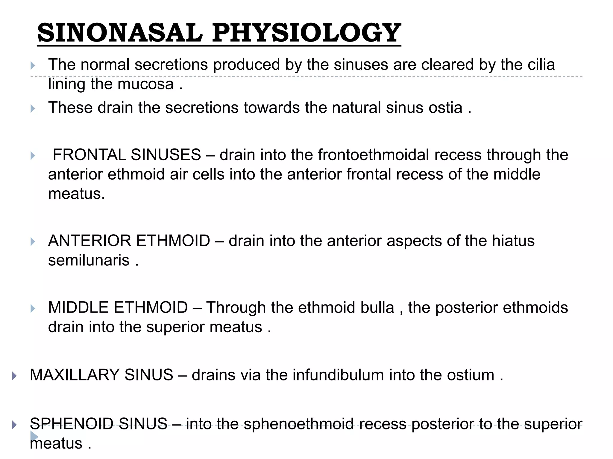


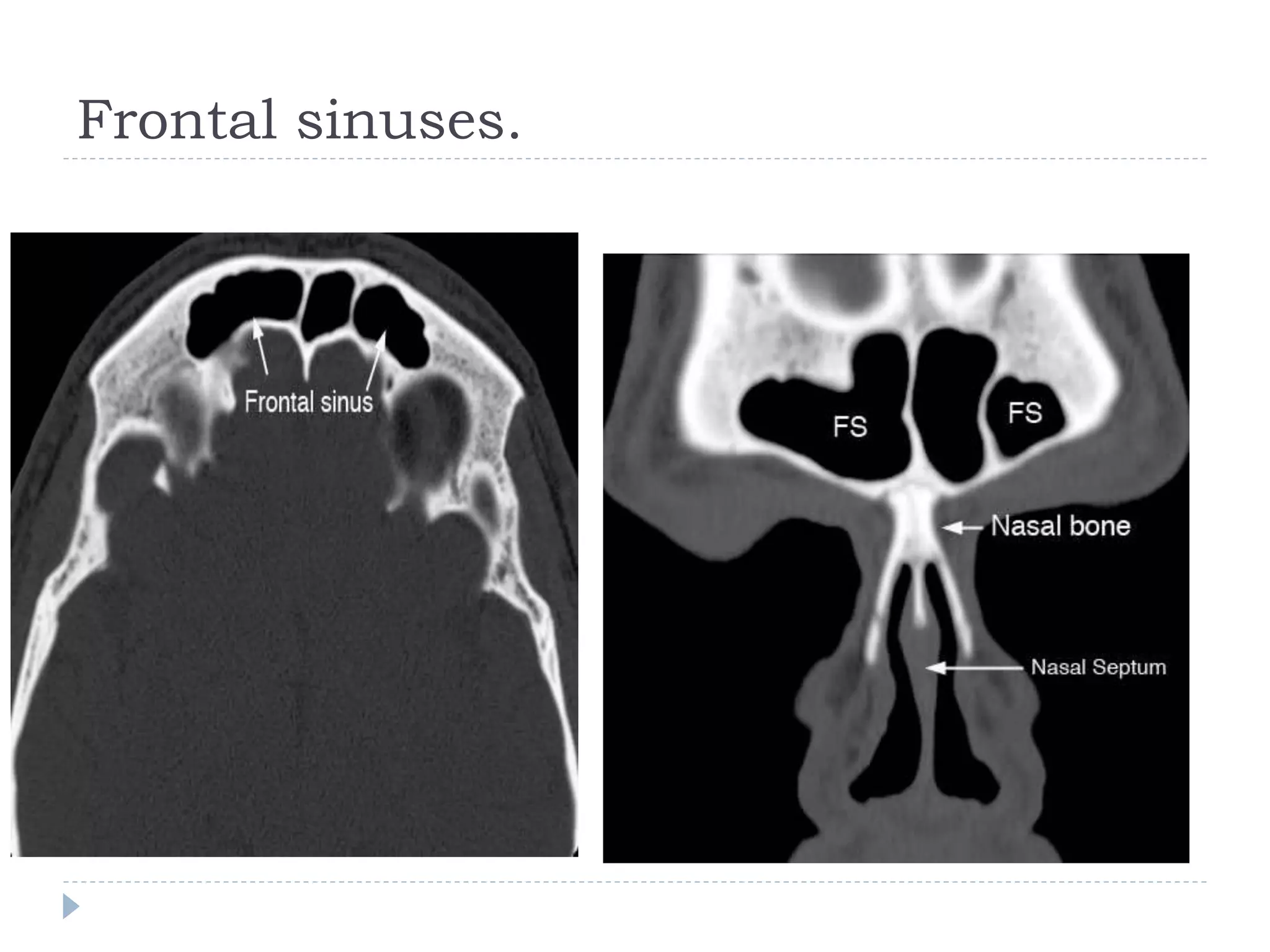

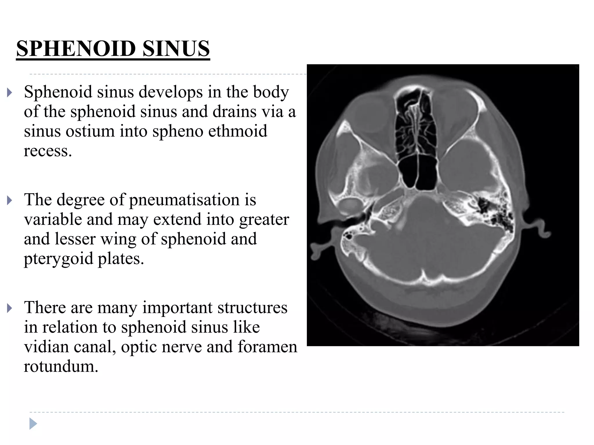


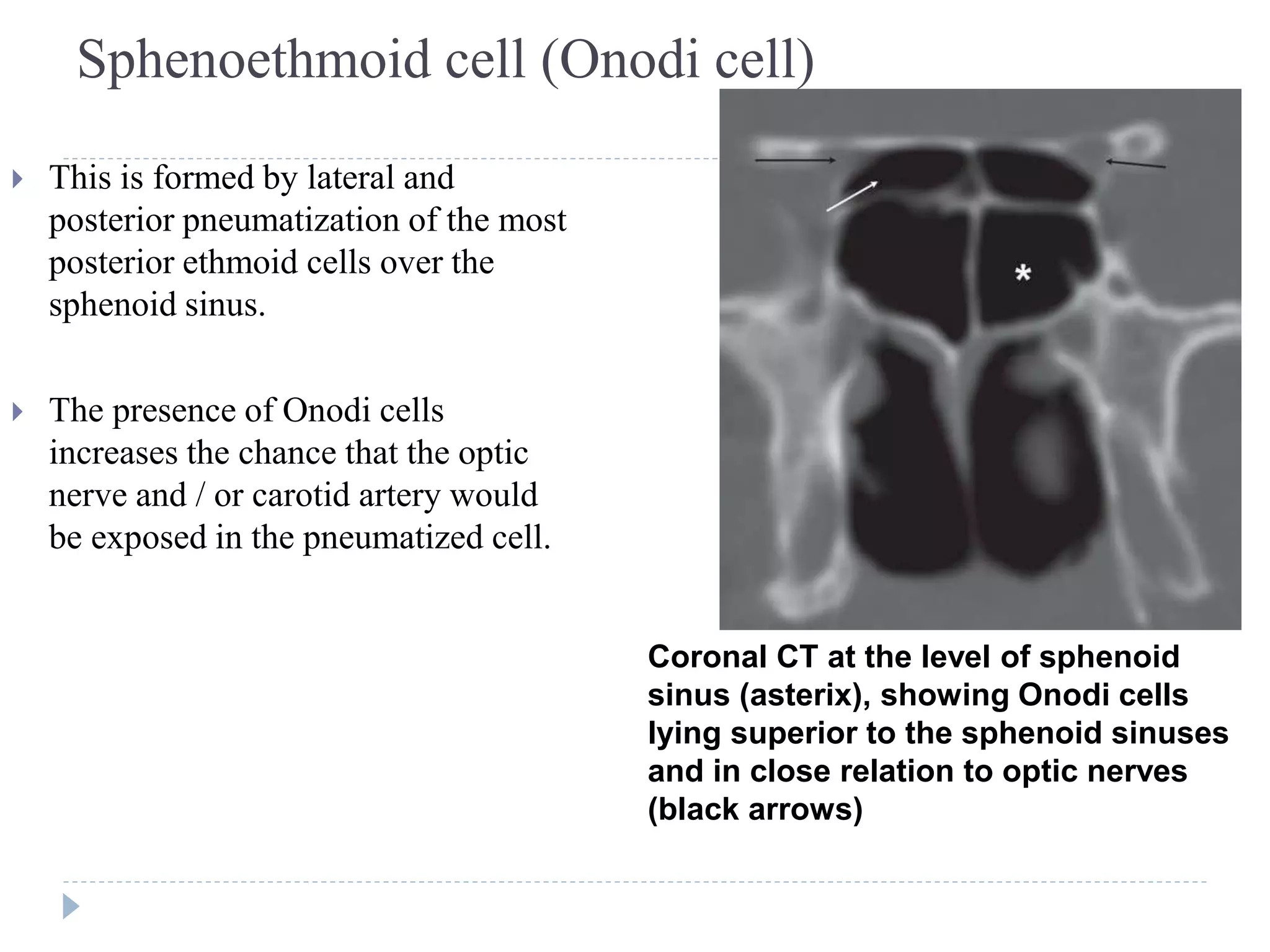


This document discusses the anatomy and imaging of the paranasal sinuses. It describes the drainage pathways of each sinus and the structures that make up the osteomeatal complex. It also covers anatomical variations that can occur like concha bullosa, Haller cells, and Onodi cells. Imaging modalities for evaluating the sinuses are described, with CT identified as the gold standard due to its ability to depict bone, soft tissues, and air. Scanning techniques for CT include coronal sections performed with the patient in a prone position and their head hyperextended.


![Radiological anatomy of_temporal_bone[1]](https://cdn.slidesharecdn.com/ss_thumbnails/radiologicalanatomyoftemporalbone1-171112100915-thumbnail.jpg?width=640&height=640&fit=bounds)


