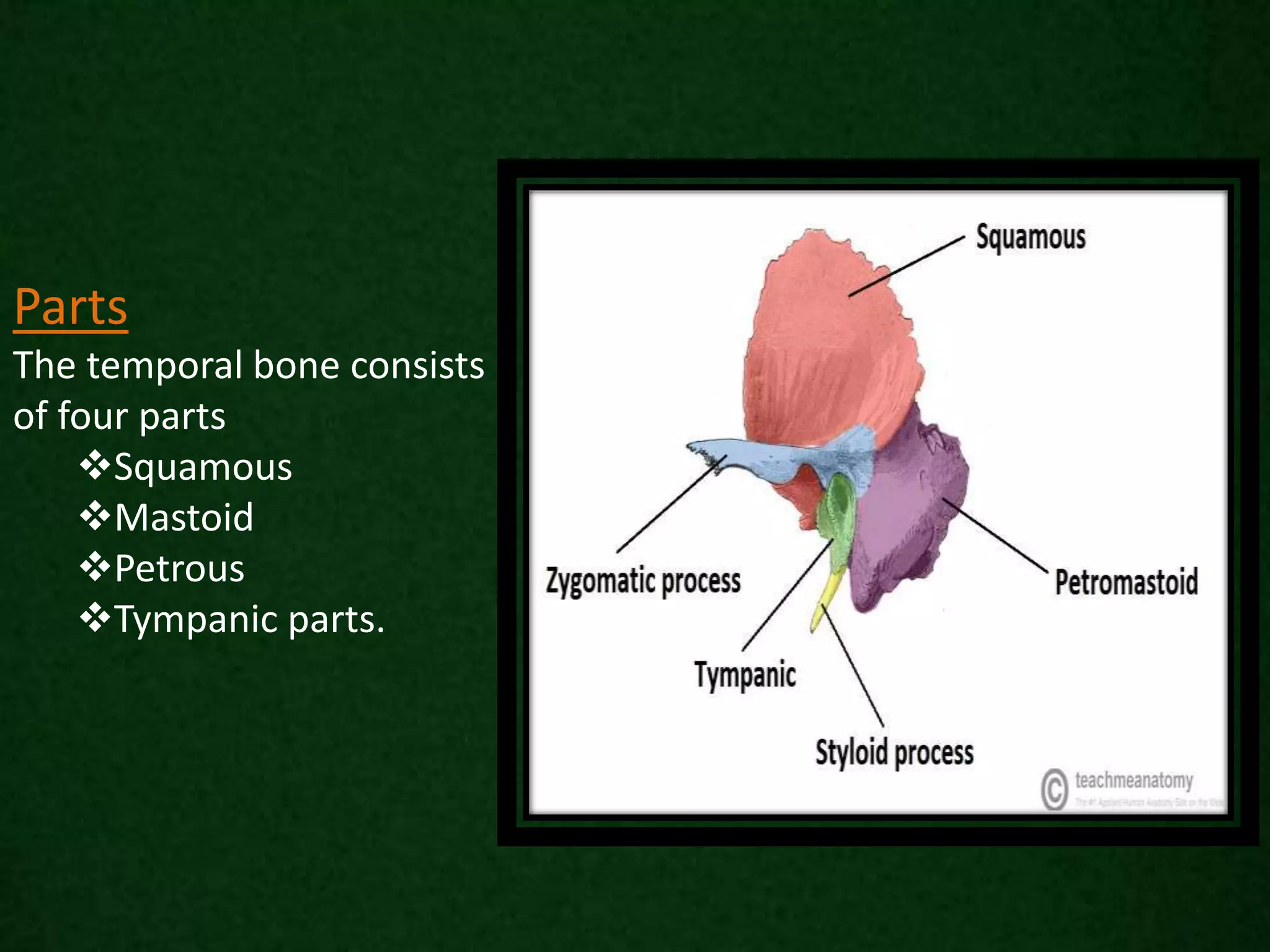
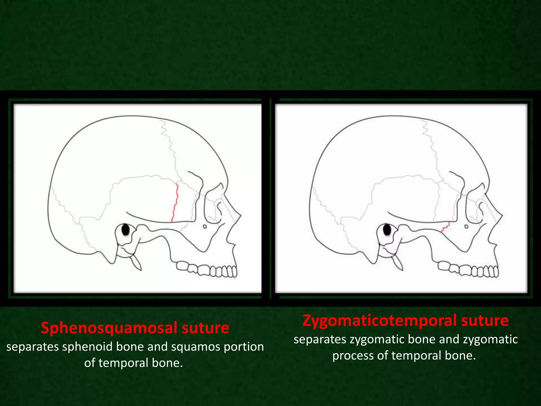
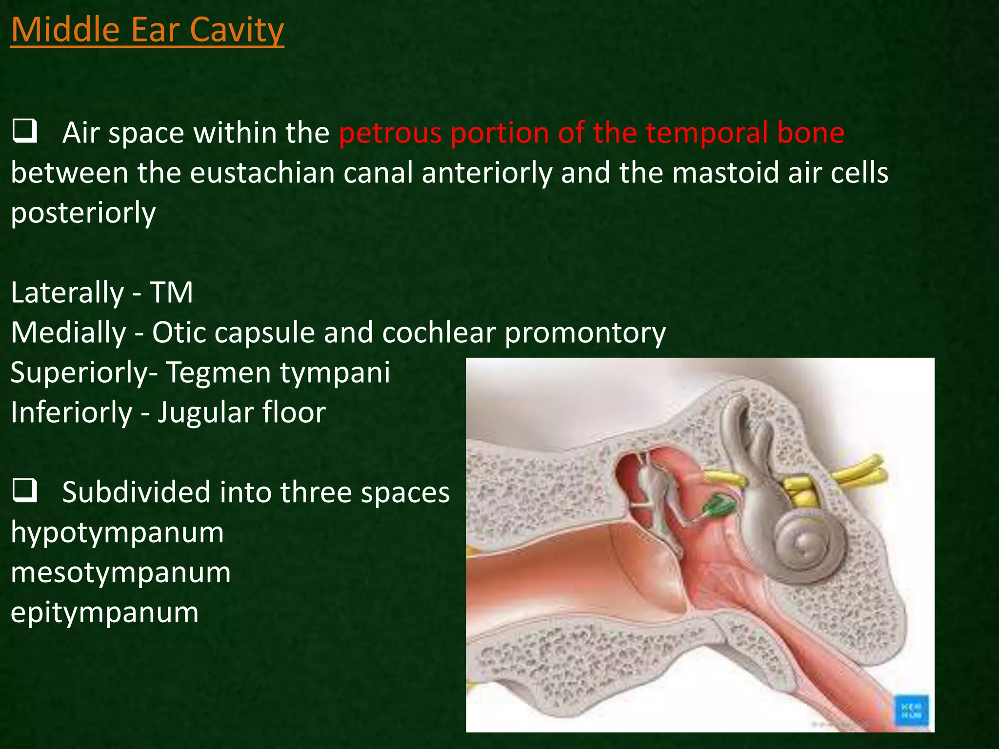
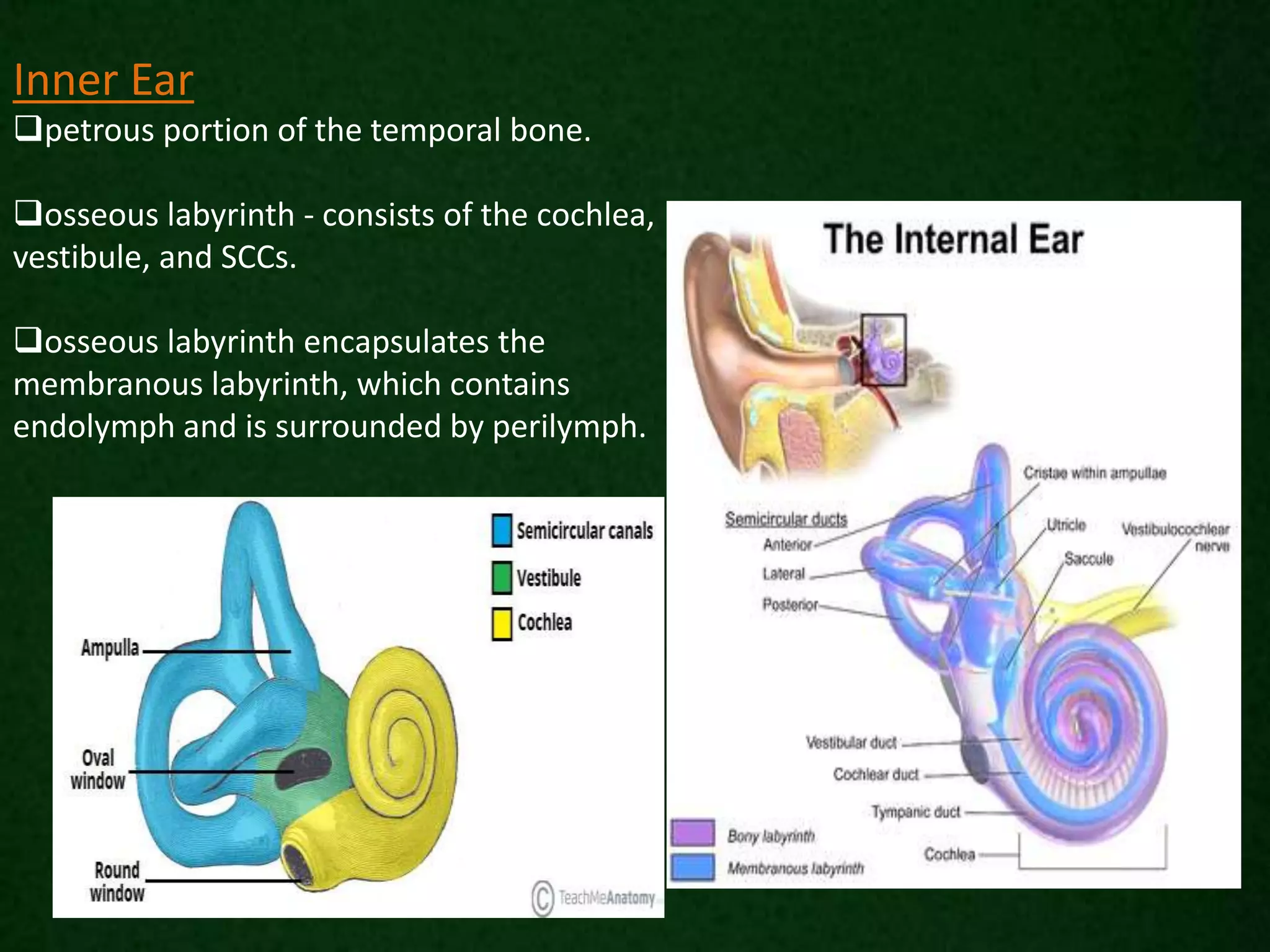
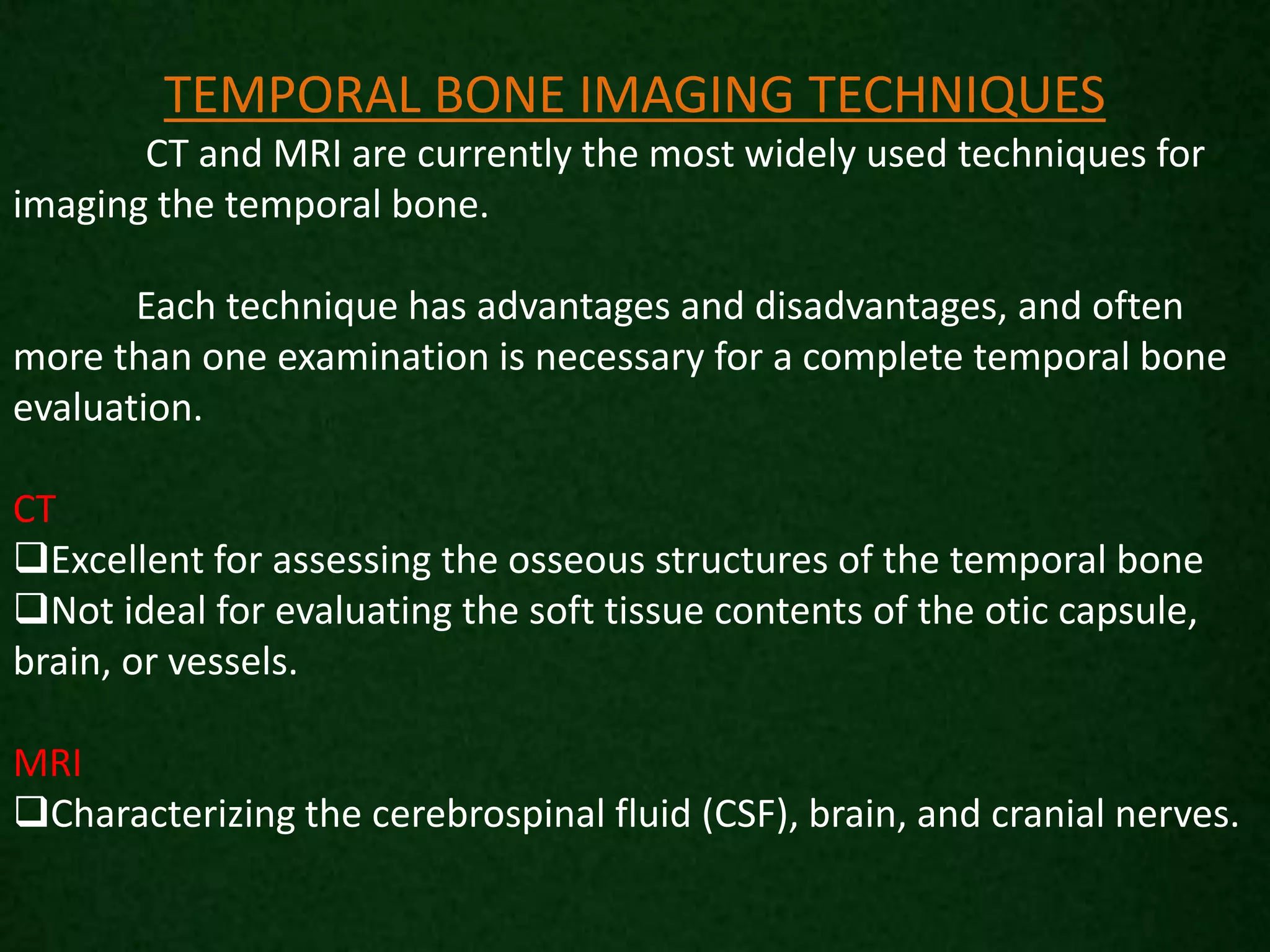
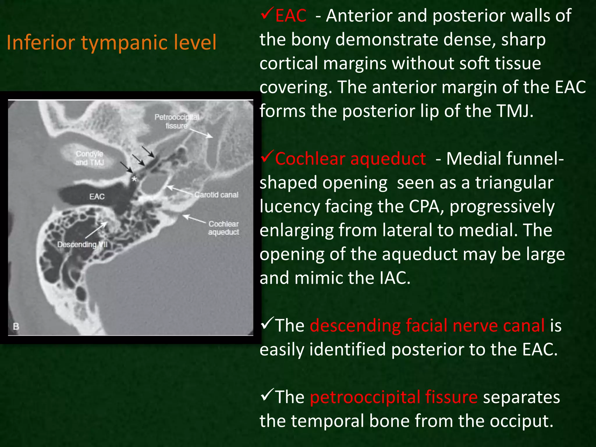
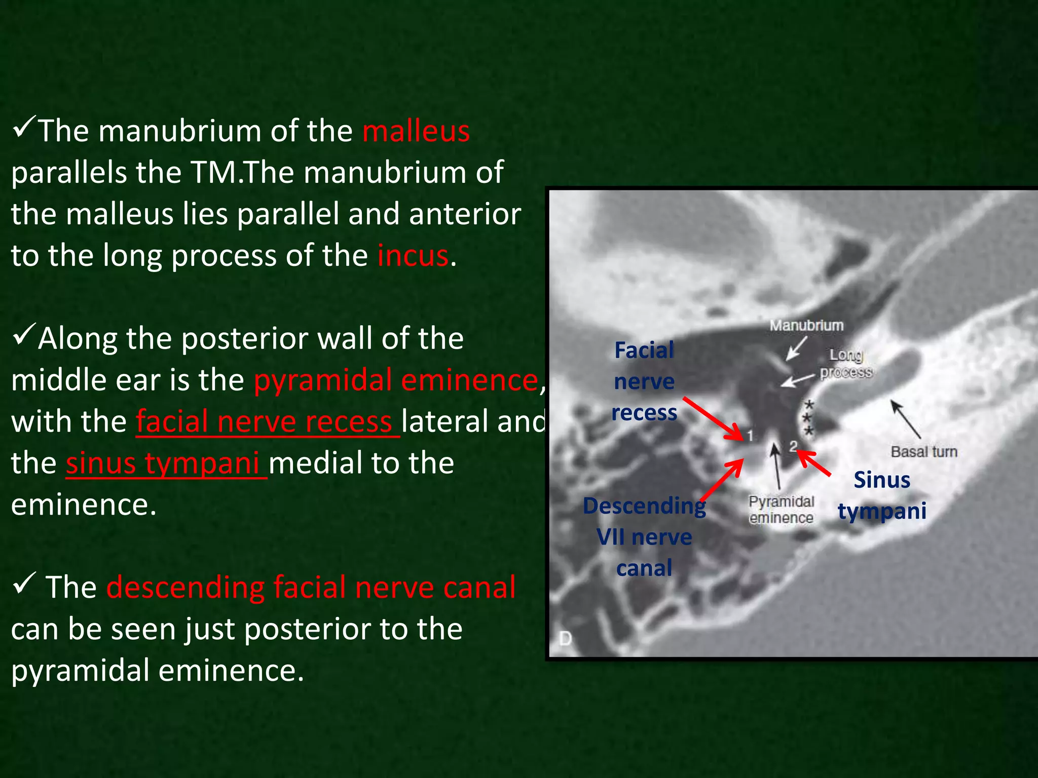
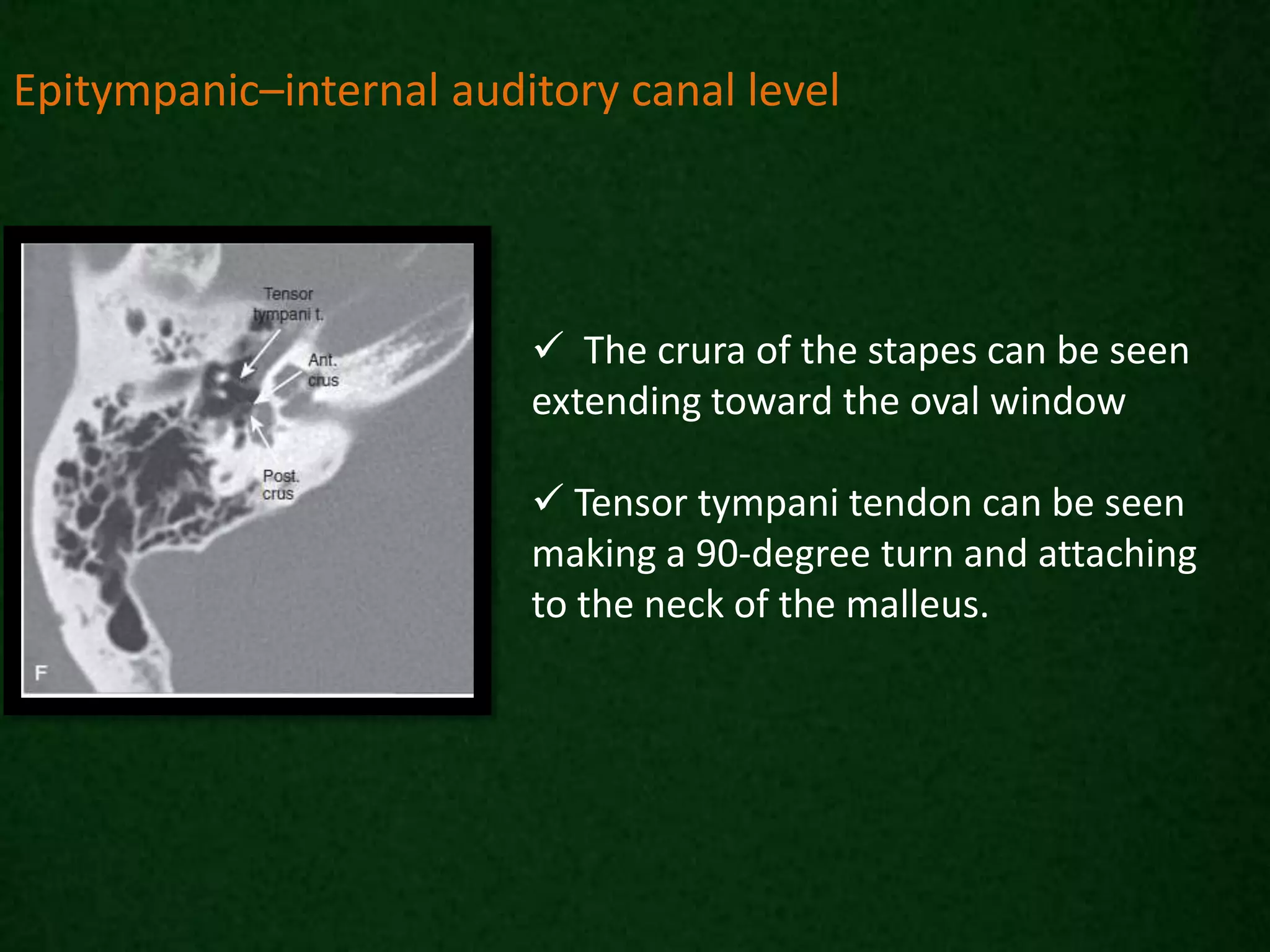
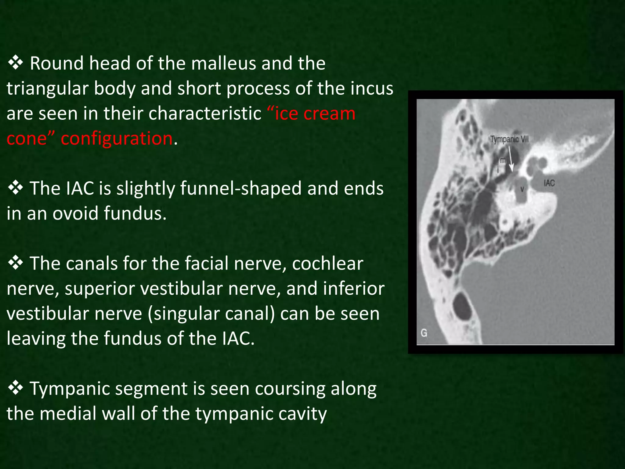
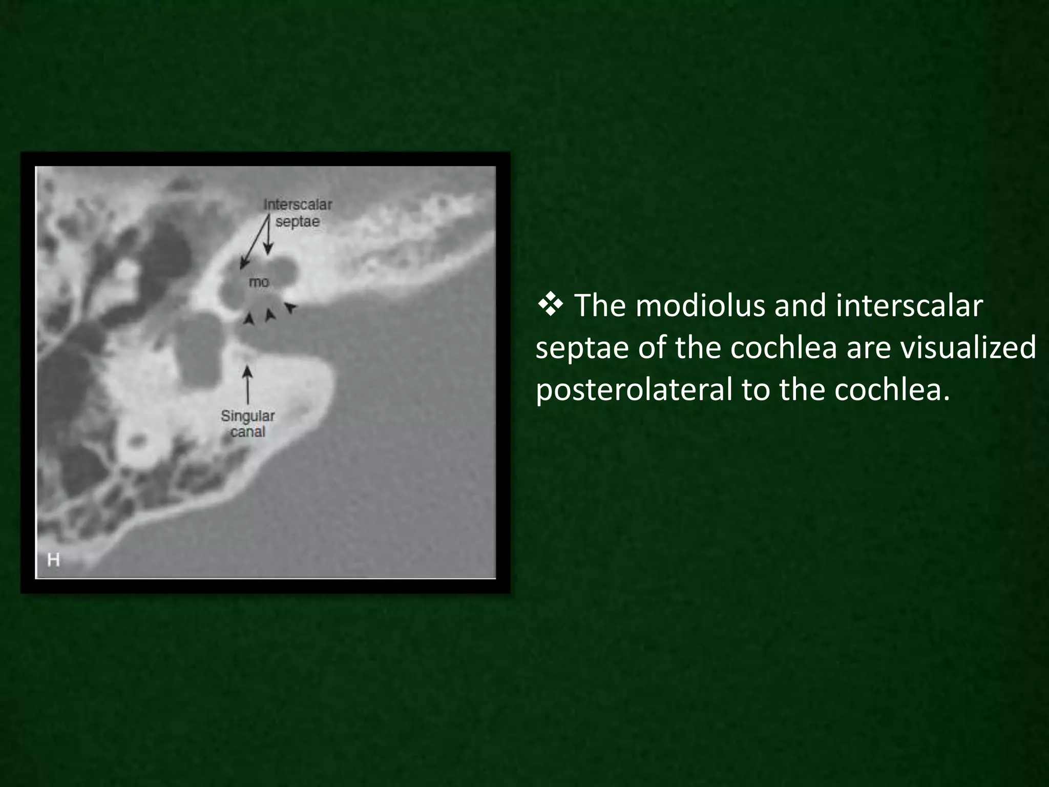
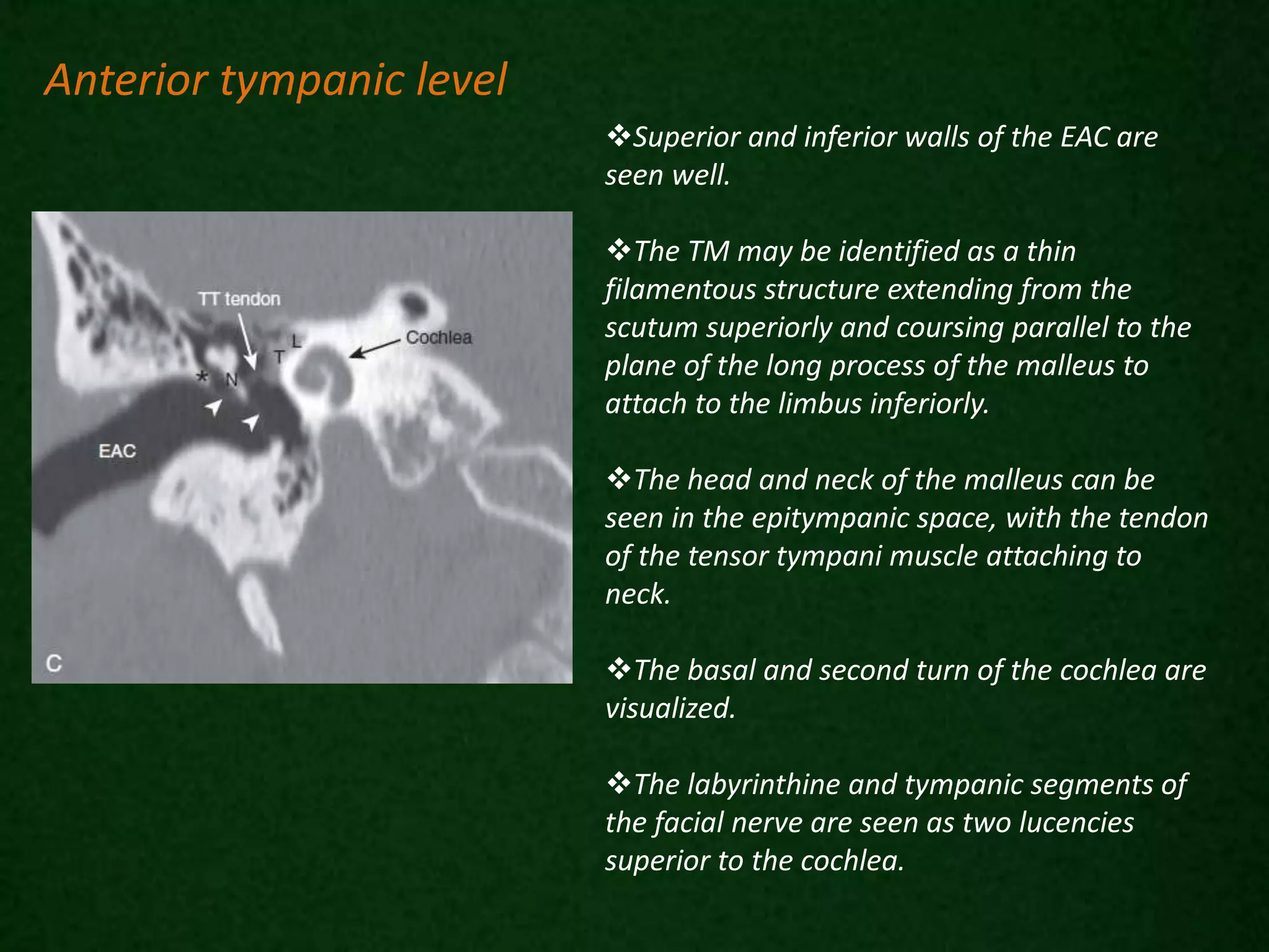
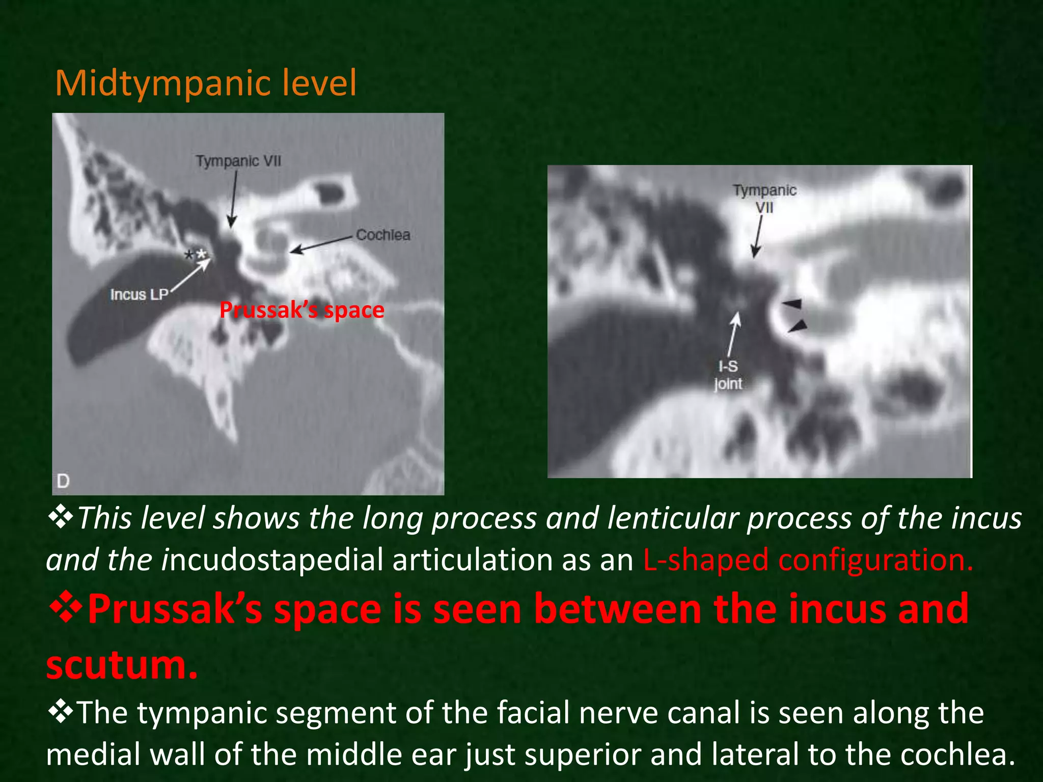
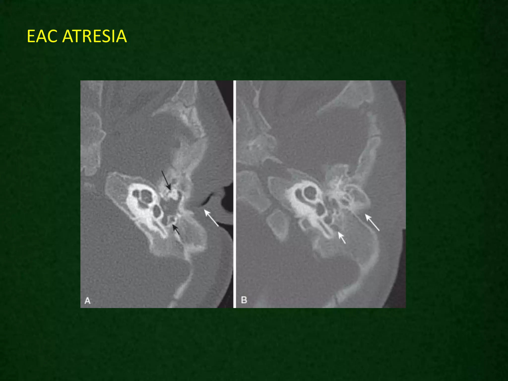
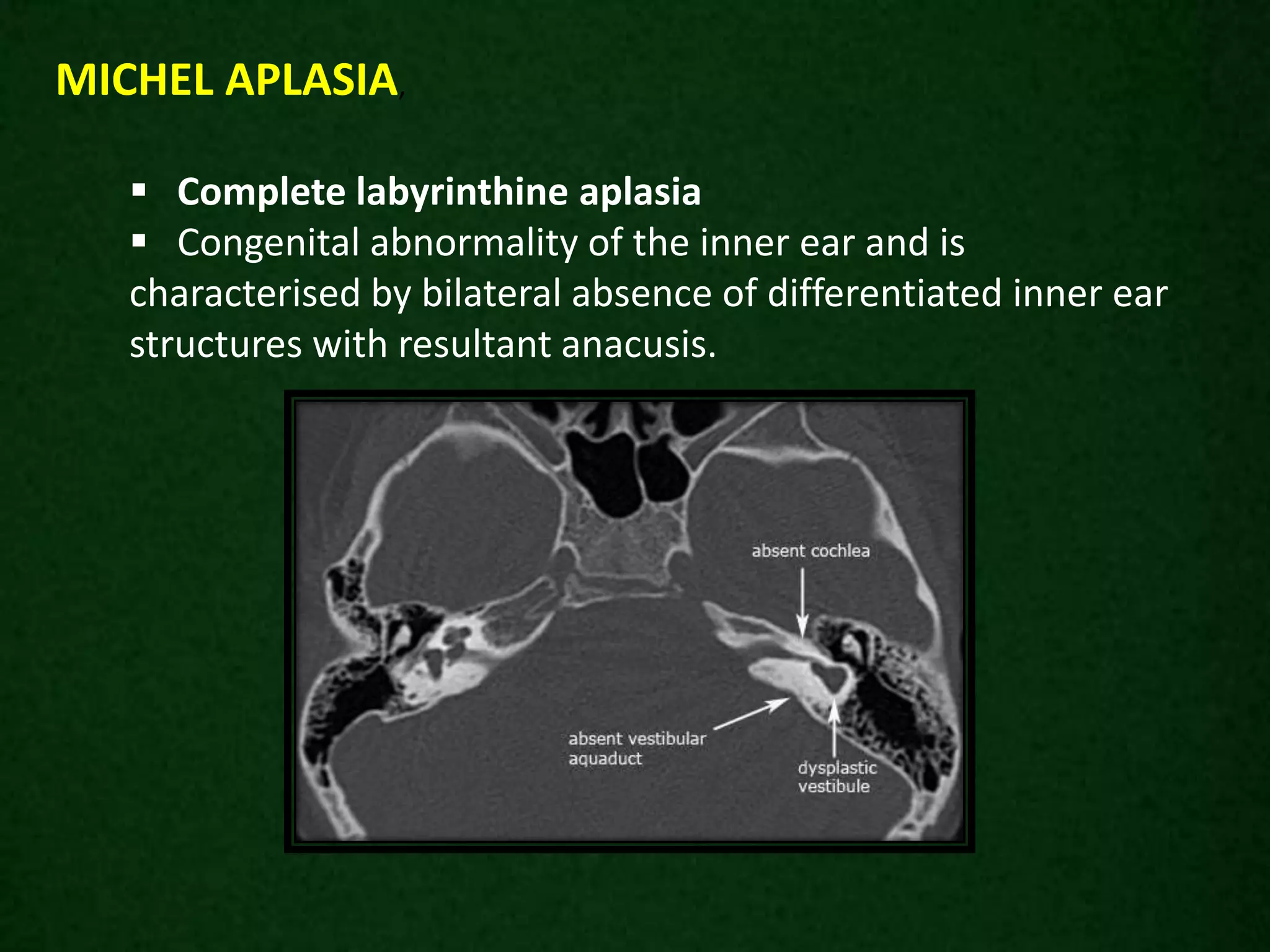
SlideShare a Scribd company logo
RADIOLOGICAL ANATOMY OF
TEMPORAL BONE
- Dr.N.Suriyaprakash
JR , Dept of RadioDiagnosis.11/12/2017
Anatomy of temporal bone
Location
The temporal bone is
situated on the sides and
the base of the cranium
and lateral to the temporal
lobe of cerebrum.
Parts
The temporal bone consists
of four parts
Squamous
Mastoid
Petrous
Tympanic parts.
Mastoid process
 The mastoid process is pyramidal bony
projection located in the posterior portion of the
temporal bone.
Styloid process
 Slender pointed part of temporal bone.
 It projects inferiorly and anteriorly from the inferior surface
of the temporal bone.
Zygomatic process
Borders / Sutures
Occipitomastoid suture
separates occipital bone and mastoid
portion of temporal bone.
Squamosal suture separates parietal
bone and squamos portion of temporal bone.
Sphenosquamosal suture
separates sphenoid bone and squamos portion
of temporal bone.
Zygomaticotemporal suture
separates zygomatic bone and zygomatic
process of temporal bone.
External acoustic meatus
Cartilaginous lateral portion
Funnel-shaped
The cartilaginous portion is
continuous with the auricle, flexible
and surrounded by fat.
Osseous medial portion
Surrounded by the tympanic
portion of the temporal bone
Two thirds of the length of the
EAC
 Covered by skin and periosteum
only.
Middle Ear Cavity
 Air space within the petrous portion of the temporal bone
between the eustachian canal anteriorly and the mastoid air cells
posteriorly
Laterally - TM
Medially - Otic capsule and cochlear promontory
Superiorly- Tegmen tympani
Inferiorly - Jugular floor
 Subdivided into three spaces
hypotympanum
mesotympanum
epitympanum
Mastoid antrum is located in the
anterosuperior part of the mastoid
portion of the temporal bone and
communicates with the epitympanum
via a narrow channel termed the
aditus ad antrum.
The antrum is surrounded by smaller
variable-sized mastoid air cells
The mastoid air cells are divided by
Koerner’s septum a thin bony structure
formed by the petrosquamous suture
that extends posteriorly from the
epitympanum, into medial and lateral
components.
Mastoid
Ossicles
 Suspended by the TM, the ossicular ligaments to the epitympanic
walls, and the oval window.
 The manubrium and lateral process of the malleus attach to the TM.
The neck of the malleus is connected to the tensor tympani tendon .The
circular head of the malleus articulates with the triangular body of the
incus.
The short process of the incus projects posteriorly and is suspended
within the epitympanic space, pointing toward the aditus ad antrum.
The long process of the incus projects inferiorly, parallel to and behind
the manubrium of the malleus. The tip of the long process bends
medially to end in the lenticular process, which articulates with the
head of the stapes
The stapes consists of the head, two crura, and a footplate .
The footplate of the stapes attaches to the oval window of the
vestibule and is secured by the annular ligament.
Inner Ear
petrous portion of the temporal bone.
osseous labyrinth - consists of the cochlea,
vestibule, and SCCs.
osseous labyrinth encapsulates the
membranous labyrinth, which contains
endolymph and is surrounded by perilymph.
Internal Auditory Canal
 Bony conduit within the petrous portion of the
temporal bone .
 Transmits VII (facial) and VIII (vestibulocochlear) from
the pontomedullary junction of the brainstem across the
cerebellopontine angle (CPA) cistern to the inner ear .
The IACs should be nearly symmetric. Although there is
wide variation in the exact shape and size of the canals,
asymmetry of greater than 2 mm suggests pathology.
The IAC is partially divided by a transverse bony crest called the
crista falciformis which runs parallel to the long axis of the canal and
divides it into a superior and inferior portion.
A vertical crest termed Bill’s bar divides the superior component into
anterior and posterior parts. The facial nerve is located in
anterosuperior compartment, the cochlear nerve in the
anteroinferior compartment, and the superior and inferior vestibular
nerves in the posterosuperior and posteroinferior compartments,
respectively
Carotid Canal
 The carotid canal enters
the base of the skull, ascends
vertically, then turns
horizontally and medially
toward the petrous apex
 This canal lies anterior
and inferior to the cochlea
and is separated from the
middle ear cavity by a thin
bony plate.
Jugular Foramen and Fossa
The jugular foramen is divided
 smaller anteromedial neural
compartment (pars nervosa) containing
cranial nerves IX, X, and XI
 larger posterolateral vascular
compartment (the pars vascularis)
containing the jugular vein.
The floor of the tympanic cavity
normally forms the roof of the jugular
fossa.
FACIAL NERVE
 Facial nerve (VII cranial) consists of a motor and
sensory root, the latter is also known as the nervous
intermedius
NUCLEI OF ORIGIN
Three brainstem nuclei contribute to the facial nerve: one motor,
one secretomotor, and one sensory:
 Facial nerve nucleus in the pons as the motor fibres of the
facial nerve loop posteriorly over the abducens nerve nucleus,
they form the facial colliculus in the floor of fourth ventricle
 Superior salivary nucleus next to the facial nucleus supplies
secretomotor parasympathetic fibres
 Nucleus of tractus solitaris lateral to the dorsal nucleus of
the vagus nerve supplies taste fibres that eventually end up in
the chorda tympani
COURSE
 Course of the facial nerve may be divided by the
stylomastoid foramen into intracranial- intrapetrous part and
extracranial part
INTRACRANIAL - INTRAPETROSAL PART
 Intracranial (cisternal) segment - zero branches
 Meatal (canalicular) segment (internal auditory canal): 8 mm
long, zero branches
 Labyrinthine segment (IAC to geniculate ganglion): 3-4 mm
long, 3 branches (from geniculate ganglion)
 Tympanic segment (from geniculate ganglion to pyramidal
eminence): 8-11 mm long, zero branches
 Mastoid segment (from pyramidal eminence to stylomastoid
foramen): 8-14 mm long, 3 branches
Intracranial (cisternal) segment
 The nerve emerges immediately
beneath the pons, lateral to
the abducens nerve and medial to
the vestibulocochlear nerve and is
joined by the nervus intermedius,
which has emerged lateral to the
main trunk.
 Together the two travel laterally
through the cerebellopontine
angle to the internal acoustic
meatus.
 The cisternal segment has no
branches.
Meatal (canalicular) segment
 Having been joined by
the nervus intermedius,they are
located in the anterior superior
quadrant of the internal auditory
canal, above the falciform crest
and anterior to Bill's bar.
The meatal segment has no
branches.
Labyrinthine segment
As the facial nerve and nervus
intermedius pass through the
anterior superior quadrant of
the internal acoustic meatusit enters
the Fallopian canal passing
anterolaterally between and superior
to the cochlea(anterior)
and vestibule(posterior), and then
bends posteriorly (the anterior or first
genu) at the geniculate
ganglionwhere the nervus
intermedius joins the facial nerve and
where fibers for tte synapse .
three branches originate:
 greater superficial petrosal nerve
 lesser petrosal nerve
 external petrosal nerve
shortest only measuring 3-4 mm
narrowest and the most susceptible to vascular compromise
Tympanic segment
As the nerve passes posteriorly
from the geniculate ganglion it
becomes the tympanic segment (8-
11 mm in length) and is
immediately beneath the lateral
semicircular canal in the medial
wall of the middle ear cavity.
The nerve passes posterior to
the cochleariform process, tensor
tympani and oval window Just
distal to the pyramidal eminence
the nerve makes a second turn
(posterior or second genu) passing
vertically downwards as the
mastoid segment.
 The tympanic segment has no
branches.
Mastoid segment
 Measures 8-14 mm in length
 Extends from the posterior genu
to the stylomastoid foramen
 It runs in the medial wall of the
aditus ad antrum of the mastoid.
 It gives off three branches
 Nerve to stapedius
 Chorda tympani terminal branch
of the nervus intermedius
 Nerve from the auricular branch
of the vagus nerve (CN X)
Extra-temporal segment
As the nerve exits the stylomastoid foramen,
 Sensory branch that supplies part of the external acoustic
meatus and tympanic membrane.
 Muscular branches - Passes between the posterior belly of
the digastric muscle and the stylohyoid muscle, supplying both
and then enters the parotid gland.
 Lying between the deep and superficial lobes of the gland the
nerve divides into two main branches at the pes anserinus -
superior temporofacial and inferior cervicofacial branches.
From the anterior border of the gland, five branches emerge
 Temporal
 Zygomatic
 Buccal
 Mandibular (marginal)
 Cervical
N
The facial nerve is the only cranial nerve that may
show normal post-contrast enhancement.
Typically enhancement is seen:
 Anterior genu (geniculate ganglion)
 Posterior genu (between tympanic and mastoid segments)
 Some enhancement can also be seen in the labrynthine,
tympanic and mastoid segments.
 Proximal greater superfical petrosal nerve
No enhancement should be seen in:
o Cisternal segment (that in the cerebellopontine angle)
o Meatal segment (that in the internal acoustic meatus)
o Extracranial segment (beyond the stylomastoid foramen)
TEMPORAL BONE IMAGING TECHNIQUES
CT and MRI are currently the most widely used techniques for
imaging the temporal bone.
Each technique has advantages and disadvantages, and often
more than one examination is necessary for a complete temporal bone
evaluation.
CT
Excellent for assessing the osseous structures of the temporal bone
Not ideal for evaluating the soft tissue contents of the otic capsule,
brain, or vessels.
MRI
Characterizing the cerebrospinal fluid (CSF), brain, and cranial nerves.
AXIAL SECTIONS
OF
TEMPORAL BONE
The carotid canal lies just anterior to the jugular fossa, forming a “snowman”-
like configuration.
Both demonstrate sharp cortical margins. Inferiorly only a small spine, the
caroticojugular spine, separates the two as they converge to enter the carotid sheath.
The descending facial nerve canal is lateral to the jugular foramen, seen as a
rounded well-corticated lucency.
The mandibular condyle and TMJ can also be seen.
Jugular foramen level.
EAC - Anterior and posterior walls of
the bony demonstrate dense, sharp
cortical margins without soft tissue
covering. The anterior margin of the EAC
forms the posterior lip of the TMJ.
Cochlear aqueduct - Medial funnel-
shaped opening seen as a triangular
lucency facing the CPA, progressively
enlarging from lateral to medial. The
opening of the aqueduct may be large
and mimic the IAC.
The descending facial nerve canal is
easily identified posterior to the EAC.
The petrooccipital fissure separates
the temporal bone from the occiput.
Inferior tympanic level
 The carotid canal can be seen early in
its anteromedial course through the skull
base, parallel and medial to the tensor
tympani muscle at this level.
The tensor tympani muscle is lateral
and parallel to the carotid canal
The normally thin TM may be visible.
The descending facial nerve canal can
be seen just posterior to the pyramidal
eminence.
 The vestibular aqueduct is seen as a
thin bony lucency along the posterior
margin of the temporal bone, near its
opening to the posterior cranial fossa.
Midtympanic level
The manubrium of the malleus
parallels the TM.The manubrium of
the malleus lies parallel and anterior
to the long process of the incus.
Along the posterior wall of the
middle ear is the pyramidal eminence,
with the facial nerve recess lateral and
the sinus tympani medial to the
eminence.
 The descending facial nerve canal
can be seen just posterior to the
pyramidal eminence.
Facial
nerve
recess
Sinus
tympaniDescending
VII nerve
canal
 The stapedial superstructure (head, crura, and tympanic portion of
the footplate) is often seen, forming an arch over the oval window.
The stapes suprastructure is medial to the incus
 The apical, middle, and basal cochlear turns are seen at this level
along with the round window niche at the basal turn.
 The crura of the stapes can be seen
extending toward the oval window
 Tensor tympani tendon can be seen
making a 90-degree turn and attaching
to the neck of the malleus.
Epitympanic–internal auditory canal level
 Round head of the malleus and the
triangular body and short process of the incus
are seen in their characteristic “ice cream
cone” configuration.
 The IAC is slightly funnel-shaped and ends
in an ovoid fundus.
 The canals for the facial nerve, cochlear
nerve, superior vestibular nerve, and inferior
vestibular nerve (singular canal) can be seen
leaving the fundus of the IAC.
 Tympanic segment is seen coursing along
the medial wall of the tympanic cavity
 The modiolus and interscalar
septae of the cochlea are visualized
posterolateral to the cochlea.
 The lateral SCC is seen protruding into the middle ear.
 The labyrinthine segment of the facial nerve canal
(fallopian canal) can be seen extending to the anterior genu,
and the tympanic segment is seen coursing along the medial
wall of the tympanic cavity.
 The aditus and antrum is situated between the epitympanum and
mastoid antrum.
 The mastoid antrum lies posterior and lateral to the aditus ad
antrum and opens into many mastoid air cells.
 The posterior SCC and common crus of the posterior and superior
SCCs can be seen.
 More superiorly the superior SCC is present.
Mastoid antrum level
CORONAL SECTIONS
OF
TEMPORAL BONE
Coronal temporomandibular joint level
 The horizontal carotid canal is seen as an oval structure just
lateral to petrooccipital suture.
 The semicanal for the tensor tympani muscle is seen as a small
lucency lateral to the carotid canal.
 The air-filled eustachian tube is inferior to the tensor tympani,
and the TMJ is seen laterally
 Tensor tympani muscle is seen
along the medial wall of the middle
ear, with the cochleariform process
separating it from the eustachian
tube below.
 The anteriormost part of the
cochlea is medial to the tensor
tympani.
 The geniculate ganglion is seen
as a small lucency superior to the
cochlea
Geniculate ganglion level
Superior and inferior walls of the EAC are
seen well.
The TM may be identified as a thin
filamentous structure extending from the
scutum superiorly and coursing parallel to the
plane of the long process of the malleus to
attach to the limbus inferiorly.
The head and neck of the malleus can be
seen in the epitympanic space, with the tendon
of the tensor tympani muscle attaching to
neck.
The basal and second turn of the cochlea are
visualized.
The labyrinthine and tympanic segments of
the facial nerve are seen as two lucencies
superior to the cochlea.
Anterior tympanic level
This level shows the long process and lenticular process of the incus
and the incudostapedial articulation as an L-shaped configuration.
Prussak’s space is seen between the incus and
scutum.
The tympanic segment of the facial nerve canal is seen along the
medial wall of the middle ear just superior and lateral to the cochlea.
Midtympanic level
Prussak’s space
The full extent of the IAC is well
visualized at this level, with the
central crista falciformis dividing
the canal into two portions.
Oval window is seen as a bony
defect in the lateral portion of the
vestibule.
Beneath the lateral SCC, the
horizontal portion of the facial
nerve canal appears as a small
circular structure.
The epitympanic space lies just
lateral to the lateral SCC.
Oval window level
Posterior tympanic level
 The facial nerve recess is lateral
to and the sinus tympani medial to
the pyramidal eminence.
 The round window niche is seen
along the basal turn of the cochlea.
The jugular foramen – dome
shaped outline.
The mastoid segment of the
facial nerve canal can be identified
lateral to the jugular foramen,
running nearly vertical and
extending toward the stylomastoid
foramen.
The mastoid antrum is seen
superiorly and laterally.
Portions of the lateral and
superior SCCs can be seen.
Jugular foramen level
SPECIAL VIEWS
Stenvers’ view
Stenvers’ view MIDSUPERIOR SCC
LEVEL. The superior SCC is seen in cross
section.
Stenvers’ view COMMON CRUS LEVEL. In
addition to common crus, portions of the
posterior SCC and superior SCC are seen.
 Oblique coronal projection.
 Superior anatomic detail.
 Used to assess electrode placement following the insertion of
a cochlear implant.
Stenvers’ view COCHLEA LEVEL. The
turns of the cochlea are seen, as is the
round window and descending facial
nerve canal .
Stenvers’ view INCUDOMALLEOLAR
JOINT LEVEL. The incudomalleolar
joint is well seen between body of
incus (I) and head of malleus (M).
Pöschl’s view
 The plane of projection is perpendicular to the long
axis of the temporal bone.
 Projection the temporal bone is imaged from its
anteromedial to posterolateral aspects.
The superior SCC is seen as a ring, and
the posterior SCC is seen in cross section.
The tympanic segment of the facial nerve
canal is seen in cross section.
PÖSCHL’S VIEW
MIDSUPERIOR
SCC LEVEL
The modiolus (m) is seen along its
axis
PÖSCHL’S VIEW MODIOLUS LEVEL
EAC ATRESIA
COCHLEAR ANOMALIES (CLASSIFICATION)
 Have different presentation depending on the timing of
developmental arrest.
Divides congenital cochlear anomalies according to the timing of
the developmental arrest.
Complete labyrinthine aplasia or Michel deformity: 3rd week
Cochlear aplasia : 4th week
Common cavity malformation to the cochlea and vestibule: early
5th week
Cochlear incomplete partition type including Cystic
cochleovestibular anomaly: late 5th week
Cochlear hypoplasia: 6th week
Cochlear incomplete partition type II including mondini
dysplasia: 7th week
 Complete labyrinthine aplasia
 Congenital abnormality of the inner ear and is
characterised by bilateral absence of differentiated inner ear
structures with resultant anacusis.
MICHEL APLASIA,
INCOMPLETE PARTITION - I
(CYSTIC COCHLEOVESTIBULAR MALFORMATION)
Abnormal cochlea
Only 1.5 turns (instead of the
normal 2.5 turns)
Normal basal turn with a cystic
apex in place of the distal 1.5
turns
Enlarged vestibule with
normal semicircular canals
Enlarged vestibular aqueduct
containing a dilated endolymphatic
sac
MONDINI MALFORMATION
incomplete partition type II anomaly with large vestibular
aqueduct.
Thank You . . .

More Related Content

What's hot

Skull base imaging
Skull base imagingSkull base imaging
Skull base imaging
Murali Chand Nallamothu
 
Ct of the larynx
Ct of the larynxCt of the larynx
Ct of the larynx
MuhammadSherwani
 
Magnetic Resonance Imaging Of Inner Ear
Magnetic Resonance Imaging Of Inner EarMagnetic Resonance Imaging Of Inner Ear
Magnetic Resonance Imaging Of Inner Ear
Sarbesh Tiwari
 
Larynx anatomy ct and mri
Larynx anatomy ct and mriLarynx anatomy ct and mri
Larynx anatomy ct and mri
Anish Choudhary
 
Presentation1.pptx, radiological anatomy of the neck.
Presentation1.pptx, radiological anatomy of the neck.Presentation1.pptx, radiological anatomy of the neck.
Presentation1.pptx, radiological anatomy of the neck.Abdellah Nazeer
 
CT anatomy of Neck Spaces RV
CT anatomy of Neck Spaces RVCT anatomy of Neck Spaces RV
CT anatomy of Neck Spaces RV
Roshan Valentine
 
Temporal bone Radiologic anatomy.. In depth
Temporal bone Radiologic anatomy.. In depthTemporal bone Radiologic anatomy.. In depth
Temporal bone Radiologic anatomy.. In depth
Ahmed Hamdy Hamdy
 
Presentation1.pptx, radiological anatomy of the petrous bone.
Presentation1.pptx, radiological anatomy of the petrous bone.Presentation1.pptx, radiological anatomy of the petrous bone.
Presentation1.pptx, radiological anatomy of the petrous bone.
Abdellah Nazeer
 
Diagnostic Imaging of Pharynx & Larynx
Diagnostic Imaging of Pharynx & LarynxDiagnostic Imaging of Pharynx & Larynx
Diagnostic Imaging of Pharynx & Larynx
Mohamed M.A. Zaitoun
 
Radiology day 3 mediastinal anatomy
Radiology day 3   mediastinal anatomyRadiology day 3   mediastinal anatomy
Radiology day 3 mediastinal anatomy
Vibhay Pareek
 
Presentation2.pptx. posterior fossa tumour
Presentation2.pptx. posterior fossa tumourPresentation2.pptx. posterior fossa tumour
Presentation2.pptx. posterior fossa tumourAbdellah Nazeer
 
Diagnostic Imaging of Deep Neck Spaces
Diagnostic Imaging of Deep Neck SpacesDiagnostic Imaging of Deep Neck Spaces
Diagnostic Imaging of Deep Neck Spaces
Mohamed M.A. Zaitoun
 
Radiological approach to neck spaces
Radiological approach to neck spacesRadiological approach to neck spaces
Radiological approach to neck spaces
Milan Silwal
 
Oral cavity, pharynx radio-anatomy
Oral cavity, pharynx radio-anatomyOral cavity, pharynx radio-anatomy
Oral cavity, pharynx radio-anatomy
Dr. Mohit Goel
 
IMAGING OF TEMPORAL BONE
IMAGING OF TEMPORAL BONEIMAGING OF TEMPORAL BONE
IMAGING OF TEMPORAL BONE
Sameer Peer
 
Imaging of the temporal bone
Imaging of the temporal bone Imaging of the temporal bone
Imaging of the temporal bone
Kanu Saha
 
SKULL BASE IMAGING
SKULL BASE IMAGINGSKULL BASE IMAGING
Radiological Imaging in Head and Neck and relevant anatomy
Radiological Imaging in Head and Neck and relevant anatomyRadiological Imaging in Head and Neck and relevant anatomy
Radiological Imaging in Head and Neck and relevant anatomy
Vibhay Pareek
 
Imaging of the neck part i
Imaging of the neck part iImaging of the neck part i
Imaging of the neck part i
Wafik Ebrahim
 
ANATOMY OF NASOPHARYNX AND STAGING CORRELATION
ANATOMY OF NASOPHARYNX AND STAGING CORRELATION ANATOMY OF NASOPHARYNX AND STAGING CORRELATION
ANATOMY OF NASOPHARYNX AND STAGING CORRELATION
Kanhu Charan
 

What's hot (20)

Skull base imaging
Skull base imagingSkull base imaging
Skull base imaging
 
Ct of the larynx
Ct of the larynxCt of the larynx
Ct of the larynx
 
Magnetic Resonance Imaging Of Inner Ear
Magnetic Resonance Imaging Of Inner EarMagnetic Resonance Imaging Of Inner Ear
Magnetic Resonance Imaging Of Inner Ear
 
Larynx anatomy ct and mri
Larynx anatomy ct and mriLarynx anatomy ct and mri
Larynx anatomy ct and mri
 
Presentation1.pptx, radiological anatomy of the neck.
Presentation1.pptx, radiological anatomy of the neck.Presentation1.pptx, radiological anatomy of the neck.
Presentation1.pptx, radiological anatomy of the neck.
 
CT anatomy of Neck Spaces RV
CT anatomy of Neck Spaces RVCT anatomy of Neck Spaces RV
CT anatomy of Neck Spaces RV
 
Temporal bone Radiologic anatomy.. In depth
Temporal bone Radiologic anatomy.. In depthTemporal bone Radiologic anatomy.. In depth
Temporal bone Radiologic anatomy.. In depth
 
Presentation1.pptx, radiological anatomy of the petrous bone.
Presentation1.pptx, radiological anatomy of the petrous bone.Presentation1.pptx, radiological anatomy of the petrous bone.
Presentation1.pptx, radiological anatomy of the petrous bone.
 
Diagnostic Imaging of Pharynx & Larynx
Diagnostic Imaging of Pharynx & LarynxDiagnostic Imaging of Pharynx & Larynx
Diagnostic Imaging of Pharynx & Larynx
 
Radiology day 3 mediastinal anatomy
Radiology day 3   mediastinal anatomyRadiology day 3   mediastinal anatomy
Radiology day 3 mediastinal anatomy
 
Presentation2.pptx. posterior fossa tumour
Presentation2.pptx. posterior fossa tumourPresentation2.pptx. posterior fossa tumour
Presentation2.pptx. posterior fossa tumour
 
Diagnostic Imaging of Deep Neck Spaces
Diagnostic Imaging of Deep Neck SpacesDiagnostic Imaging of Deep Neck Spaces
Diagnostic Imaging of Deep Neck Spaces
 
Radiological approach to neck spaces
Radiological approach to neck spacesRadiological approach to neck spaces
Radiological approach to neck spaces
 
Oral cavity, pharynx radio-anatomy
Oral cavity, pharynx radio-anatomyOral cavity, pharynx radio-anatomy
Oral cavity, pharynx radio-anatomy
 
IMAGING OF TEMPORAL BONE
IMAGING OF TEMPORAL BONEIMAGING OF TEMPORAL BONE
IMAGING OF TEMPORAL BONE
 
Imaging of the temporal bone
Imaging of the temporal bone Imaging of the temporal bone
Imaging of the temporal bone
 
SKULL BASE IMAGING
SKULL BASE IMAGINGSKULL BASE IMAGING
SKULL BASE IMAGING
 
Radiological Imaging in Head and Neck and relevant anatomy
Radiological Imaging in Head and Neck and relevant anatomyRadiological Imaging in Head and Neck and relevant anatomy
Radiological Imaging in Head and Neck and relevant anatomy
 
Imaging of the neck part i
Imaging of the neck part iImaging of the neck part i
Imaging of the neck part i
 
ANATOMY OF NASOPHARYNX AND STAGING CORRELATION
ANATOMY OF NASOPHARYNX AND STAGING CORRELATION ANATOMY OF NASOPHARYNX AND STAGING CORRELATION
ANATOMY OF NASOPHARYNX AND STAGING CORRELATION
 

Similar to Radiological anatomy of_temporal_bone[1]

Anatomy of temporal bone By Dr.Vijay kumar , AMU
Anatomy of temporal bone By Dr.Vijay kumar , AMUAnatomy of temporal bone By Dr.Vijay kumar , AMU
Anatomy of temporal bone By Dr.Vijay kumar , AMU
vijaymgims
 
TEMPORAL BONE.pptx
TEMPORAL BONE.pptxTEMPORAL BONE.pptx
TEMPORAL BONE.pptx
DrRashidKaziMahbubur
 
Anatomy of skull base
Anatomy of skull baseAnatomy of skull base
Anatomy of skull base
Dr. Shilpa M J
 
Middle ear anatomy
Middle ear anatomyMiddle ear anatomy
Middle ear anatomy
Amro1988
 
Anatomy of temporal bone final
Anatomy of temporal bone finalAnatomy of temporal bone final
Anatomy of temporal bone final
DR. KANIKA BHARGAVA
 
Anatomy of human ear
Anatomy of human earAnatomy of human ear
Anatomy of human ear
Dr Safika Zaman
 
Skull base anatomy by Dr. Aditya Tiwari
Skull base anatomy by Dr. Aditya TiwariSkull base anatomy by Dr. Aditya Tiwari
Skull base anatomy by Dr. Aditya Tiwari
Aditya Tiwari
 
997610 anatomy-head
997610 anatomy-head997610 anatomy-head
997610 anatomy-headabctutor
 
The Head - Human Anatomy
The Head - Human AnatomyThe Head - Human Anatomy
The Head - Human Anatomy
Examville.com LLC
 
997610 anatomy-head
997610 anatomy-head997610 anatomy-head
997610 anatomy-headYoAmoNYC
 
Anatomy facial nerve
Anatomy facial nerveAnatomy facial nerve
Anatomy facial nerve
محمد حسين خميس
 
Cranial bones
Cranial bonesCranial bones
Cranial bones
AnibalSoledispa
 
Blood supply of head and neck
Blood supply of head and neckBlood supply of head and neck
Blood supply of head and neck
Dr.Vamsi Reddy
 
Trigeminal and facial nerve.pptx
Trigeminal and facial nerve.pptxTrigeminal and facial nerve.pptx
Trigeminal and facial nerve.pptx
Ankur Rathaur
 
Trigeminal and facial nerve.pptx
Trigeminal and facial nerve.pptxTrigeminal and facial nerve.pptx
Trigeminal and facial nerve.pptx
Ankur Rathaur
 
Facial Nerve Presentation by Dr Salison Salim Panicker ENT surgeon.pptx
Facial Nerve Presentation by Dr Salison Salim Panicker ENT surgeon.pptxFacial Nerve Presentation by Dr Salison Salim Panicker ENT surgeon.pptx
Facial Nerve Presentation by Dr Salison Salim Panicker ENT surgeon.pptx
salisonsalim1
 
Lec 5 skull
Lec 5 skullLec 5 skull
Lec 5 skull
Meinah Razak
 
Cranial fossae in base of the human skull
Cranial fossae in base of the human skullCranial fossae in base of the human skull
Cranial fossae in base of the human skull
WajahatFaiz4
 
Lateral skull base anatomy and applied science by Dr, bomkar bam
Lateral skull base anatomy and applied science by Dr, bomkar bamLateral skull base anatomy and applied science by Dr, bomkar bam
Lateral skull base anatomy and applied science by Dr, bomkar bam
Bomkar Bam
 
Group 2 Medsurg ppt on Anatomy and physiology of ear-1.pptx
Group 2 Medsurg ppt on Anatomy and physiology of ear-1.pptxGroup 2 Medsurg ppt on Anatomy and physiology of ear-1.pptx
Group 2 Medsurg ppt on Anatomy and physiology of ear-1.pptx
IjeomaNwokoro
 

Similar to Radiological anatomy of_temporal_bone[1] (20)

Anatomy of temporal bone By Dr.Vijay kumar , AMU
Anatomy of temporal bone By Dr.Vijay kumar , AMUAnatomy of temporal bone By Dr.Vijay kumar , AMU
Anatomy of temporal bone By Dr.Vijay kumar , AMU
 
TEMPORAL BONE.pptx
TEMPORAL BONE.pptxTEMPORAL BONE.pptx
TEMPORAL BONE.pptx
 
Anatomy of skull base
Anatomy of skull baseAnatomy of skull base
Anatomy of skull base
 
Middle ear anatomy
Middle ear anatomyMiddle ear anatomy
Middle ear anatomy
 
Anatomy of temporal bone final
Anatomy of temporal bone finalAnatomy of temporal bone final
Anatomy of temporal bone final
 
Anatomy of human ear
Anatomy of human earAnatomy of human ear
Anatomy of human ear
 
Skull base anatomy by Dr. Aditya Tiwari
Skull base anatomy by Dr. Aditya TiwariSkull base anatomy by Dr. Aditya Tiwari
Skull base anatomy by Dr. Aditya Tiwari
 
997610 anatomy-head
997610 anatomy-head997610 anatomy-head
997610 anatomy-head
 
The Head - Human Anatomy
The Head - Human AnatomyThe Head - Human Anatomy
The Head - Human Anatomy
 
997610 anatomy-head
997610 anatomy-head997610 anatomy-head
997610 anatomy-head
 
Anatomy facial nerve
Anatomy facial nerveAnatomy facial nerve
Anatomy facial nerve
 
Cranial bones
Cranial bonesCranial bones
Cranial bones
 
Blood supply of head and neck
Blood supply of head and neckBlood supply of head and neck
Blood supply of head and neck
 
Trigeminal and facial nerve.pptx
Trigeminal and facial nerve.pptxTrigeminal and facial nerve.pptx
Trigeminal and facial nerve.pptx
 
Trigeminal and facial nerve.pptx
Trigeminal and facial nerve.pptxTrigeminal and facial nerve.pptx
Trigeminal and facial nerve.pptx
 
Facial Nerve Presentation by Dr Salison Salim Panicker ENT surgeon.pptx
Facial Nerve Presentation by Dr Salison Salim Panicker ENT surgeon.pptxFacial Nerve Presentation by Dr Salison Salim Panicker ENT surgeon.pptx
Facial Nerve Presentation by Dr Salison Salim Panicker ENT surgeon.pptx
 
Lec 5 skull
Lec 5 skullLec 5 skull
Lec 5 skull
 
Cranial fossae in base of the human skull
Cranial fossae in base of the human skullCranial fossae in base of the human skull
Cranial fossae in base of the human skull
 
Lateral skull base anatomy and applied science by Dr, bomkar bam
Lateral skull base anatomy and applied science by Dr, bomkar bamLateral skull base anatomy and applied science by Dr, bomkar bam
Lateral skull base anatomy and applied science by Dr, bomkar bam
 
Group 2 Medsurg ppt on Anatomy and physiology of ear-1.pptx
Group 2 Medsurg ppt on Anatomy and physiology of ear-1.pptxGroup 2 Medsurg ppt on Anatomy and physiology of ear-1.pptx
Group 2 Medsurg ppt on Anatomy and physiology of ear-1.pptx
 

More from suriyaprakash nagarajan

Ultrasound evaluation of fetal thorax
Ultrasound evaluation of fetal thoraxUltrasound evaluation of fetal thorax
Ultrasound evaluation of fetal thorax
suriyaprakash nagarajan
 
Multiple sclerosis
Multiple sclerosisMultiple sclerosis
Multiple sclerosis
suriyaprakash nagarajan
 
Metabolic & endocrine disorders of skeletal system
Metabolic & endocrine disorders of skeletal systemMetabolic & endocrine disorders of skeletal system
Metabolic & endocrine disorders of skeletal system
suriyaprakash nagarajan
 
Benign bone tumors
Benign bone tumorsBenign bone tumors
Benign bone tumors
suriyaprakash nagarajan
 
Prostate carcinoma
Prostate carcinomaProstate carcinoma
Prostate carcinoma
suriyaprakash nagarajan
 
Renal malignancy
Renal malignancyRenal malignancy
Renal malignancy
suriyaprakash nagarajan
 
Retroperitoneum
RetroperitoneumRetroperitoneum
Retroperitoneum
suriyaprakash nagarajan
 
Spleen[1]
Spleen[1]Spleen[1]
Stomach and duodenum_-_benign_lesions[2]
Stomach and duodenum_-_benign_lesions[2]Stomach and duodenum_-_benign_lesions[2]
Stomach and duodenum_-_benign_lesions[2]
suriyaprakash nagarajan
 
Enteroclysis
EnteroclysisEnteroclysis
Radiological anatomy of_abdomen[1]
Radiological anatomy of_abdomen[1]Radiological anatomy of_abdomen[1]
Radiological anatomy of_abdomen[1]
suriyaprakash nagarajan
 

More from suriyaprakash nagarajan (11)

Ultrasound evaluation of fetal thorax
Ultrasound evaluation of fetal thoraxUltrasound evaluation of fetal thorax
Ultrasound evaluation of fetal thorax
 
Multiple sclerosis
Multiple sclerosisMultiple sclerosis
Multiple sclerosis
 
Metabolic & endocrine disorders of skeletal system
Metabolic & endocrine disorders of skeletal systemMetabolic & endocrine disorders of skeletal system
Metabolic & endocrine disorders of skeletal system
 
Benign bone tumors
Benign bone tumorsBenign bone tumors
Benign bone tumors
 
Prostate carcinoma
Prostate carcinomaProstate carcinoma
Prostate carcinoma
 
Renal malignancy
Renal malignancyRenal malignancy
Renal malignancy
 
Retroperitoneum
RetroperitoneumRetroperitoneum
Retroperitoneum
 
Spleen[1]
Spleen[1]Spleen[1]
Spleen[1]
 
Stomach and duodenum_-_benign_lesions[2]
Stomach and duodenum_-_benign_lesions[2]Stomach and duodenum_-_benign_lesions[2]
Stomach and duodenum_-_benign_lesions[2]
 
Enteroclysis
EnteroclysisEnteroclysis
Enteroclysis
 
Radiological anatomy of_abdomen[1]
Radiological anatomy of_abdomen[1]Radiological anatomy of_abdomen[1]
Radiological anatomy of_abdomen[1]
 

Recently uploaded

CLASS 11 CBSE B.St Project AIDS TO TRADE - INSURANCE
CLASS 11 CBSE B.St Project AIDS TO TRADE - INSURANCECLASS 11 CBSE B.St Project AIDS TO TRADE - INSURANCE
CLASS 11 CBSE B.St Project AIDS TO TRADE - INSURANCE
BhavyaRajput3
 
The approach at University of Liverpool.pptx
The approach at University of Liverpool.pptxThe approach at University of Liverpool.pptx
The approach at University of Liverpool.pptx
Jisc
 
How to Create Map Views in the Odoo 17 ERP
How to Create Map Views in the Odoo 17 ERPHow to Create Map Views in the Odoo 17 ERP
How to Create Map Views in the Odoo 17 ERP
Celine George
 
PART A. Introduction to Costumer Service
PART A. Introduction to Costumer ServicePART A. Introduction to Costumer Service
PART A. Introduction to Costumer Service
PedroFerreira53928
 
Sha'Carri Richardson Presentation 202345
Sha'Carri Richardson Presentation 202345Sha'Carri Richardson Presentation 202345
Sha'Carri Richardson Presentation 202345
beazzy04
 
aaaaaaaaaaaaaaaaaaaaaaaaaaaaaaaaaaaaaaaaaaaaaaaaaaaaaaa
aaaaaaaaaaaaaaaaaaaaaaaaaaaaaaaaaaaaaaaaaaaaaaaaaaaaaaaaaaaaaaaaaaaaaaaaaaaaaaaaaaaaaaaaaaaaaaaaaaaaaaaaaaaaaa
aaaaaaaaaaaaaaaaaaaaaaaaaaaaaaaaaaaaaaaaaaaaaaaaaaaaaaa
siemaillard
 
aaaaaaaaaaaaaaaaaaaaaaaaaaaaaaaaaaaaaaaaa
aaaaaaaaaaaaaaaaaaaaaaaaaaaaaaaaaaaaaaaaaaaaaaaaaaaaaaaaaaaaaaaaaaaaaaaaaaaaaaaaaa
aaaaaaaaaaaaaaaaaaaaaaaaaaaaaaaaaaaaaaaaa
siemaillard
 
The Challenger.pdf DNHS Official Publication
The Challenger.pdf DNHS Official PublicationThe Challenger.pdf DNHS Official Publication
The Challenger.pdf DNHS Official Publication
Delapenabediema
 
Unit 8 - Information and Communication Technology (Paper I).pdf
Unit 8 - Information and Communication Technology (Paper I).pdfUnit 8 - Information and Communication Technology (Paper I).pdf
Unit 8 - Information and Communication Technology (Paper I).pdf
Thiyagu K
 
The Art Pastor's Guide to Sabbath | Steve Thomason
The Art Pastor's Guide to Sabbath | Steve ThomasonThe Art Pastor's Guide to Sabbath | Steve Thomason
The Art Pastor's Guide to Sabbath | Steve Thomason
Steve Thomason
 
Operation Blue Star - Saka Neela Tara
Operation Blue Star   -  Saka Neela TaraOperation Blue Star   -  Saka Neela Tara
Operation Blue Star - Saka Neela Tara
Balvir Singh
 
The geography of Taylor Swift - some ideas
The geography of Taylor Swift - some ideasThe geography of Taylor Swift - some ideas
The geography of Taylor Swift - some ideas
GeoBlogs
 
Phrasal Verbs.XXXXXXXXXXXXXXXXXXXXXXXXXX
Phrasal Verbs.XXXXXXXXXXXXXXXXXXXXXXXXXXPhrasal Verbs.XXXXXXXXXXXXXXXXXXXXXXXXXX
Phrasal Verbs.XXXXXXXXXXXXXXXXXXXXXXXXXX
MIRIAMSALINAS13
 
TESDA TM1 REVIEWER FOR NATIONAL ASSESSMENT WRITTEN AND ORAL QUESTIONS WITH A...
TESDA TM1 REVIEWER  FOR NATIONAL ASSESSMENT WRITTEN AND ORAL QUESTIONS WITH A...TESDA TM1 REVIEWER  FOR NATIONAL ASSESSMENT WRITTEN AND ORAL QUESTIONS WITH A...
TESDA TM1 REVIEWER FOR NATIONAL ASSESSMENT WRITTEN AND ORAL QUESTIONS WITH A...
EugeneSaldivar
 
Welcome to TechSoup New Member Orientation and Q&A (May 2024).pdf
Welcome to TechSoup   New Member Orientation and Q&A (May 2024).pdfWelcome to TechSoup   New Member Orientation and Q&A (May 2024).pdf
Welcome to TechSoup New Member Orientation and Q&A (May 2024).pdf
TechSoup
 
Model Attribute Check Company Auto Property
Model Attribute  Check Company Auto PropertyModel Attribute  Check Company Auto Property
Model Attribute Check Company Auto Property
Celine George
 
Template Jadual Bertugas Kelas (Boleh Edit)
Template Jadual Bertugas Kelas (Boleh Edit)Template Jadual Bertugas Kelas (Boleh Edit)
Template Jadual Bertugas Kelas (Boleh Edit)
rosedainty
 
MARUTI SUZUKI- A Successful Joint Venture in India.pptx
MARUTI SUZUKI- A Successful Joint Venture in India.pptxMARUTI SUZUKI- A Successful Joint Venture in India.pptx
MARUTI SUZUKI- A Successful Joint Venture in India.pptx
bennyroshan06
 
Digital Tools and AI for Teaching Learning and Research
Digital Tools and AI for Teaching Learning and ResearchDigital Tools and AI for Teaching Learning and Research
Digital Tools and AI for Teaching Learning and Research
Vikramjit Singh
 
Overview on Edible Vaccine: Pros & Cons with Mechanism
Overview on Edible Vaccine: Pros & Cons with MechanismOverview on Edible Vaccine: Pros & Cons with Mechanism
Overview on Edible Vaccine: Pros & Cons with Mechanism
DeeptiGupta154
 

Recently uploaded (20)

CLASS 11 CBSE B.St Project AIDS TO TRADE - INSURANCE
CLASS 11 CBSE B.St Project AIDS TO TRADE - INSURANCECLASS 11 CBSE B.St Project AIDS TO TRADE - INSURANCE
CLASS 11 CBSE B.St Project AIDS TO TRADE - INSURANCE
 
The approach at University of Liverpool.pptx
The approach at University of Liverpool.pptxThe approach at University of Liverpool.pptx
The approach at University of Liverpool.pptx
 
How to Create Map Views in the Odoo 17 ERP
How to Create Map Views in the Odoo 17 ERPHow to Create Map Views in the Odoo 17 ERP
How to Create Map Views in the Odoo 17 ERP
 
PART A. Introduction to Costumer Service
PART A. Introduction to Costumer ServicePART A. Introduction to Costumer Service
PART A. Introduction to Costumer Service
 
Sha'Carri Richardson Presentation 202345
Sha'Carri Richardson Presentation 202345Sha'Carri Richardson Presentation 202345
Sha'Carri Richardson Presentation 202345
 
aaaaaaaaaaaaaaaaaaaaaaaaaaaaaaaaaaaaaaaaaaaaaaaaaaaaaaa
aaaaaaaaaaaaaaaaaaaaaaaaaaaaaaaaaaaaaaaaaaaaaaaaaaaaaaaaaaaaaaaaaaaaaaaaaaaaaaaaaaaaaaaaaaaaaaaaaaaaaaaaaaaaaa
aaaaaaaaaaaaaaaaaaaaaaaaaaaaaaaaaaaaaaaaaaaaaaaaaaaaaaa
 
aaaaaaaaaaaaaaaaaaaaaaaaaaaaaaaaaaaaaaaaa
aaaaaaaaaaaaaaaaaaaaaaaaaaaaaaaaaaaaaaaaaaaaaaaaaaaaaaaaaaaaaaaaaaaaaaaaaaaaaaaaaa
aaaaaaaaaaaaaaaaaaaaaaaaaaaaaaaaaaaaaaaaa
 
The Challenger.pdf DNHS Official Publication
The Challenger.pdf DNHS Official PublicationThe Challenger.pdf DNHS Official Publication
The Challenger.pdf DNHS Official Publication
 
Unit 8 - Information and Communication Technology (Paper I).pdf
Unit 8 - Information and Communication Technology (Paper I).pdfUnit 8 - Information and Communication Technology (Paper I).pdf
Unit 8 - Information and Communication Technology (Paper I).pdf
 
The Art Pastor's Guide to Sabbath | Steve Thomason
The Art Pastor's Guide to Sabbath | Steve ThomasonThe Art Pastor's Guide to Sabbath | Steve Thomason
The Art Pastor's Guide to Sabbath | Steve Thomason
 
Operation Blue Star - Saka Neela Tara
Operation Blue Star   -  Saka Neela TaraOperation Blue Star   -  Saka Neela Tara
Operation Blue Star - Saka Neela Tara
 
The geography of Taylor Swift - some ideas
The geography of Taylor Swift - some ideasThe geography of Taylor Swift - some ideas
The geography of Taylor Swift - some ideas
 
Phrasal Verbs.XXXXXXXXXXXXXXXXXXXXXXXXXX
Phrasal Verbs.XXXXXXXXXXXXXXXXXXXXXXXXXXPhrasal Verbs.XXXXXXXXXXXXXXXXXXXXXXXXXX
Phrasal Verbs.XXXXXXXXXXXXXXXXXXXXXXXXXX
 
TESDA TM1 REVIEWER FOR NATIONAL ASSESSMENT WRITTEN AND ORAL QUESTIONS WITH A...
TESDA TM1 REVIEWER  FOR NATIONAL ASSESSMENT WRITTEN AND ORAL QUESTIONS WITH A...TESDA TM1 REVIEWER  FOR NATIONAL ASSESSMENT WRITTEN AND ORAL QUESTIONS WITH A...
TESDA TM1 REVIEWER FOR NATIONAL ASSESSMENT WRITTEN AND ORAL QUESTIONS WITH A...
 
Welcome to TechSoup New Member Orientation and Q&A (May 2024).pdf
Welcome to TechSoup   New Member Orientation and Q&A (May 2024).pdfWelcome to TechSoup   New Member Orientation and Q&A (May 2024).pdf
Welcome to TechSoup New Member Orientation and Q&A (May 2024).pdf
 
Model Attribute Check Company Auto Property
Model Attribute  Check Company Auto PropertyModel Attribute  Check Company Auto Property
Model Attribute Check Company Auto Property
 
Template Jadual Bertugas Kelas (Boleh Edit)
Template Jadual Bertugas Kelas (Boleh Edit)Template Jadual Bertugas Kelas (Boleh Edit)
Template Jadual Bertugas Kelas (Boleh Edit)
 
MARUTI SUZUKI- A Successful Joint Venture in India.pptx
MARUTI SUZUKI- A Successful Joint Venture in India.pptxMARUTI SUZUKI- A Successful Joint Venture in India.pptx
MARUTI SUZUKI- A Successful Joint Venture in India.pptx
 
Digital Tools and AI for Teaching Learning and Research
Digital Tools and AI for Teaching Learning and ResearchDigital Tools and AI for Teaching Learning and Research
Digital Tools and AI for Teaching Learning and Research
 
Overview on Edible Vaccine: Pros & Cons with Mechanism
Overview on Edible Vaccine: Pros & Cons with MechanismOverview on Edible Vaccine: Pros & Cons with Mechanism
Overview on Edible Vaccine: Pros & Cons with Mechanism
 

Radiological anatomy of_temporal_bone[1]

  • 1. RADIOLOGICAL ANATOMY OF TEMPORAL BONE - Dr.N.Suriyaprakash JR , Dept of RadioDiagnosis.11/12/2017
  • 2. Anatomy of temporal bone Location The temporal bone is situated on the sides and the base of the cranium and lateral to the temporal lobe of cerebrum.
  • 3. Parts The temporal bone consists of four parts Squamous Mastoid Petrous Tympanic parts.
  • 4. Mastoid process  The mastoid process is pyramidal bony projection located in the posterior portion of the temporal bone.
  • 5. Styloid process  Slender pointed part of temporal bone.  It projects inferiorly and anteriorly from the inferior surface of the temporal bone.
  • 7. Borders / Sutures Occipitomastoid suture separates occipital bone and mastoid portion of temporal bone. Squamosal suture separates parietal bone and squamos portion of temporal bone.
  • 8. Sphenosquamosal suture separates sphenoid bone and squamos portion of temporal bone. Zygomaticotemporal suture separates zygomatic bone and zygomatic process of temporal bone.
  • 9. External acoustic meatus Cartilaginous lateral portion Funnel-shaped The cartilaginous portion is continuous with the auricle, flexible and surrounded by fat. Osseous medial portion Surrounded by the tympanic portion of the temporal bone Two thirds of the length of the EAC  Covered by skin and periosteum only.
  • 10. Middle Ear Cavity  Air space within the petrous portion of the temporal bone between the eustachian canal anteriorly and the mastoid air cells posteriorly Laterally - TM Medially - Otic capsule and cochlear promontory Superiorly- Tegmen tympani Inferiorly - Jugular floor  Subdivided into three spaces hypotympanum mesotympanum epitympanum
  • 11. Mastoid antrum is located in the anterosuperior part of the mastoid portion of the temporal bone and communicates with the epitympanum via a narrow channel termed the aditus ad antrum. The antrum is surrounded by smaller variable-sized mastoid air cells The mastoid air cells are divided by Koerner’s septum a thin bony structure formed by the petrosquamous suture that extends posteriorly from the epitympanum, into medial and lateral components. Mastoid
  • 12. Ossicles  Suspended by the TM, the ossicular ligaments to the epitympanic walls, and the oval window.  The manubrium and lateral process of the malleus attach to the TM. The neck of the malleus is connected to the tensor tympani tendon .The circular head of the malleus articulates with the triangular body of the incus. The short process of the incus projects posteriorly and is suspended within the epitympanic space, pointing toward the aditus ad antrum. The long process of the incus projects inferiorly, parallel to and behind the manubrium of the malleus. The tip of the long process bends medially to end in the lenticular process, which articulates with the head of the stapes
  • 13. The stapes consists of the head, two crura, and a footplate . The footplate of the stapes attaches to the oval window of the vestibule and is secured by the annular ligament.
  • 14. Inner Ear petrous portion of the temporal bone. osseous labyrinth - consists of the cochlea, vestibule, and SCCs. osseous labyrinth encapsulates the membranous labyrinth, which contains endolymph and is surrounded by perilymph.
  • 15. Internal Auditory Canal  Bony conduit within the petrous portion of the temporal bone .  Transmits VII (facial) and VIII (vestibulocochlear) from the pontomedullary junction of the brainstem across the cerebellopontine angle (CPA) cistern to the inner ear . The IACs should be nearly symmetric. Although there is wide variation in the exact shape and size of the canals, asymmetry of greater than 2 mm suggests pathology.
  • 16. The IAC is partially divided by a transverse bony crest called the crista falciformis which runs parallel to the long axis of the canal and divides it into a superior and inferior portion. A vertical crest termed Bill’s bar divides the superior component into anterior and posterior parts. The facial nerve is located in anterosuperior compartment, the cochlear nerve in the anteroinferior compartment, and the superior and inferior vestibular nerves in the posterosuperior and posteroinferior compartments, respectively
  • 17. Carotid Canal  The carotid canal enters the base of the skull, ascends vertically, then turns horizontally and medially toward the petrous apex  This canal lies anterior and inferior to the cochlea and is separated from the middle ear cavity by a thin bony plate.
  • 18. Jugular Foramen and Fossa The jugular foramen is divided  smaller anteromedial neural compartment (pars nervosa) containing cranial nerves IX, X, and XI  larger posterolateral vascular compartment (the pars vascularis) containing the jugular vein. The floor of the tympanic cavity normally forms the roof of the jugular fossa.
  • 19. FACIAL NERVE  Facial nerve (VII cranial) consists of a motor and sensory root, the latter is also known as the nervous intermedius NUCLEI OF ORIGIN Three brainstem nuclei contribute to the facial nerve: one motor, one secretomotor, and one sensory:  Facial nerve nucleus in the pons as the motor fibres of the facial nerve loop posteriorly over the abducens nerve nucleus, they form the facial colliculus in the floor of fourth ventricle  Superior salivary nucleus next to the facial nucleus supplies secretomotor parasympathetic fibres  Nucleus of tractus solitaris lateral to the dorsal nucleus of the vagus nerve supplies taste fibres that eventually end up in the chorda tympani
  • 20. COURSE  Course of the facial nerve may be divided by the stylomastoid foramen into intracranial- intrapetrous part and extracranial part
  • 21.
  • 22. INTRACRANIAL - INTRAPETROSAL PART  Intracranial (cisternal) segment - zero branches  Meatal (canalicular) segment (internal auditory canal): 8 mm long, zero branches  Labyrinthine segment (IAC to geniculate ganglion): 3-4 mm long, 3 branches (from geniculate ganglion)  Tympanic segment (from geniculate ganglion to pyramidal eminence): 8-11 mm long, zero branches  Mastoid segment (from pyramidal eminence to stylomastoid foramen): 8-14 mm long, 3 branches
  • 23. Intracranial (cisternal) segment  The nerve emerges immediately beneath the pons, lateral to the abducens nerve and medial to the vestibulocochlear nerve and is joined by the nervus intermedius, which has emerged lateral to the main trunk.  Together the two travel laterally through the cerebellopontine angle to the internal acoustic meatus.  The cisternal segment has no branches.
  • 24. Meatal (canalicular) segment  Having been joined by the nervus intermedius,they are located in the anterior superior quadrant of the internal auditory canal, above the falciform crest and anterior to Bill's bar. The meatal segment has no branches.
  • 25. Labyrinthine segment As the facial nerve and nervus intermedius pass through the anterior superior quadrant of the internal acoustic meatusit enters the Fallopian canal passing anterolaterally between and superior to the cochlea(anterior) and vestibule(posterior), and then bends posteriorly (the anterior or first genu) at the geniculate ganglionwhere the nervus intermedius joins the facial nerve and where fibers for tte synapse .
  • 26. three branches originate:  greater superficial petrosal nerve  lesser petrosal nerve  external petrosal nerve shortest only measuring 3-4 mm narrowest and the most susceptible to vascular compromise
  • 27.
  • 28. Tympanic segment As the nerve passes posteriorly from the geniculate ganglion it becomes the tympanic segment (8- 11 mm in length) and is immediately beneath the lateral semicircular canal in the medial wall of the middle ear cavity. The nerve passes posterior to the cochleariform process, tensor tympani and oval window Just distal to the pyramidal eminence the nerve makes a second turn (posterior or second genu) passing vertically downwards as the mastoid segment.  The tympanic segment has no branches.
  • 29. Mastoid segment  Measures 8-14 mm in length  Extends from the posterior genu to the stylomastoid foramen  It runs in the medial wall of the aditus ad antrum of the mastoid.  It gives off three branches  Nerve to stapedius  Chorda tympani terminal branch of the nervus intermedius  Nerve from the auricular branch of the vagus nerve (CN X)
  • 30.
  • 31. Extra-temporal segment As the nerve exits the stylomastoid foramen,  Sensory branch that supplies part of the external acoustic meatus and tympanic membrane.  Muscular branches - Passes between the posterior belly of the digastric muscle and the stylohyoid muscle, supplying both and then enters the parotid gland.  Lying between the deep and superficial lobes of the gland the nerve divides into two main branches at the pes anserinus - superior temporofacial and inferior cervicofacial branches. From the anterior border of the gland, five branches emerge  Temporal  Zygomatic  Buccal  Mandibular (marginal)  Cervical
  • 32. N
  • 33. The facial nerve is the only cranial nerve that may show normal post-contrast enhancement. Typically enhancement is seen:  Anterior genu (geniculate ganglion)  Posterior genu (between tympanic and mastoid segments)  Some enhancement can also be seen in the labrynthine, tympanic and mastoid segments.  Proximal greater superfical petrosal nerve No enhancement should be seen in: o Cisternal segment (that in the cerebellopontine angle) o Meatal segment (that in the internal acoustic meatus) o Extracranial segment (beyond the stylomastoid foramen)
  • 34. TEMPORAL BONE IMAGING TECHNIQUES CT and MRI are currently the most widely used techniques for imaging the temporal bone. Each technique has advantages and disadvantages, and often more than one examination is necessary for a complete temporal bone evaluation. CT Excellent for assessing the osseous structures of the temporal bone Not ideal for evaluating the soft tissue contents of the otic capsule, brain, or vessels. MRI Characterizing the cerebrospinal fluid (CSF), brain, and cranial nerves.
  • 36. The carotid canal lies just anterior to the jugular fossa, forming a “snowman”- like configuration. Both demonstrate sharp cortical margins. Inferiorly only a small spine, the caroticojugular spine, separates the two as they converge to enter the carotid sheath. The descending facial nerve canal is lateral to the jugular foramen, seen as a rounded well-corticated lucency. The mandibular condyle and TMJ can also be seen. Jugular foramen level.
  • 37. EAC - Anterior and posterior walls of the bony demonstrate dense, sharp cortical margins without soft tissue covering. The anterior margin of the EAC forms the posterior lip of the TMJ. Cochlear aqueduct - Medial funnel- shaped opening seen as a triangular lucency facing the CPA, progressively enlarging from lateral to medial. The opening of the aqueduct may be large and mimic the IAC. The descending facial nerve canal is easily identified posterior to the EAC. The petrooccipital fissure separates the temporal bone from the occiput. Inferior tympanic level
  • 38.  The carotid canal can be seen early in its anteromedial course through the skull base, parallel and medial to the tensor tympani muscle at this level. The tensor tympani muscle is lateral and parallel to the carotid canal The normally thin TM may be visible. The descending facial nerve canal can be seen just posterior to the pyramidal eminence.  The vestibular aqueduct is seen as a thin bony lucency along the posterior margin of the temporal bone, near its opening to the posterior cranial fossa. Midtympanic level
  • 39. The manubrium of the malleus parallels the TM.The manubrium of the malleus lies parallel and anterior to the long process of the incus. Along the posterior wall of the middle ear is the pyramidal eminence, with the facial nerve recess lateral and the sinus tympani medial to the eminence.  The descending facial nerve canal can be seen just posterior to the pyramidal eminence. Facial nerve recess Sinus tympaniDescending VII nerve canal
  • 40.  The stapedial superstructure (head, crura, and tympanic portion of the footplate) is often seen, forming an arch over the oval window. The stapes suprastructure is medial to the incus  The apical, middle, and basal cochlear turns are seen at this level along with the round window niche at the basal turn.
  • 41.  The crura of the stapes can be seen extending toward the oval window  Tensor tympani tendon can be seen making a 90-degree turn and attaching to the neck of the malleus. Epitympanic–internal auditory canal level
  • 42.  Round head of the malleus and the triangular body and short process of the incus are seen in their characteristic “ice cream cone” configuration.  The IAC is slightly funnel-shaped and ends in an ovoid fundus.  The canals for the facial nerve, cochlear nerve, superior vestibular nerve, and inferior vestibular nerve (singular canal) can be seen leaving the fundus of the IAC.  Tympanic segment is seen coursing along the medial wall of the tympanic cavity
  • 43.  The modiolus and interscalar septae of the cochlea are visualized posterolateral to the cochlea.
  • 44.  The lateral SCC is seen protruding into the middle ear.  The labyrinthine segment of the facial nerve canal (fallopian canal) can be seen extending to the anterior genu, and the tympanic segment is seen coursing along the medial wall of the tympanic cavity.
  • 45.  The aditus and antrum is situated between the epitympanum and mastoid antrum.  The mastoid antrum lies posterior and lateral to the aditus ad antrum and opens into many mastoid air cells.  The posterior SCC and common crus of the posterior and superior SCCs can be seen.  More superiorly the superior SCC is present. Mastoid antrum level
  • 47. Coronal temporomandibular joint level  The horizontal carotid canal is seen as an oval structure just lateral to petrooccipital suture.  The semicanal for the tensor tympani muscle is seen as a small lucency lateral to the carotid canal.  The air-filled eustachian tube is inferior to the tensor tympani, and the TMJ is seen laterally
  • 48.  Tensor tympani muscle is seen along the medial wall of the middle ear, with the cochleariform process separating it from the eustachian tube below.  The anteriormost part of the cochlea is medial to the tensor tympani.  The geniculate ganglion is seen as a small lucency superior to the cochlea Geniculate ganglion level
  • 49. Superior and inferior walls of the EAC are seen well. The TM may be identified as a thin filamentous structure extending from the scutum superiorly and coursing parallel to the plane of the long process of the malleus to attach to the limbus inferiorly. The head and neck of the malleus can be seen in the epitympanic space, with the tendon of the tensor tympani muscle attaching to neck. The basal and second turn of the cochlea are visualized. The labyrinthine and tympanic segments of the facial nerve are seen as two lucencies superior to the cochlea. Anterior tympanic level
  • 50. This level shows the long process and lenticular process of the incus and the incudostapedial articulation as an L-shaped configuration. Prussak’s space is seen between the incus and scutum. The tympanic segment of the facial nerve canal is seen along the medial wall of the middle ear just superior and lateral to the cochlea. Midtympanic level Prussak’s space
  • 51. The full extent of the IAC is well visualized at this level, with the central crista falciformis dividing the canal into two portions. Oval window is seen as a bony defect in the lateral portion of the vestibule. Beneath the lateral SCC, the horizontal portion of the facial nerve canal appears as a small circular structure. The epitympanic space lies just lateral to the lateral SCC. Oval window level
  • 52. Posterior tympanic level  The facial nerve recess is lateral to and the sinus tympani medial to the pyramidal eminence.  The round window niche is seen along the basal turn of the cochlea.
  • 53. The jugular foramen – dome shaped outline. The mastoid segment of the facial nerve canal can be identified lateral to the jugular foramen, running nearly vertical and extending toward the stylomastoid foramen. The mastoid antrum is seen superiorly and laterally. Portions of the lateral and superior SCCs can be seen. Jugular foramen level
  • 55. Stenvers’ view Stenvers’ view MIDSUPERIOR SCC LEVEL. The superior SCC is seen in cross section. Stenvers’ view COMMON CRUS LEVEL. In addition to common crus, portions of the posterior SCC and superior SCC are seen.  Oblique coronal projection.  Superior anatomic detail.  Used to assess electrode placement following the insertion of a cochlear implant.
  • 56. Stenvers’ view COCHLEA LEVEL. The turns of the cochlea are seen, as is the round window and descending facial nerve canal . Stenvers’ view INCUDOMALLEOLAR JOINT LEVEL. The incudomalleolar joint is well seen between body of incus (I) and head of malleus (M).
  • 57. Pöschl’s view  The plane of projection is perpendicular to the long axis of the temporal bone.  Projection the temporal bone is imaged from its anteromedial to posterolateral aspects. The superior SCC is seen as a ring, and the posterior SCC is seen in cross section. The tympanic segment of the facial nerve canal is seen in cross section. PÖSCHL’S VIEW MIDSUPERIOR SCC LEVEL The modiolus (m) is seen along its axis PÖSCHL’S VIEW MODIOLUS LEVEL
  • 59. COCHLEAR ANOMALIES (CLASSIFICATION)  Have different presentation depending on the timing of developmental arrest. Divides congenital cochlear anomalies according to the timing of the developmental arrest. Complete labyrinthine aplasia or Michel deformity: 3rd week Cochlear aplasia : 4th week Common cavity malformation to the cochlea and vestibule: early 5th week Cochlear incomplete partition type including Cystic cochleovestibular anomaly: late 5th week Cochlear hypoplasia: 6th week Cochlear incomplete partition type II including mondini dysplasia: 7th week
  • 60.  Complete labyrinthine aplasia  Congenital abnormality of the inner ear and is characterised by bilateral absence of differentiated inner ear structures with resultant anacusis. MICHEL APLASIA,
  • 61. INCOMPLETE PARTITION - I (CYSTIC COCHLEOVESTIBULAR MALFORMATION)
  • 62. Abnormal cochlea Only 1.5 turns (instead of the normal 2.5 turns) Normal basal turn with a cystic apex in place of the distal 1.5 turns Enlarged vestibule with normal semicircular canals Enlarged vestibular aqueduct containing a dilated endolymphatic sac MONDINI MALFORMATION incomplete partition type II anomaly with large vestibular aqueduct.
  • 63. Thank You . . .