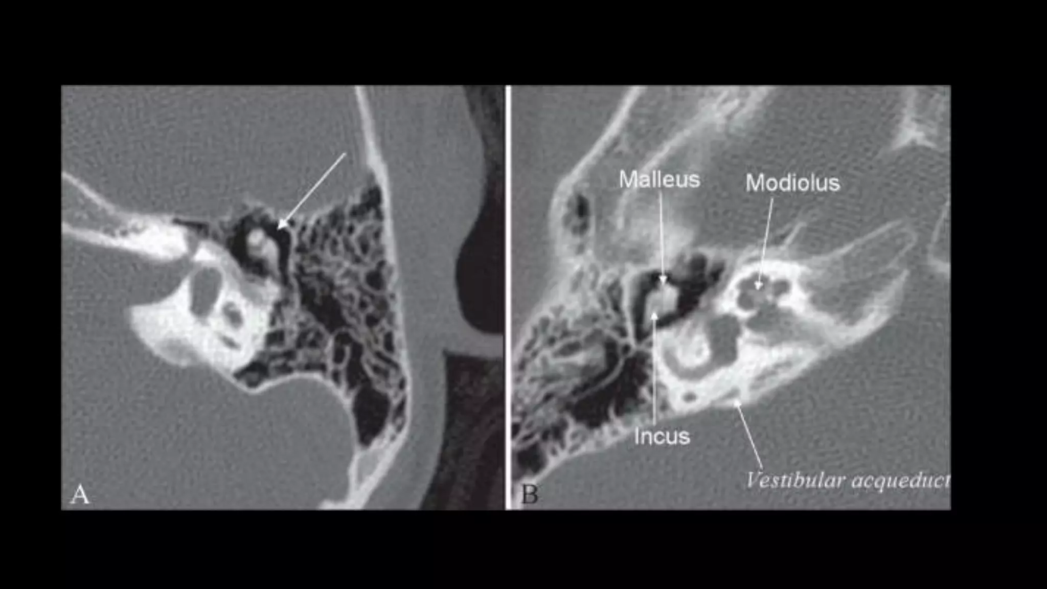
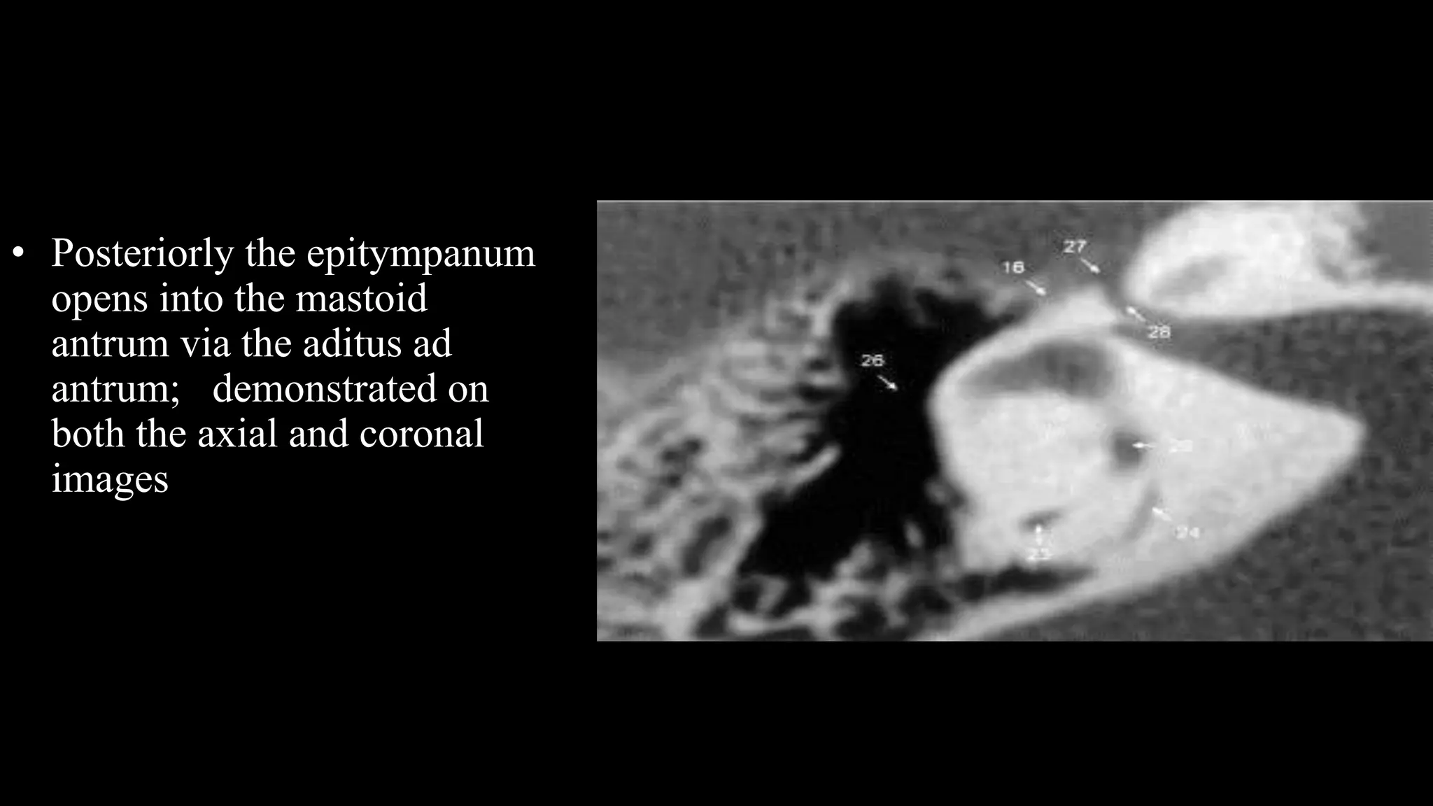
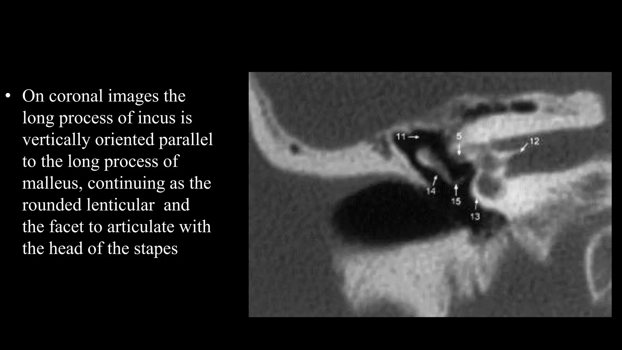
Moderator: Dr Pankaj Kumar
Presenter: Dr Yasha Gupta
Department of ENT
Dr Baba Saheb Ambedkar Hospital.
WHY??
• Identify anatomy
• Operative planning
• Risk assessment
• Informed consent
Informations
• Aeration
• Position of dura, Facial Nerve and Vessels
• Labyrinth
• Extension of disease
• Evidence of previous surgery
Planes of scanning
 Axial
 30 Degrees to anthropological base line
 Parallel to lateral SCC.
 Best displays inner & middle ear.
 Sections parallel to the hard Palate
 Coronal plane
 patient head extended in prone or supine with 105 degree
 plane is perpendicular to the lateral SCC
 Sections are parallel to posterior wall of maxillary sinus
 Saggital plane
4
105
0
30
Planes
• Pöschl plane approximates the plane of the sSCC
• Stenvers plane is perpendicular to both the axial plane
and the Pöschl plane
AXIAL CUTS
• Superior SCC
• Posterior SCC
• Vestibular aqueduct
• IAC
• Facial
• LSCC
Superior SCC
• Start Superiorly
• Superior SCC comes into
view
• Seen perpendicular to long
axis of temporal bone
Posterior SCC
• Posterior SCC appears
Parallel to
Petrous temporal bone
Vestibular Aqueduct
• Vestibular aqueduct
Veers Ventrally
Onto the posterior cranial fossa surface of
Temporal bone
Inernal Auditory Canal
• It is next to appear
• All structures anterior to it are cochlear
• Posterior to it are vestibular
Lateral SCC
• Signet ring appearance
Ossicles
• Ice cream cone appearance
• The ice-cream cone sign represents the normal appearance of
the malleus and incus on an axial high-resolution CT scan
(HRCT) image of the temporal bone .
• The ball (scoop) of the ice cream is formed by the head of
malleus and the cone is formed by the body of the incus.
• The space between the ice-cream cone and the scutum is
called Prussak's space.
Cochlea
• Stack of coins
Cochlear aqueduct
CORONAL
Middle Ear
• Three compartments in coronal
plane.
• A line drawn from lower edge of
the scutum to the tympanic
portion of the facial nerve -
epi/mesotympanum.
• parallel to the floor of the
external auditory canal
meso/hypotympanum
Epitympanum
It contains :
• Head of the malleus
• Malleo–incudal articulation
• Body and short process of the
incus,
• Short process of Incus projects
posteriorly towards aditus.
These structures are best
demonstrated on axial images.
• Tegment tympani – roof of
epitympanum – barrier
between middle cranial
fossa and middle ear cavity.
• Best evaluated on coronal
images
• Posteriorly the epitympanum
opens into the mastoid
antrum via the aditus ad
antrum; demonstrated on
both the axial and coronal
images
Mesotympanum
• The mesotympanum contains the
ossicular chain
• Oval and round window
• Long process of the Incus
• Stapes
• Handle of Malleus
• Facial nerve Canal
• On coronal images the
long process of incus is
vertically oriented parallel
to the long process of
malleus, continuing as the
rounded lenticular and
the facet to articulate with
the head of the stapes
• The stapes hub and crura
are best demonstrated on
axial images at the level
of the oval window
• The stapes footplate sits
in the oval window niche
and cannot always be
discretely identified on
CT.
Ct temporal bone

Ct temporal bone

  • 1.
    Moderator: Dr PankajKumar Presenter: Dr Yasha Gupta Department of ENT Dr Baba Saheb Ambedkar Hospital.
  • 2.
    WHY?? • Identify anatomy •Operative planning • Risk assessment • Informed consent
  • 3.
    Informations • Aeration • Positionof dura, Facial Nerve and Vessels • Labyrinth • Extension of disease • Evidence of previous surgery
  • 4.
    Planes of scanning Axial  30 Degrees to anthropological base line  Parallel to lateral SCC.  Best displays inner & middle ear.  Sections parallel to the hard Palate  Coronal plane  patient head extended in prone or supine with 105 degree  plane is perpendicular to the lateral SCC  Sections are parallel to posterior wall of maxillary sinus  Saggital plane 4 105 0 30
  • 5.
    Planes • Pöschl planeapproximates the plane of the sSCC • Stenvers plane is perpendicular to both the axial plane and the Pöschl plane
  • 6.
    AXIAL CUTS • SuperiorSCC • Posterior SCC • Vestibular aqueduct • IAC • Facial • LSCC
  • 7.
    Superior SCC • StartSuperiorly • Superior SCC comes into view • Seen perpendicular to long axis of temporal bone
  • 8.
    Posterior SCC • PosteriorSCC appears Parallel to Petrous temporal bone
  • 9.
    Vestibular Aqueduct • Vestibularaqueduct Veers Ventrally Onto the posterior cranial fossa surface of Temporal bone
  • 11.
    Inernal Auditory Canal •It is next to appear • All structures anterior to it are cochlear • Posterior to it are vestibular
  • 14.
    Lateral SCC • Signetring appearance
  • 17.
    Ossicles • Ice creamcone appearance
  • 19.
    • The ice-creamcone sign represents the normal appearance of the malleus and incus on an axial high-resolution CT scan (HRCT) image of the temporal bone . • The ball (scoop) of the ice cream is formed by the head of malleus and the cone is formed by the body of the incus. • The space between the ice-cream cone and the scutum is called Prussak's space.
  • 21.
  • 22.
  • 23.
  • 24.
    Middle Ear • Threecompartments in coronal plane. • A line drawn from lower edge of the scutum to the tympanic portion of the facial nerve - epi/mesotympanum. • parallel to the floor of the external auditory canal meso/hypotympanum
  • 25.
    Epitympanum It contains : •Head of the malleus • Malleo–incudal articulation • Body and short process of the incus, • Short process of Incus projects posteriorly towards aditus. These structures are best demonstrated on axial images.
  • 26.
    • Tegment tympani– roof of epitympanum – barrier between middle cranial fossa and middle ear cavity. • Best evaluated on coronal images
  • 27.
    • Posteriorly theepitympanum opens into the mastoid antrum via the aditus ad antrum; demonstrated on both the axial and coronal images
  • 28.
    Mesotympanum • The mesotympanumcontains the ossicular chain • Oval and round window • Long process of the Incus • Stapes • Handle of Malleus • Facial nerve Canal
  • 29.
    • On coronalimages the long process of incus is vertically oriented parallel to the long process of malleus, continuing as the rounded lenticular and the facet to articulate with the head of the stapes
  • 30.
    • The stapeshub and crura are best demonstrated on axial images at the level of the oval window • The stapes footplate sits in the oval window niche and cannot always be discretely identified on CT.