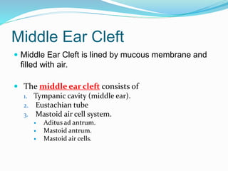
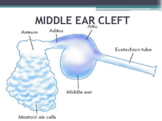
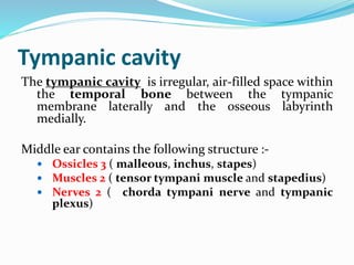
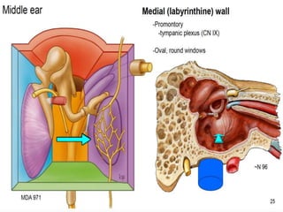
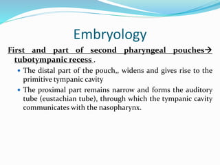
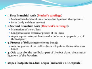
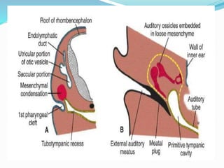
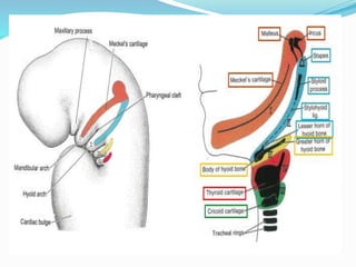
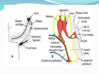
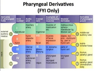
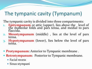
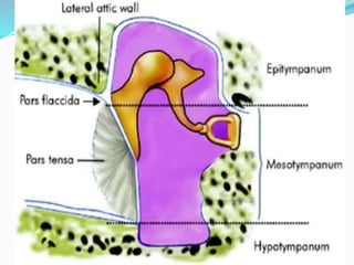
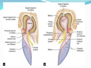
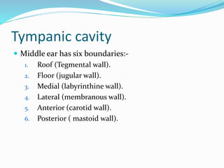
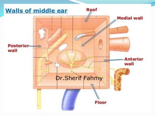
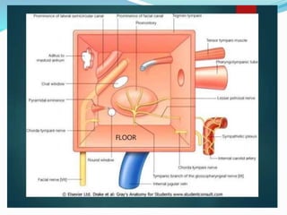
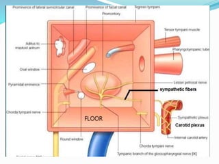
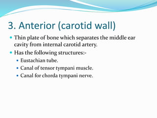
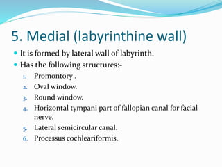
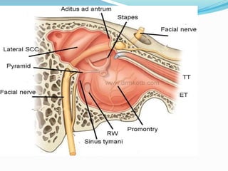
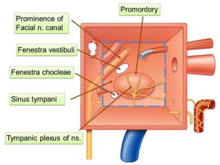
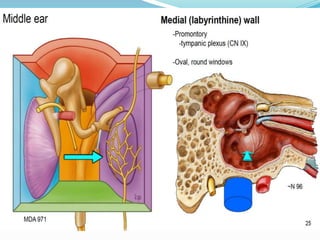
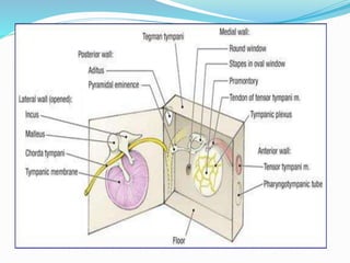
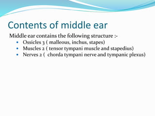
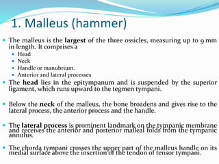
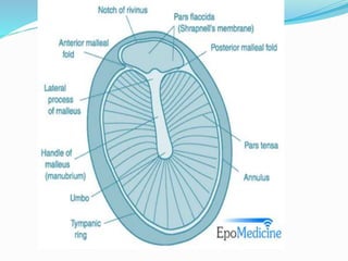
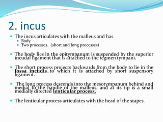
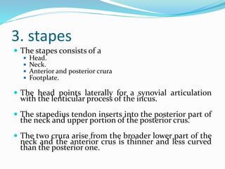
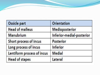
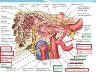
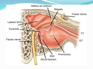
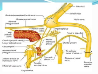
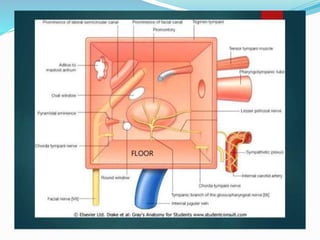
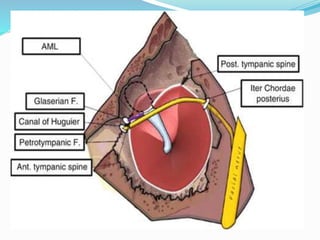
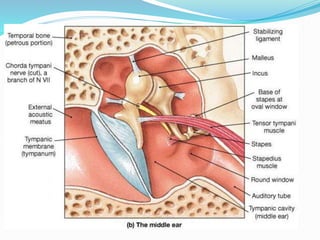
Dr. Amro Al-amleh : ENT resident
Middle Ear Cleft
 Middle Ear Cleft is lined by mucous membrane and
filled with air.
 The middle ear cleft consists of
1. Tympanic cavity (middle ear).
2. Eustachian tube
3. Mastoid air cell system.
 Aditus ad antrum.
 Mastoid antrum.
 Mastoid air cells.
Tympanic cavity
The tympanic cavity is irregular, air-filled space within
the temporal bone between the tympanic
membrane laterally and the osseous labyrinth
medially.
Middle ear contains the following structure :-
 Ossicles 3 ( malleous, inchus, stapes)
 Muscles 2 ( tensor tympani muscle and stapedius)
 Nerves 2 ( chorda tympani nerve and tympanic
plexus)
Dimensions of the Tympanic cavity
 Anterio-posterior (width): 15 mm
 Vertical (height): 15 mm
 Transverse (depth):
 Epitympanum: 6mm
 Mesotympanum: 2mm
 Hypotympanum: 4mm
Embryology
First and part of second pharyngeal pouches
tubotympanic recess .
 The distal part of the pouch,, widens and gives rise to the
primitive tympanic cavity
 The proximal part remains narrow and forms the auditory
tube (eustachian tube), through which the tympanic cavity
communicates with the nasopharynx.
 1. First Branchial Arch (Meckel’s cartilage)
 Malleus( head and neck ,anterior malleal ligament, short process)
 incus (body and short process)
 2. Second Branchial Arch (Reichert’s cartilage):
 Manubrium of the malleus
 Long process and lenticular process of the incus
 stapes suprastructures ( head+ neck+ both cura + tympanic part of
the foot plate )
 3. Process of folius (mesenchyme bone):
 Anterior process of the malleus (so develops from the membranous
bone)
 4. Otic capsule: the vestibular part of the foot plate ; the annular
ligament of the footplate.
 stapes footplate has dual origin (2nd arch + otic capsule)
The tympanic cavity (Tympanum)
The tympanic cavity is divided into three compartments:
1. Epitympanum or attic (upper), lies above the level of
the malleolar folds and pars tensa, and medial to pars
flaccida.
2. Mesotympanum (middle) , lies at the level of pars
tensa.
3. Hypotympanum (lower), lies below the level of pars
tensa.
 Protympanum: Anterior to Tympanic membrane .
 Retrotympanum: Posterior to Tympanic membrane.
 Facial recess
 Sinus stympani
 Epitympanum is separated from the mesotympanum
and hypotympanum by series of mucosal folds.
 Mesotympanum + hypotympanum are continuous
with each other (tympanic cavity proper)
Content of the epitympanum:
1. Head & neck of the malleus
2. Body & short process of the incus
3. Lateral semicircular canal
4. Tympanic part of the fallopian canal of facial nerve
5. Aditus
Content of the Mesotympanum:
1. Handle of the malleus
2. Long process of the incus
3. Stapes
4. Round, oval windows
Middle ear mucosa
 Wraps ossicles, muscles, ligaments and nerves like
peritoneum.
 Mucosal folds divide middle ear into various
compartments:-
Compartments
 Compartments of epitympanum:-
 Prussak’s space
 Attic compartments :-
 Anterior attic compartment
 Posterior attic compartment
 Compartments of mesotympanum
 Inferior incudal space
 Anterior pouch of von Troeltch
 Posterior pouch of von Troeltch
Pouch of von Tröltsch
Anterior pouch of von Tröltsch: Lies between the
 Anterior malleal fold
 The portion of the tympanic membrane anterior to the
handle of the malleus.
Posterior pouch of von Tröltsch: Lies between
 Posterior malleal fold
 The portion of the tympanic membrane posterior to the
handle of the malleus
Mucosal folds
 Anterior malleal fold : Neck of the malleus to
anteriosuperior margin of the tympanic sulcus.
 Posterior malleal fold : Neck of the malleus to
posteriosuperior margin of the tympanic sulcus.
 Lateral malleolar fold : Neck of the malleus to rim of
notch of Rivinus.
Tympanic cavity
 Middle ear has six boundaries:-
1. Roof (Tegmental wall).
2. Floor (jugular wall).
3. Medial (labyrinthine wall).
4. Lateral (membranous wall).
5. Anterior (carotid wall).
6. Posterior ( mastoid wall).
1. Roof ( Tegmental wall)
 It is formed by thin plate of bone called tegmen
tympani.
 Tegmen tympani extends posteriorly to form roof of
auditus and mastod antrum ( tegmen antri).
 Tegmen tympani seperates middle ear from the middle
cranial fossa.
 Both the petrous and squamous portions of the
temporal bone form it; and the petrosquamous
suture line, which does not close until adult life, can
provide a route of access for infection into the
extradural space in children.
 Petrosquamous fissure :-
 Superior tympanic canaliculus  Superior petrosal
nerve.
 Veins from the tympanic cavity running to the
superior petrosal sinus pass through this suture line.
2. Floor ( jugular wall)
 Thin plate of bone which separates Hypotympanum from
the jugular Fossa.
 At the junction of the floor and the medial wall of the
cavity  inferior tympanic canaliculus  the entry of
the tympanic branch of the glossopharyngeal nerve
into the middle ear.
 IX, X, XI are nerve that emerge through jugular foramen,
so they are found beneath the floor of the middle ear.
Inferior tympanic canaliculus
 Is small passage of
 Tympanic branch of glossopharyngeal nerve (Jacobson’s
nerve).
 Inferior tympanic artery.
 It is bony ridge dividing carotid canal from jugular
fossa .
 In lateral part of jugular fossa is mastoid canaliculus
 auricular branch of vagus nerve (Arnold’s nerve).
3. Anterior (carotid wall)
 Thin plate of bone which separates the middle ear
cavity from internal carotid artery.
 Has the following structures:-
 Eustachian tube.
 Canal of tensor tympani muscle.
 Canal for chorda tympani nerve.
 The lower-third of the anterior wall consists of a thin
plate of bone covering the carotid artery.
 This plate is perforated by caroticotympanic nerves
carrying sympathetic fibres to the tympanic plexus
Caroticotympani branches of internal carotid artery.
 The middle-third comprises the tympanic orifice of
the Eustachian tube, which is oval and 5 × 2 mm in
size.
 Just above this is canal containing the tensor tympani
muscle that subsequently runs along the medial wall
of the tympanic cavity.
The caroticotympanic arteries (tympanic branch) are
small branches of the internal carotid artery; they enter
the tympanic cavity through a minute foramen in
the carotid canal, and anastomose with the anterior
tympanic branch of the internal maxillary, and with
the posterior tympanic branch of the stylomastoid artery.
The caroticotympanic nerves are nerves which supply
the eardrum ("tympanum") and carotid canal. They are the
postganglionic sympathetic fibers from internal carotid
plexus which enter the tympanic cavity via
the caroticotympanic artery. These fibers join the tympanic
plexus which are located on the surface of
the promontory.
5. Medial (labyrinthine wall)
 It is formed by lateral wall of labyrinth.
 Has the following structures:-
1. Promontory .
2. Oval window.
3. Round window.
4. Horizontal tympani part of fallopian canal for facial
nerve.
5. Lateral semicircular canal.
6. Processus cochleariformis.
 The promontory is bony bulge which is due to basal coil of
the cochlea and has small grooves on its surface containing
the nerves which form the tympanic plexus.
 Behind and above the promontory is the oval window
(fenestra vestibuli) ; nearly kidney-shaped opening that
connects the tympanic cavity with the vestibule, but which
is closed by the footplate of the stapes.
 The facial nerve canal runs above the promontory and oval
window in an anteroposterior direction and is marked
anteriorly by the processus cochleariformis.
 Processus cochleariformis is curved projection of bone,
which houses the tendon of the tensor tympani muscle.
 Behind the oval window, the facial canal starts to turn
inferiorly as it begins its descent in the posterior wall of the
tympanic cavity.
 Round window (Fenestra cochlea ) is coved overed by
secondary TM.
 It lies below and behind oval window from which it is
separated by posterior extension of the promontory called
the subiculum.
 Ponticulum is bridge of bone from the pyramidal
eminence to the promontory.
4. Posterior ( mastoid wall)
 It lies close to mastoid air cells.
 Has the following structures:-
1. Aditus ad antrum.
2. Fossa incudis
3. Pyramid.
4. Facial nerve (vertical mastoid part).
5. Facial ( suprapyramidal ) recess.
6. Sinus (infrapyranidal) tympani.
 Aditus ad antrum is large irregular opening through
which mastoid antrum opens into attic.
 Below the aditus is a small depression, the fossa
incudis, which houses the short process of the incus
and its suspensory ligament.
 Below the fossa incudis and medial to the opening of
the chorda tympani nerve is the pyramid, a small
hollow conical projection with its apex pointing
anteriorly.
 This houses the stapedius muscle and tendon,
which inserts into the posterior aspect of the head of
stapes.
 Vertical mastoid part of the fallopian canal for facial nerve
runs in posterior wall just behind the pyramid.
 Facial ( suprapyramidal ) recess is depression in posterior
wall lateral to the pyramid.
Boundaries:
 Posterio-Medial: Facial nerve + pyramid
 Anterio-Lateral: tympanic annulus + chorda tympani
 Superior: fossa incudis (near the short process of
incus)
 Sinus (infrapyranidal) tympani is deep recess lies mediak
to pyramid . It is bounded by the subiculum below and the
ponticulus aboce
6.Lateral ( membranous wall)
 Tympanic membrane.
 Scutum.
Contents of middle ear
Middle ear contains the following structure :-
 Ossicles 3 ( malleous, inchus, stapes)
 Muscles 2 ( tensor tympani muscle and stapedius)
 Nerves 2 ( chorda tympani nerve and tympanic plexus)
Ossicles
 There are three middle ear ossicles :-
 Malleus.
 Incus.
 Stapes.
1. Malleus (hammer)
 The malleus is the largest of the three ossicles, measuring up to 9 mm
in length. It comprises a
 Head
 Neck
 Handle or manubrium.
 Anterior and lateral processes
 The head lies in the epitympanum and is suspended by the superior
ligament, which runs upward to the tegmen tympani.
 Below the neck of the malleus, the bone broadens and gives rise to the
lateral process, the anterior process and the handle.
 The lateral process is prominent landmark on the tympanic membrane
and receives the anterior and posterior malleal folds from the tympanic
annulus.
 The chorda tympani crosses the upper part of the malleus handle on its
medial surface above the insertion of the tendon of tensor tympani.
2. incus
 The incus articulates with the malleus and has
 Body.
 Two processes. (short and long processes)
 The body lies in the epitympanum is suspended by the superior
incudal ligament that is attached to the tegmen tympani.
 The short process projects backwards from the body to lie in the
fossa incudis to which it is attached by short suspensory
ligament.
 The long process descends into the mesotympanum behind and
medial to the handle of the malleus, and at its tip is a small
medially directed lenticular process.
 The lenticular process articulates with the head of the stapes.
3. stapes
 The stapes consists of a
 Head.
 Neck.
 Anterior and posterior crura
 Footplate.
 The head points laterally for a synovial articulation
with the lenticular process of the incus.
 The stapedius tendon inserts into the posterior part of
the neck and upper portion of the posterior crus.
 The two crura arise from the broader lower part of the
neck and the anterior crus is thinner and less curved
than the posterior one.
Muscles
1. Stapedius
2. Tensor tympani muscle
1. stapedius
 Origin :- The conical cavity and canal within the
pyramid.
 Inerstion :- The posterior part of the neck and upper
portion of the posterior crus.
 Nerve supply:- Nerve to stapedius (branch of facial nerve ).
 Blood supply :- stapedial artery.
Function :- dampening of middle ear mechanics ( loud
sound).
Tensor tympani muscle
 This is a long slender muscle arising from
 Origin:-
 The walls of the bony canal lying above the Eustachian tube.
 The cartilaginous portion of the Eustachian tube.
 The greater wing of the sphenoid.
 Insertion :- The medial aspect of the upper end of the
malleus handle hust below the neck..
 From its origins, the muscle passes backwards into the
tympanic cavity where it lies on the medial wall, a little
below the level of the facial nerve.
 The bony covering of the canal is often deficient in its
tympanic segment where the muscle is replaced by slender
tendon. This enters the processus cochleariformis .
 The muscle is supplied from the mandibular nerve by way
of a branch from the medial pterygoid nerve.
Nerve
1. Tympanic plexus
2. Chorda tympani nerve
Tympanic plexus
 Tympanic plexus lies on the promontory and it
supplies to :-
 Medial surface of TM
 Tympanic cavity
 Mastoid air cells
 Bony eustachian tube
1. Tympanic plexus
 It is formed by the following nerves:-
1. Tympanic branch of glossopharyngeal nerve
2. Sympathetic fibers :- caroticotympanic nerves come
form the sympathetic plexus, which is present round
the internal carotid artery.
2. Chorda tympani nerve
 The branch of facial nerve enters the middle ear
through posterior canaliculus.
 It runs on medial surface of TM, lies b/w the malleous
and long process of incus.
 Above the tendon of tensor tympani to continue
forwards and leave by way of the anterior canaliculus.
 It carries gustatory fibers from ant. 2/3 of tongue and
parasympathetic fibers to submandibular and
sublingual salivary glands.
Blood supply to middle ear
External carotid artery
 Maxillary artery
 Anterior tympani artery. (glaserian artery)  branch of the first
part of the maxillary artery.
 Middle meningeal artery  superior tympanic artery runs in the
canal for the tensor tympani muscle and supplies this muscle and the
lining membrane of the canal.
 Superficial petrosal artery
 Artery to pterygoid canal.
 Posterior auricular artery
 Sylomastoid artery  posterior tympanic artery.
 Ascending pharyngeal artery
 Tympanic branch (inferior tympanic artery)
Internal carotid artery
 Caroticotympani branches.
Thank you

Middle ear anatomy

  • 1.
    Dr. Amro Al-amleh: ENT resident
  • 3.
    Middle Ear Cleft Middle Ear Cleft is lined by mucous membrane and filled with air.  The middle ear cleft consists of 1. Tympanic cavity (middle ear). 2. Eustachian tube 3. Mastoid air cell system.  Aditus ad antrum.  Mastoid antrum.  Mastoid air cells.
  • 5.
    Tympanic cavity The tympaniccavity is irregular, air-filled space within the temporal bone between the tympanic membrane laterally and the osseous labyrinth medially. Middle ear contains the following structure :-  Ossicles 3 ( malleous, inchus, stapes)  Muscles 2 ( tensor tympani muscle and stapedius)  Nerves 2 ( chorda tympani nerve and tympanic plexus)
  • 7.
    Dimensions of theTympanic cavity  Anterio-posterior (width): 15 mm  Vertical (height): 15 mm  Transverse (depth):  Epitympanum: 6mm  Mesotympanum: 2mm  Hypotympanum: 4mm
  • 9.
    Embryology First and partof second pharyngeal pouches tubotympanic recess .  The distal part of the pouch,, widens and gives rise to the primitive tympanic cavity  The proximal part remains narrow and forms the auditory tube (eustachian tube), through which the tympanic cavity communicates with the nasopharynx.
  • 10.
     1. FirstBranchial Arch (Meckel’s cartilage)  Malleus( head and neck ,anterior malleal ligament, short process)  incus (body and short process)  2. Second Branchial Arch (Reichert’s cartilage):  Manubrium of the malleus  Long process and lenticular process of the incus  stapes suprastructures ( head+ neck+ both cura + tympanic part of the foot plate )  3. Process of folius (mesenchyme bone):  Anterior process of the malleus (so develops from the membranous bone)  4. Otic capsule: the vestibular part of the foot plate ; the annular ligament of the footplate.  stapes footplate has dual origin (2nd arch + otic capsule)
  • 15.
    The tympanic cavity(Tympanum) The tympanic cavity is divided into three compartments: 1. Epitympanum or attic (upper), lies above the level of the malleolar folds and pars tensa, and medial to pars flaccida. 2. Mesotympanum (middle) , lies at the level of pars tensa. 3. Hypotympanum (lower), lies below the level of pars tensa.  Protympanum: Anterior to Tympanic membrane .  Retrotympanum: Posterior to Tympanic membrane.  Facial recess  Sinus stympani
  • 17.
     Epitympanum isseparated from the mesotympanum and hypotympanum by series of mucosal folds.  Mesotympanum + hypotympanum are continuous with each other (tympanic cavity proper)
  • 18.
    Content of theepitympanum: 1. Head & neck of the malleus 2. Body & short process of the incus 3. Lateral semicircular canal 4. Tympanic part of the fallopian canal of facial nerve 5. Aditus Content of the Mesotympanum: 1. Handle of the malleus 2. Long process of the incus 3. Stapes 4. Round, oval windows
  • 20.
    Middle ear mucosa Wraps ossicles, muscles, ligaments and nerves like peritoneum.  Mucosal folds divide middle ear into various compartments:-
  • 21.
    Compartments  Compartments ofepitympanum:-  Prussak’s space  Attic compartments :-  Anterior attic compartment  Posterior attic compartment  Compartments of mesotympanum  Inferior incudal space  Anterior pouch of von Troeltch  Posterior pouch of von Troeltch
  • 22.
    Pouch of vonTröltsch Anterior pouch of von Tröltsch: Lies between the  Anterior malleal fold  The portion of the tympanic membrane anterior to the handle of the malleus. Posterior pouch of von Tröltsch: Lies between  Posterior malleal fold  The portion of the tympanic membrane posterior to the handle of the malleus
  • 26.
    Mucosal folds  Anteriormalleal fold : Neck of the malleus to anteriosuperior margin of the tympanic sulcus.  Posterior malleal fold : Neck of the malleus to posteriosuperior margin of the tympanic sulcus.  Lateral malleolar fold : Neck of the malleus to rim of notch of Rivinus.
  • 27.
    Tympanic cavity  Middleear has six boundaries:- 1. Roof (Tegmental wall). 2. Floor (jugular wall). 3. Medial (labyrinthine wall). 4. Lateral (membranous wall). 5. Anterior (carotid wall). 6. Posterior ( mastoid wall).
  • 29.
    1. Roof (Tegmental wall)  It is formed by thin plate of bone called tegmen tympani.  Tegmen tympani extends posteriorly to form roof of auditus and mastod antrum ( tegmen antri).  Tegmen tympani seperates middle ear from the middle cranial fossa.
  • 31.
     Both thepetrous and squamous portions of the temporal bone form it; and the petrosquamous suture line, which does not close until adult life, can provide a route of access for infection into the extradural space in children.  Petrosquamous fissure :-  Superior tympanic canaliculus  Superior petrosal nerve.  Veins from the tympanic cavity running to the superior petrosal sinus pass through this suture line.
  • 32.
    2. Floor (jugular wall)  Thin plate of bone which separates Hypotympanum from the jugular Fossa.  At the junction of the floor and the medial wall of the cavity  inferior tympanic canaliculus  the entry of the tympanic branch of the glossopharyngeal nerve into the middle ear.  IX, X, XI are nerve that emerge through jugular foramen, so they are found beneath the floor of the middle ear.
  • 33.
    Inferior tympanic canaliculus Is small passage of  Tympanic branch of glossopharyngeal nerve (Jacobson’s nerve).  Inferior tympanic artery.  It is bony ridge dividing carotid canal from jugular fossa .  In lateral part of jugular fossa is mastoid canaliculus  auricular branch of vagus nerve (Arnold’s nerve).
  • 37.
    3. Anterior (carotidwall)  Thin plate of bone which separates the middle ear cavity from internal carotid artery.  Has the following structures:-  Eustachian tube.  Canal of tensor tympani muscle.  Canal for chorda tympani nerve.
  • 39.
     The lower-thirdof the anterior wall consists of a thin plate of bone covering the carotid artery.  This plate is perforated by caroticotympanic nerves carrying sympathetic fibres to the tympanic plexus Caroticotympani branches of internal carotid artery.  The middle-third comprises the tympanic orifice of the Eustachian tube, which is oval and 5 × 2 mm in size.  Just above this is canal containing the tensor tympani muscle that subsequently runs along the medial wall of the tympanic cavity.
  • 40.
    The caroticotympanic arteries(tympanic branch) are small branches of the internal carotid artery; they enter the tympanic cavity through a minute foramen in the carotid canal, and anastomose with the anterior tympanic branch of the internal maxillary, and with the posterior tympanic branch of the stylomastoid artery. The caroticotympanic nerves are nerves which supply the eardrum ("tympanum") and carotid canal. They are the postganglionic sympathetic fibers from internal carotid plexus which enter the tympanic cavity via the caroticotympanic artery. These fibers join the tympanic plexus which are located on the surface of the promontory.
  • 41.
    5. Medial (labyrinthinewall)  It is formed by lateral wall of labyrinth.  Has the following structures:- 1. Promontory . 2. Oval window. 3. Round window. 4. Horizontal tympani part of fallopian canal for facial nerve. 5. Lateral semicircular canal. 6. Processus cochleariformis.
  • 45.
     The promontoryis bony bulge which is due to basal coil of the cochlea and has small grooves on its surface containing the nerves which form the tympanic plexus.  Behind and above the promontory is the oval window (fenestra vestibuli) ; nearly kidney-shaped opening that connects the tympanic cavity with the vestibule, but which is closed by the footplate of the stapes.  The facial nerve canal runs above the promontory and oval window in an anteroposterior direction and is marked anteriorly by the processus cochleariformis.  Processus cochleariformis is curved projection of bone, which houses the tendon of the tensor tympani muscle.
  • 46.
     Behind theoval window, the facial canal starts to turn inferiorly as it begins its descent in the posterior wall of the tympanic cavity.  Round window (Fenestra cochlea ) is coved overed by secondary TM.  It lies below and behind oval window from which it is separated by posterior extension of the promontory called the subiculum.  Ponticulum is bridge of bone from the pyramidal eminence to the promontory.
  • 48.
    4. Posterior (mastoid wall)  It lies close to mastoid air cells.  Has the following structures:- 1. Aditus ad antrum. 2. Fossa incudis 3. Pyramid. 4. Facial nerve (vertical mastoid part). 5. Facial ( suprapyramidal ) recess. 6. Sinus (infrapyranidal) tympani.
  • 51.
     Aditus adantrum is large irregular opening through which mastoid antrum opens into attic.  Below the aditus is a small depression, the fossa incudis, which houses the short process of the incus and its suspensory ligament.  Below the fossa incudis and medial to the opening of the chorda tympani nerve is the pyramid, a small hollow conical projection with its apex pointing anteriorly.  This houses the stapedius muscle and tendon, which inserts into the posterior aspect of the head of stapes.
  • 52.
     Vertical mastoidpart of the fallopian canal for facial nerve runs in posterior wall just behind the pyramid.  Facial ( suprapyramidal ) recess is depression in posterior wall lateral to the pyramid. Boundaries:  Posterio-Medial: Facial nerve + pyramid  Anterio-Lateral: tympanic annulus + chorda tympani  Superior: fossa incudis (near the short process of incus)  Sinus (infrapyranidal) tympani is deep recess lies mediak to pyramid . It is bounded by the subiculum below and the ponticulus aboce
  • 56.
    6.Lateral ( membranouswall)  Tympanic membrane.  Scutum.
  • 57.
    Contents of middleear Middle ear contains the following structure :-  Ossicles 3 ( malleous, inchus, stapes)  Muscles 2 ( tensor tympani muscle and stapedius)  Nerves 2 ( chorda tympani nerve and tympanic plexus)
  • 58.
    Ossicles  There arethree middle ear ossicles :-  Malleus.  Incus.  Stapes.
  • 59.
    1. Malleus (hammer) The malleus is the largest of the three ossicles, measuring up to 9 mm in length. It comprises a  Head  Neck  Handle or manubrium.  Anterior and lateral processes  The head lies in the epitympanum and is suspended by the superior ligament, which runs upward to the tegmen tympani.  Below the neck of the malleus, the bone broadens and gives rise to the lateral process, the anterior process and the handle.  The lateral process is prominent landmark on the tympanic membrane and receives the anterior and posterior malleal folds from the tympanic annulus.  The chorda tympani crosses the upper part of the malleus handle on its medial surface above the insertion of the tendon of tensor tympani.
  • 63.
    2. incus  Theincus articulates with the malleus and has  Body.  Two processes. (short and long processes)  The body lies in the epitympanum is suspended by the superior incudal ligament that is attached to the tegmen tympani.  The short process projects backwards from the body to lie in the fossa incudis to which it is attached by short suspensory ligament.  The long process descends into the mesotympanum behind and medial to the handle of the malleus, and at its tip is a small medially directed lenticular process.  The lenticular process articulates with the head of the stapes.
  • 64.
    3. stapes  Thestapes consists of a  Head.  Neck.  Anterior and posterior crura  Footplate.  The head points laterally for a synovial articulation with the lenticular process of the incus.  The stapedius tendon inserts into the posterior part of the neck and upper portion of the posterior crus.  The two crura arise from the broader lower part of the neck and the anterior crus is thinner and less curved than the posterior one.
  • 67.
  • 68.
    1. stapedius  Origin:- The conical cavity and canal within the pyramid.  Inerstion :- The posterior part of the neck and upper portion of the posterior crus.  Nerve supply:- Nerve to stapedius (branch of facial nerve ).  Blood supply :- stapedial artery. Function :- dampening of middle ear mechanics ( loud sound).
  • 71.
    Tensor tympani muscle This is a long slender muscle arising from  Origin:-  The walls of the bony canal lying above the Eustachian tube.  The cartilaginous portion of the Eustachian tube.  The greater wing of the sphenoid.  Insertion :- The medial aspect of the upper end of the malleus handle hust below the neck..  From its origins, the muscle passes backwards into the tympanic cavity where it lies on the medial wall, a little below the level of the facial nerve.  The bony covering of the canal is often deficient in its tympanic segment where the muscle is replaced by slender tendon. This enters the processus cochleariformis .  The muscle is supplied from the mandibular nerve by way of a branch from the medial pterygoid nerve.
  • 73.
    Nerve 1. Tympanic plexus 2.Chorda tympani nerve
  • 74.
    Tympanic plexus  Tympanicplexus lies on the promontory and it supplies to :-  Medial surface of TM  Tympanic cavity  Mastoid air cells  Bony eustachian tube
  • 75.
    1. Tympanic plexus It is formed by the following nerves:- 1. Tympanic branch of glossopharyngeal nerve 2. Sympathetic fibers :- caroticotympanic nerves come form the sympathetic plexus, which is present round the internal carotid artery.
  • 78.
    2. Chorda tympaninerve  The branch of facial nerve enters the middle ear through posterior canaliculus.  It runs on medial surface of TM, lies b/w the malleous and long process of incus.  Above the tendon of tensor tympani to continue forwards and leave by way of the anterior canaliculus.  It carries gustatory fibers from ant. 2/3 of tongue and parasympathetic fibers to submandibular and sublingual salivary glands.
  • 81.
    Blood supply tomiddle ear External carotid artery  Maxillary artery  Anterior tympani artery. (glaserian artery)  branch of the first part of the maxillary artery.  Middle meningeal artery  superior tympanic artery runs in the canal for the tensor tympani muscle and supplies this muscle and the lining membrane of the canal.  Superficial petrosal artery  Artery to pterygoid canal.  Posterior auricular artery  Sylomastoid artery  posterior tympanic artery.  Ascending pharyngeal artery  Tympanic branch (inferior tympanic artery) Internal carotid artery  Caroticotympani branches.
  • 85.