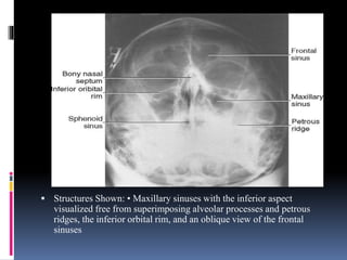
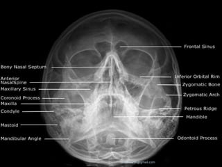
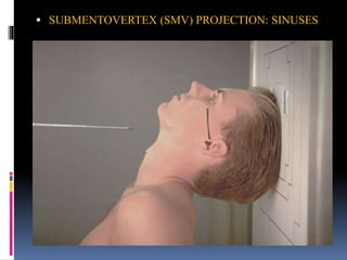
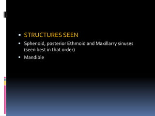
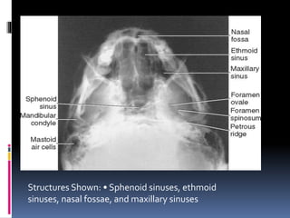
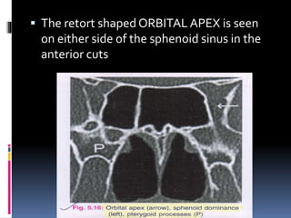
IMAGING IN ENT
Moderator: Dr.Karthik
Presenter:- Dr.Trisha
Imaging of the ear
Superior surface of petrous bone
Medial surface of petrous
bone
FACIAL NERVE:-
RADIOLOGY OF NOSE AND
PARANASAL SINUSES
 PARANASAL SINUSES
 Technical Factors
 A medium kV range of 70 to 80 is commonly
used to provide sufficient contrast of the air-
filled paranasal sinuses. Optimum density as
controlled by the mAs is especially important
for sinus radiography to visualize pathology
within the sinus cavities. A small focal spot
should be used for maximum detail.
LATERAL VIEW
 Lateral side of the skull lies against the film
and x-ray beam is projected perpendicular
from the other side.
 Center CR to a point midway between
outer canthus and EAM.
 LATERAL POSITION—RIGHT OR LEFT
LATERAL: SINUSES
 Respiration
 Suspend respiration during exposure.
 Notes:To visualize air-fluid levels, an erect position
with a horizontal beam is required. Fluid within the
paranasal sinus cavities is thick and gelatin-like,
causing it to cling to the cavity walls.To visualize
this fluid, allow a short time (at least 5 minutes) for
the fluid to settle after a patient's position has
been changed (i.e., from recumbent to erect
 STRUCTURES SEEN-
 ANTERIORAND POSTERIOR EXTENT OF SPHENOID, FRONTALAND
MAXILLARY SINUSES
 SELLATURCICA
 ETHMOID SINUSES
 CONDYLE AND NECK OF MANDIBLE
 CALDWELLVIEW
 A/K/A OCCIPITOFRONTAL VIEW OR NOSE
FOREHEAD POSITION
 Part Position
 Place patient's nose and forehead against upright
table with neck extended to elevate the OML 15°
from horizontal. A radiolucent support between
forehead and upright Bucky or table may be used
to maintain this position. CR remains horizontal.
(alternate method if Bucky can be tilted 15°.)
 Center X-RAY to CR and to nasion, ensuring no
rotation.
 Align CR horizontal, parallel to floor.
POSITION: SINUSES Caldwell Method
STRUCTURES SEEN
1. FRONTAL SINUSES (SEEN BEST)
2. ETHMOID SINUSES
3. MAXILLARY SINUSES
4. FRONTAL PROCESSOF ZYGOMA AND
ZYGOMATIC PROCESSOF FRONTAL BONE
5. SUPERIOR MARGIN OF ORBIT AND LAMINA
PAPYRACEA
6. SUPERIOR ORBITAL FISSURE
 WATER’SVIEW
A.K.A OCCIPITOMENTAL VIEW OR NOSE CHIN
POSITION
IT IS TAKEN IN SUCH A WAY THAT NOSE AND
CHIN OF THE PATIENT TOUCH THE FILM WHILE
X-RAY BEAM IS PROJECTED FROM BEHIND.
Part Position
Extend neck, placing chin and nose against table/film.
• Adjust head until MML is perpendicular to film; OML will form a
37° angle with the plane of the film.
• Ensure that no rotation or tilt exists.
• Center film to CR and to acanthion.
 PARIETOACANTHIAL PROJECTION: SINUSESWaters Method
 STRUCTURES SEEN
 Maxillary sinuses (seen best)
 Frontal sinuses
 Sphenoid sinuses (if the film is taken with
open mouth)
 Zygoma
 Zygomatic arch
 Nasal bone
 Frontal process of maxilla
 Structures Shown: • Maxillary sinuses with the inferior aspect
visualized free from superimposing alveolar processes and petrous
ridges, the inferior orbital rim, and an oblique view of the frontal
sinuses
-drdhiru456@gmail.com
 SUBMENTOVERTICAL (BASAL)VIEW
 THEVIEW ISTAKEN WITHVERTEX NEARTHE FILM AND
X-RAY BEAM PROJECTED AT RIGHT ANGLESTOTHE
FILM FROMTHE SUBMENTAL AREA.
 Part Position
 Raise chin, hyperextend neck if possible until OML is
parallel to table/film.
 Head rests on vertex of skull.
 Ensure no rotation or tilt
 SUBMENTOVERTEX (SMV) PROJECTION: SINUSES
 STRUCTURES SEEN
 Sphenoid, posterior Ethmoid and Maxillarry sinuses
(seen best in that order)
 Mandible
Structures Shown: • Sphenoid sinuses, ethmoid
sinuses, nasal fossae, and maxillary sinuses
CT NOSE AND PNS
BASIC CONCEPTS
 CT scans typically obtained for visualizing the
paranasal sinus should include coronal and axial (3-
mm) cross
sections.
 Soft tissue and bony windows facilitate evaluation
of disease processes and the bony architecture.
 The use of intravenous contrast material just prior
to scanning can help define soft tissue lesions and
delineate vascularized structures, such as vascular
tumors.
 Contrast-enhanced CT is particularly useful in
evaluating neoplastic, chronic, and inflammatory
processes.
 The CT scan is the GOLD STANDARD
investigation in all preoperative cases as it gives
detailed bony anatomy of the area and serves as
a ‘road map’ for the operating surgeon.
 CT scans are best done after a course of
antibiotics, so that acute inflammation is not
mistaken for chronic mucosal disease.
CORONAL CUTS
 One should study the scout film first
The coronal cuts should be read from
anterior to posterior.
The most anterior cuts show frontal sinus
and nasal bone.
The interfrontal septum is in midline
inferiorly, but may deviate to either side.
The interfrontal sinus septum may at
times be pneumatised.
The multiple frontal septae show a
‘classical scalloping’ of the frontal sinus,
which is lost in cases of mucoceles.
The inferior turbinate is visualised, any
hypertrophy of the inferior turbinate is looked
for.
A mucosal swelling is seen in the anterior part of
the septum.This is the SEPTALTUBERCLE.
The septum should be studied for deviations and
spurs.
The middle turbinate is visualised, any
anatomical variations like concha bullosa or a
paradoxically curved middle turbinate should be
looked for.
The attachment of MT at the junction of the
medial and lateral lamellae of the cribriform
plate is seen.
The level of the cribriform plate and the depth
of the olfactory fossa should be assessed and
classified according to KEROS classification.
 The ethmoidal bulla is seen lateral to the
middle turbinate.
 A cell extending above the orbit, behind
the frontal sinus is seen here.This cell is
supraobital cell.
 Uncinate process:This is a 3-dimensional
sickle-shaped (also described as a hook- or L-
shaped) bone of the lateral nasal wall.
Anteriorly, the uncinate process attaches to
the lacrimal bone; inferiorly, the uncinate
process attaches to the ethmoidal process of
the inferior turbinate.The posterior edge lies
in the hiatus semilunaris inferioris. Superiorly,
the uncinate process may attach to the
middle turbinate, lamina papyracea, and/or
the skull base
The uncinate process is seen below the
bulla.
The groove between the uncinate process
and the bulla is HIATUS SEMILUNARIS.
Hiatus semilunaris and infundibulum are
seen leading into the normal maxillary
ostium
The mode of attachment of the uncinate process
should be carefully studied so as to ascertain the
pathway of drainage of frontal sinus.
Variations in the anatomy of the uncinate
process, and the presence of Haller cell should be
looked for
 2-3 mm behind the bulla, the anterior ethmoidal artery is seen
as a classical ‘BEAKING’ of the medial orbital wall.
 Once branching from the ophthalmic artery, it accompanies
the nasociliary nerve through the anterior ethmoidal canal to
supply the anterior and middle ethmoidal cells, frontal sinus,
and anterosuperior aspect of the lateral nasal wall.
 Ethmoidal artery is an important anatomical structure to be
recognized during endoscopic sinus surgery.
 The anterior ethmoidal artery is the best landmark for the
roof of the ethmoid sinus or the anterior base of the skull.
 After reaching the medial wall of the orbit, the Ophthalmic
Artery turns anteriorly. The posterior ethmoidal arteries
enters the nose via the posterior ethmoidal canal and
supplies the posterior ethmoidal sinuses and enters the skull
to supply the meninges.
 The Ophthalmic Artery continues anteriorly, giving off
the anterior ethmoidal artery which enters the nose after
traversing the anterior ethmoidal canal and supplies the
anterior and middle ethmoidal sinuses as well as the frontal
sinus and also enters the cranium to supply the meninges
 The middle turbinate is attached to lamina
papyracea by its ground lamella.This
lamella separates anterior ethmoid cells
from posterior ethmoid cells.
 The posterior ethmoidal cells are larger and fewer
than the anterior ethmoidal cells.
 The posterior ethmoid artery may occasionally be
identified in the region of the skull base.
 The maxillary sinus changes shape from triangular
to ovoid in its posterior cuts.
 The orbit changes from a circular outline to a
triangular shape.
 The posterior most attachment of middle
turbinate to the palatine bone is seen
Posterior part of the orbit with the
extraocular muscles and the optic nerve is
seen.
 The fissure between the orbit and the
maxillary sinus i.e. the INFERIOR ORBITAL
FISSURE is seen in this cut.
 The INFERIOR ORBITAL FISSURE opens into
the INFRATEMPORAL FOSSA
 Sphenoid sinus is seen.
 The sphenoid dominance should be noted when
the intersphenoid septum is asymmetrical.
 Sphenoid sinus ostium may also be visualised ,
though it is better seen in saggital cuts.
 The retort shaped ORBITAL APEX is seen
on either side of the sphenoid sinus in the
anterior cuts
 The maxillary nerve passes through and exits
the skull via the pterygopalatine fossa and the
foramen rotundum.
 Vidian canal transmits the nerve of pterygoid
canal (vidian nerve), artery of the pterygoid
canal and vein of the pterygoid canal)
 A canal may be seen below the sphenoid
sinus between the Pterygopalatine fossa
and the posterior choana, this is
SPHENOPALATINE FORAMEN.
 It transmits the sphenopalatine artery and
vein and the superior
nasal and nasopalatine nerves
 Coronal sections of the nasopharynx show the-
eustachian tube opening, torus tubaris. Fossa of
rosenmuller and the adenoids, if present.
 Asymmetry of the Fossa of rosenmuller should
be looked for.
 The foramen ovale is seen laterally in the
greater wing of sphenoid
Contents of Foramen Ovale –
1. Mandibular nerve
2. Accessory meningeal artery
3. Lesser petrosal nerve.
4.Emissary veins
Widening of Foramen Ovale may be seen in
nasopharyngeal angiofibroma.
 Destruction of Foramen Ovale may be seen in
carcinoma nasopharynx.
AXIAL SCANS
 Axial scans are best read from inferior to
superior.
 N.L.D., anteroposterior deviations of septum
and nasopharynx can be studied well in Axial cuts
SAGGITAL SECTIONS
 Best for studying details of the lateral nasal wall anatomy
ANATOMICAL VARITATIONS
 Agger nasi: This is a bony prominence that is often
pneumatized in the ascending process of the maxilla. Its
location below the frontal sinus also defines the anterior
limit of the frontal recess
 Uncinate process: This is a 3-dimensional sickle-shaped
(also described as a hook- or L-shaped) bone of the
lateral nasal wall. Anteriorly, the uncinate process attaches
to the lacrimal bone; inferiorly, the uncinate process
attaches to the ethmoidal process of the inferior turbinate.
The posterior edge lies in the hiatus semilunaris
inferioris. Superiorly, the uncinate process may attach to the
middle turbinate, lamina papyracea, and/or the skull base.
-drdhiru456@gmail.com
Concha bullosa: The concha bullosa is a pneumatized middle turbinate. An
enlarged middle turbinate may obstruct the middle meatus and the infundibulum
causing recurrent disease. It may also serve as a focal area of sinus disease
-drdhiru456@gmail.com
 Paradoxical middle turbinate:The major curvature of the
middle turbinate may project laterally, leading to
narrowing of the middle meatus
-drdhiru456@gmail.com
Haller cell (infraorbital cell):The Haller cell is usually situated below the
orbit in the roof of the maxillary sinus. It is a pneumatized ethmoid cell
that projects along the medial roof of the maxillary sinus. Enlarged Haller
cells may contribute to narrowing of the ethmoidal infundibulum and
recurrent sinus disease, despite previous (incomplete)
surgery.
 The anterior ethmoid cells may migrate into
frontal recess area where they are then named
Frontal cells.
 I –Type I frontal cell (a single air cell above agger
nasi)
 II –Type II frontal cell (a series of air cells above
agger nasi but below the orbital roof)
 III –Type III frontal cell (this cell extends into the
frontal sinus but is contiguous with agger nasi )
 IV –Type IV frontal cell lies completely within
the frontal sinus (Loner cell)
TYPES OF FRONTAL CELLS-
-drdhiru456@gmail.com
KEROS CLASSIFICATION-
 The Keros classification is a method of classifying the
depth of the olfactory fossa.
 In adults, the olfactory recess is a variable depression in
the cribriform plate that medially is bounded by the
perpendicular plate and laterally by the lateral lamella. It
contains olfactory nerves and a small artery
 The depth of the olfactory fossa is determined by the
height of the lateral lamella of the cribriform plate. Keros
in 19621, classified the depth into three categories.
 type 1 : has a depth of 1 - 3 mm (26.3% of population)
 type 2 : has a depth of 4 - 7mm (73.3% of population)
 type 3 : has a depth of 8 - 16mm (0.5% of population)
-drdhiru456@gmail.com
IMAGING OF THE PHARYNX
IMAGING OF LARYNX
THANKYOU !!

Imaging in ent

  • 1.
    IMAGING IN ENT Moderator:Dr.Karthik Presenter:- Dr.Trisha
  • 2.
  • 3.
    Superior surface ofpetrous bone
  • 5.
    Medial surface ofpetrous bone
  • 25.
  • 56.
    RADIOLOGY OF NOSEAND PARANASAL SINUSES
  • 57.
     PARANASAL SINUSES Technical Factors  A medium kV range of 70 to 80 is commonly used to provide sufficient contrast of the air- filled paranasal sinuses. Optimum density as controlled by the mAs is especially important for sinus radiography to visualize pathology within the sinus cavities. A small focal spot should be used for maximum detail.
  • 58.
    LATERAL VIEW  Lateralside of the skull lies against the film and x-ray beam is projected perpendicular from the other side.  Center CR to a point midway between outer canthus and EAM.
  • 59.
     LATERAL POSITION—RIGHTOR LEFT LATERAL: SINUSES
  • 60.
     Respiration  Suspendrespiration during exposure.  Notes:To visualize air-fluid levels, an erect position with a horizontal beam is required. Fluid within the paranasal sinus cavities is thick and gelatin-like, causing it to cling to the cavity walls.To visualize this fluid, allow a short time (at least 5 minutes) for the fluid to settle after a patient's position has been changed (i.e., from recumbent to erect
  • 61.
     STRUCTURES SEEN- ANTERIORAND POSTERIOR EXTENT OF SPHENOID, FRONTALAND MAXILLARY SINUSES  SELLATURCICA  ETHMOID SINUSES  CONDYLE AND NECK OF MANDIBLE
  • 63.
     CALDWELLVIEW  A/K/AOCCIPITOFRONTAL VIEW OR NOSE FOREHEAD POSITION
  • 64.
     Part Position Place patient's nose and forehead against upright table with neck extended to elevate the OML 15° from horizontal. A radiolucent support between forehead and upright Bucky or table may be used to maintain this position. CR remains horizontal. (alternate method if Bucky can be tilted 15°.)  Center X-RAY to CR and to nasion, ensuring no rotation.  Align CR horizontal, parallel to floor.
  • 65.
  • 66.
    STRUCTURES SEEN 1. FRONTALSINUSES (SEEN BEST) 2. ETHMOID SINUSES 3. MAXILLARY SINUSES 4. FRONTAL PROCESSOF ZYGOMA AND ZYGOMATIC PROCESSOF FRONTAL BONE 5. SUPERIOR MARGIN OF ORBIT AND LAMINA PAPYRACEA 6. SUPERIOR ORBITAL FISSURE
  • 68.
     WATER’SVIEW A.K.A OCCIPITOMENTALVIEW OR NOSE CHIN POSITION IT IS TAKEN IN SUCH A WAY THAT NOSE AND CHIN OF THE PATIENT TOUCH THE FILM WHILE X-RAY BEAM IS PROJECTED FROM BEHIND.
  • 69.
    Part Position Extend neck,placing chin and nose against table/film. • Adjust head until MML is perpendicular to film; OML will form a 37° angle with the plane of the film. • Ensure that no rotation or tilt exists. • Center film to CR and to acanthion.
  • 70.
  • 71.
     STRUCTURES SEEN Maxillary sinuses (seen best)  Frontal sinuses  Sphenoid sinuses (if the film is taken with open mouth)  Zygoma  Zygomatic arch  Nasal bone  Frontal process of maxilla
  • 72.
     Structures Shown:• Maxillary sinuses with the inferior aspect visualized free from superimposing alveolar processes and petrous ridges, the inferior orbital rim, and an oblique view of the frontal sinuses
  • 73.
  • 74.
     SUBMENTOVERTICAL (BASAL)VIEW THEVIEW ISTAKEN WITHVERTEX NEARTHE FILM AND X-RAY BEAM PROJECTED AT RIGHT ANGLESTOTHE FILM FROMTHE SUBMENTAL AREA.
  • 75.
     Part Position Raise chin, hyperextend neck if possible until OML is parallel to table/film.  Head rests on vertex of skull.  Ensure no rotation or tilt
  • 76.
     SUBMENTOVERTEX (SMV)PROJECTION: SINUSES
  • 77.
     STRUCTURES SEEN Sphenoid, posterior Ethmoid and Maxillarry sinuses (seen best in that order)  Mandible
  • 78.
    Structures Shown: •Sphenoid sinuses, ethmoid sinuses, nasal fossae, and maxillary sinuses
  • 79.
  • 80.
    BASIC CONCEPTS  CTscans typically obtained for visualizing the paranasal sinus should include coronal and axial (3- mm) cross sections.  Soft tissue and bony windows facilitate evaluation of disease processes and the bony architecture.  The use of intravenous contrast material just prior to scanning can help define soft tissue lesions and delineate vascularized structures, such as vascular tumors.  Contrast-enhanced CT is particularly useful in evaluating neoplastic, chronic, and inflammatory processes.
  • 81.
     The CTscan is the GOLD STANDARD investigation in all preoperative cases as it gives detailed bony anatomy of the area and serves as a ‘road map’ for the operating surgeon.  CT scans are best done after a course of antibiotics, so that acute inflammation is not mistaken for chronic mucosal disease.
  • 82.
    CORONAL CUTS  Oneshould study the scout film first
  • 83.
    The coronal cutsshould be read from anterior to posterior. The most anterior cuts show frontal sinus and nasal bone.
  • 85.
    The interfrontal septumis in midline inferiorly, but may deviate to either side. The interfrontal sinus septum may at times be pneumatised. The multiple frontal septae show a ‘classical scalloping’ of the frontal sinus, which is lost in cases of mucoceles.
  • 87.
    The inferior turbinateis visualised, any hypertrophy of the inferior turbinate is looked for. A mucosal swelling is seen in the anterior part of the septum.This is the SEPTALTUBERCLE. The septum should be studied for deviations and spurs.
  • 89.
    The middle turbinateis visualised, any anatomical variations like concha bullosa or a paradoxically curved middle turbinate should be looked for. The attachment of MT at the junction of the medial and lateral lamellae of the cribriform plate is seen. The level of the cribriform plate and the depth of the olfactory fossa should be assessed and classified according to KEROS classification.
  • 91.
     The ethmoidalbulla is seen lateral to the middle turbinate.  A cell extending above the orbit, behind the frontal sinus is seen here.This cell is supraobital cell.
  • 93.
     Uncinate process:Thisis a 3-dimensional sickle-shaped (also described as a hook- or L- shaped) bone of the lateral nasal wall. Anteriorly, the uncinate process attaches to the lacrimal bone; inferiorly, the uncinate process attaches to the ethmoidal process of the inferior turbinate.The posterior edge lies in the hiatus semilunaris inferioris. Superiorly, the uncinate process may attach to the middle turbinate, lamina papyracea, and/or the skull base
  • 94.
    The uncinate processis seen below the bulla. The groove between the uncinate process and the bulla is HIATUS SEMILUNARIS. Hiatus semilunaris and infundibulum are seen leading into the normal maxillary ostium
  • 96.
    The mode ofattachment of the uncinate process should be carefully studied so as to ascertain the pathway of drainage of frontal sinus. Variations in the anatomy of the uncinate process, and the presence of Haller cell should be looked for
  • 99.
     2-3 mmbehind the bulla, the anterior ethmoidal artery is seen as a classical ‘BEAKING’ of the medial orbital wall.  Once branching from the ophthalmic artery, it accompanies the nasociliary nerve through the anterior ethmoidal canal to supply the anterior and middle ethmoidal cells, frontal sinus, and anterosuperior aspect of the lateral nasal wall.  Ethmoidal artery is an important anatomical structure to be recognized during endoscopic sinus surgery.  The anterior ethmoidal artery is the best landmark for the roof of the ethmoid sinus or the anterior base of the skull.
  • 101.
     After reachingthe medial wall of the orbit, the Ophthalmic Artery turns anteriorly. The posterior ethmoidal arteries enters the nose via the posterior ethmoidal canal and supplies the posterior ethmoidal sinuses and enters the skull to supply the meninges.  The Ophthalmic Artery continues anteriorly, giving off the anterior ethmoidal artery which enters the nose after traversing the anterior ethmoidal canal and supplies the anterior and middle ethmoidal sinuses as well as the frontal sinus and also enters the cranium to supply the meninges
  • 103.
     The middleturbinate is attached to lamina papyracea by its ground lamella.This lamella separates anterior ethmoid cells from posterior ethmoid cells.
  • 105.
     The posteriorethmoidal cells are larger and fewer than the anterior ethmoidal cells.  The posterior ethmoid artery may occasionally be identified in the region of the skull base.  The maxillary sinus changes shape from triangular to ovoid in its posterior cuts.  The orbit changes from a circular outline to a triangular shape.
  • 106.
     The posteriormost attachment of middle turbinate to the palatine bone is seen
  • 108.
    Posterior part ofthe orbit with the extraocular muscles and the optic nerve is seen.  The fissure between the orbit and the maxillary sinus i.e. the INFERIOR ORBITAL FISSURE is seen in this cut.  The INFERIOR ORBITAL FISSURE opens into the INFRATEMPORAL FOSSA
  • 110.
     Sphenoid sinusis seen.  The sphenoid dominance should be noted when the intersphenoid septum is asymmetrical.  Sphenoid sinus ostium may also be visualised , though it is better seen in saggital cuts.
  • 112.
     The retortshaped ORBITAL APEX is seen on either side of the sphenoid sinus in the anterior cuts
  • 113.
     The maxillarynerve passes through and exits the skull via the pterygopalatine fossa and the foramen rotundum.  Vidian canal transmits the nerve of pterygoid canal (vidian nerve), artery of the pterygoid canal and vein of the pterygoid canal)
  • 114.
     A canalmay be seen below the sphenoid sinus between the Pterygopalatine fossa and the posterior choana, this is SPHENOPALATINE FORAMEN.  It transmits the sphenopalatine artery and vein and the superior nasal and nasopalatine nerves
  • 116.
     Coronal sectionsof the nasopharynx show the- eustachian tube opening, torus tubaris. Fossa of rosenmuller and the adenoids, if present.  Asymmetry of the Fossa of rosenmuller should be looked for.
  • 118.
     The foramenovale is seen laterally in the greater wing of sphenoid
  • 119.
    Contents of ForamenOvale – 1. Mandibular nerve 2. Accessory meningeal artery 3. Lesser petrosal nerve. 4.Emissary veins
  • 120.
    Widening of ForamenOvale may be seen in nasopharyngeal angiofibroma.  Destruction of Foramen Ovale may be seen in carcinoma nasopharynx.
  • 121.
    AXIAL SCANS  Axialscans are best read from inferior to superior.  N.L.D., anteroposterior deviations of septum and nasopharynx can be studied well in Axial cuts
  • 125.
    SAGGITAL SECTIONS  Bestfor studying details of the lateral nasal wall anatomy
  • 126.
  • 127.
     Agger nasi:This is a bony prominence that is often pneumatized in the ascending process of the maxilla. Its location below the frontal sinus also defines the anterior limit of the frontal recess
  • 128.
     Uncinate process:This is a 3-dimensional sickle-shaped (also described as a hook- or L-shaped) bone of the lateral nasal wall. Anteriorly, the uncinate process attaches to the lacrimal bone; inferiorly, the uncinate process attaches to the ethmoidal process of the inferior turbinate. The posterior edge lies in the hiatus semilunaris inferioris. Superiorly, the uncinate process may attach to the middle turbinate, lamina papyracea, and/or the skull base.
  • 130.
    -drdhiru456@gmail.com Concha bullosa: Theconcha bullosa is a pneumatized middle turbinate. An enlarged middle turbinate may obstruct the middle meatus and the infundibulum causing recurrent disease. It may also serve as a focal area of sinus disease
  • 131.
    -drdhiru456@gmail.com  Paradoxical middleturbinate:The major curvature of the middle turbinate may project laterally, leading to narrowing of the middle meatus
  • 132.
    -drdhiru456@gmail.com Haller cell (infraorbitalcell):The Haller cell is usually situated below the orbit in the roof of the maxillary sinus. It is a pneumatized ethmoid cell that projects along the medial roof of the maxillary sinus. Enlarged Haller cells may contribute to narrowing of the ethmoidal infundibulum and recurrent sinus disease, despite previous (incomplete) surgery.
  • 133.
     The anteriorethmoid cells may migrate into frontal recess area where they are then named Frontal cells.  I –Type I frontal cell (a single air cell above agger nasi)  II –Type II frontal cell (a series of air cells above agger nasi but below the orbital roof)  III –Type III frontal cell (this cell extends into the frontal sinus but is contiguous with agger nasi )  IV –Type IV frontal cell lies completely within the frontal sinus (Loner cell)
  • 134.
  • 136.
  • 137.
    KEROS CLASSIFICATION-  TheKeros classification is a method of classifying the depth of the olfactory fossa.  In adults, the olfactory recess is a variable depression in the cribriform plate that medially is bounded by the perpendicular plate and laterally by the lateral lamella. It contains olfactory nerves and a small artery  The depth of the olfactory fossa is determined by the height of the lateral lamella of the cribriform plate. Keros in 19621, classified the depth into three categories.  type 1 : has a depth of 1 - 3 mm (26.3% of population)  type 2 : has a depth of 4 - 7mm (73.3% of population)  type 3 : has a depth of 8 - 16mm (0.5% of population)
  • 138.
  • 139.
  • 170.
  • 210.