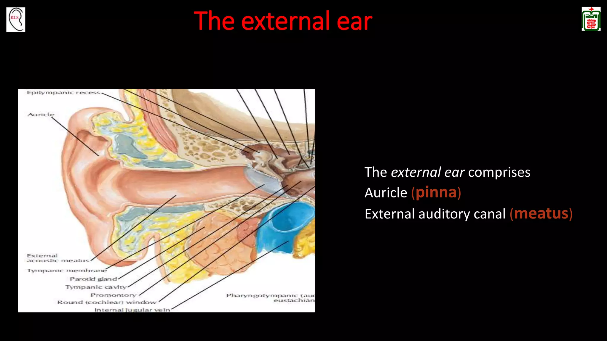
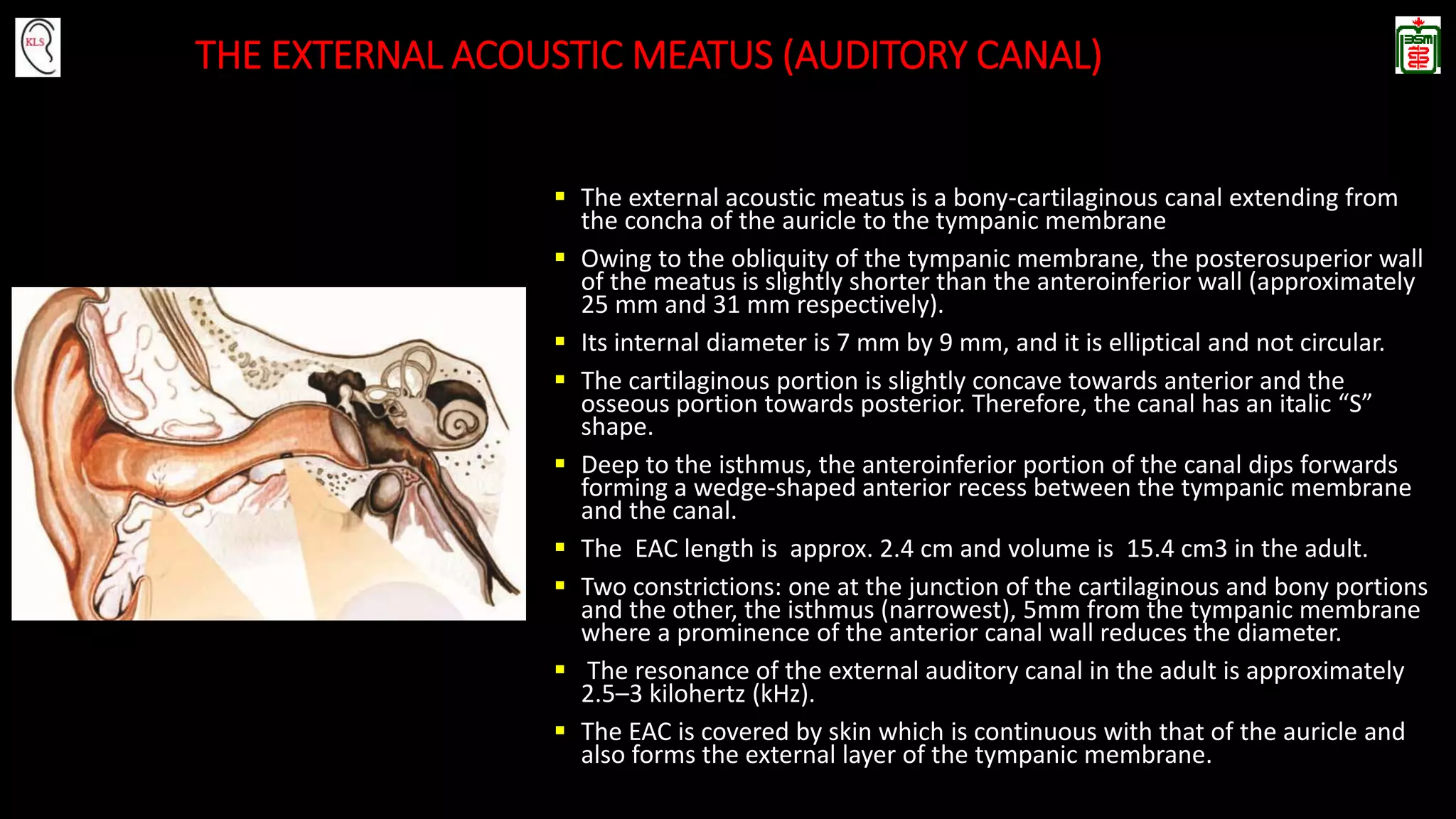
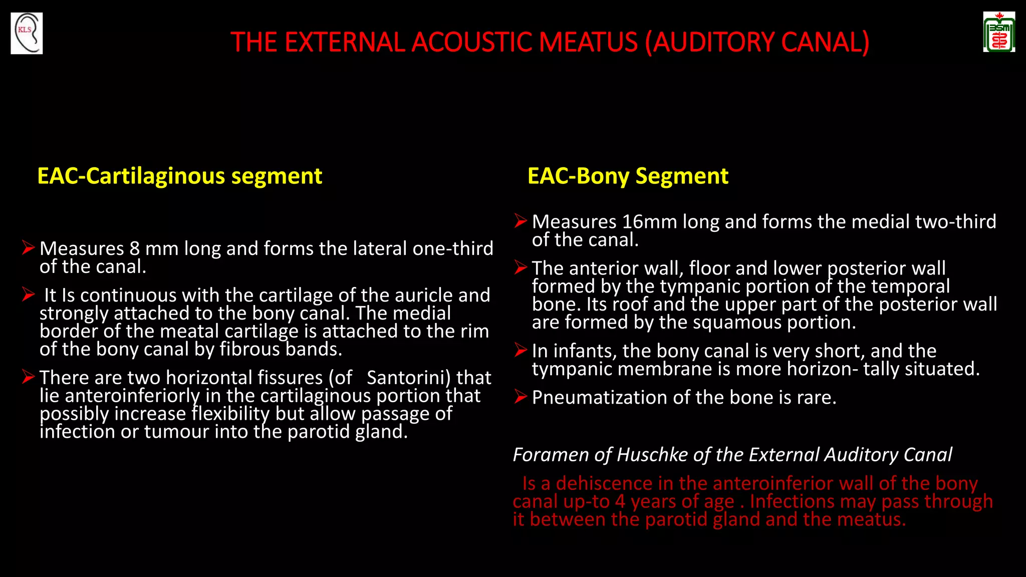
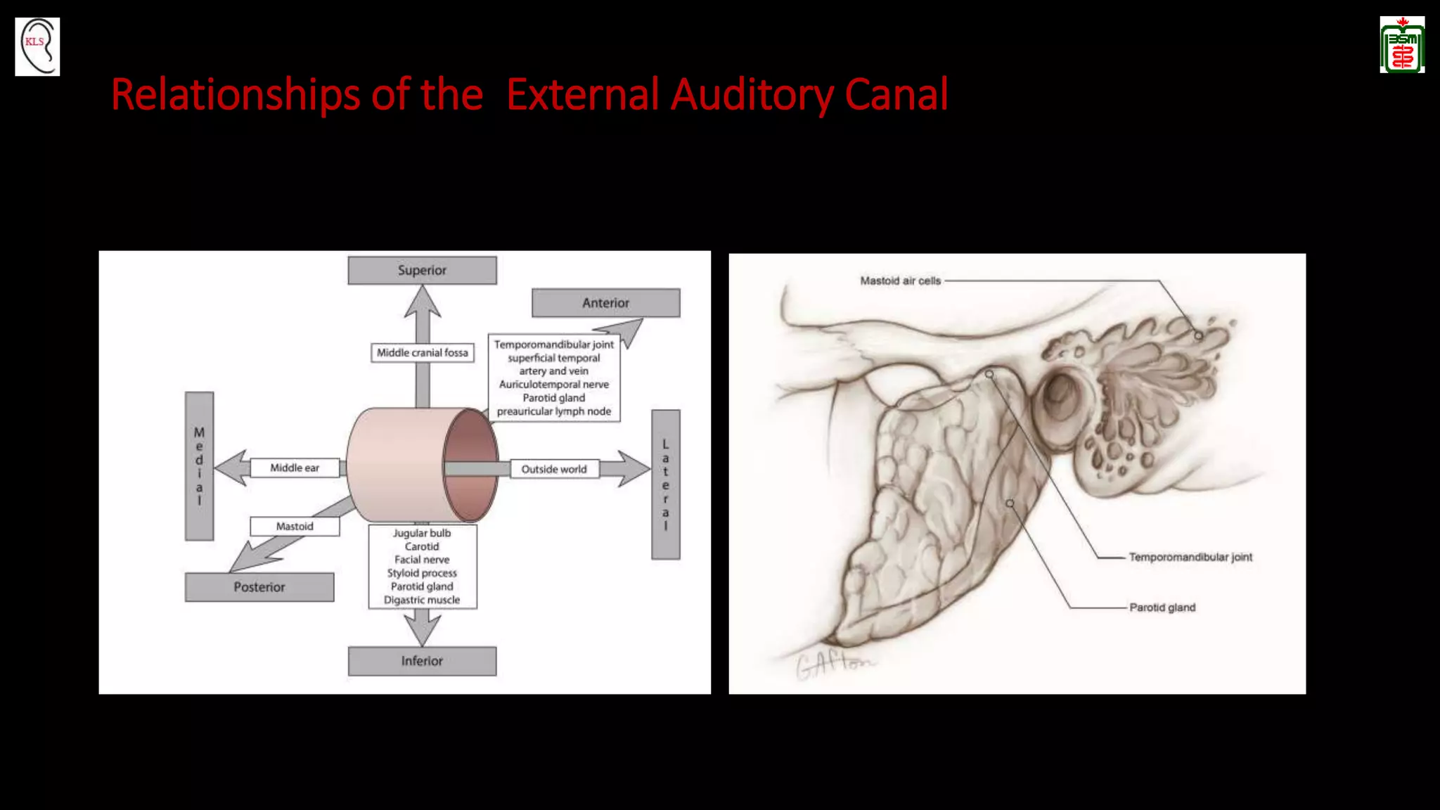
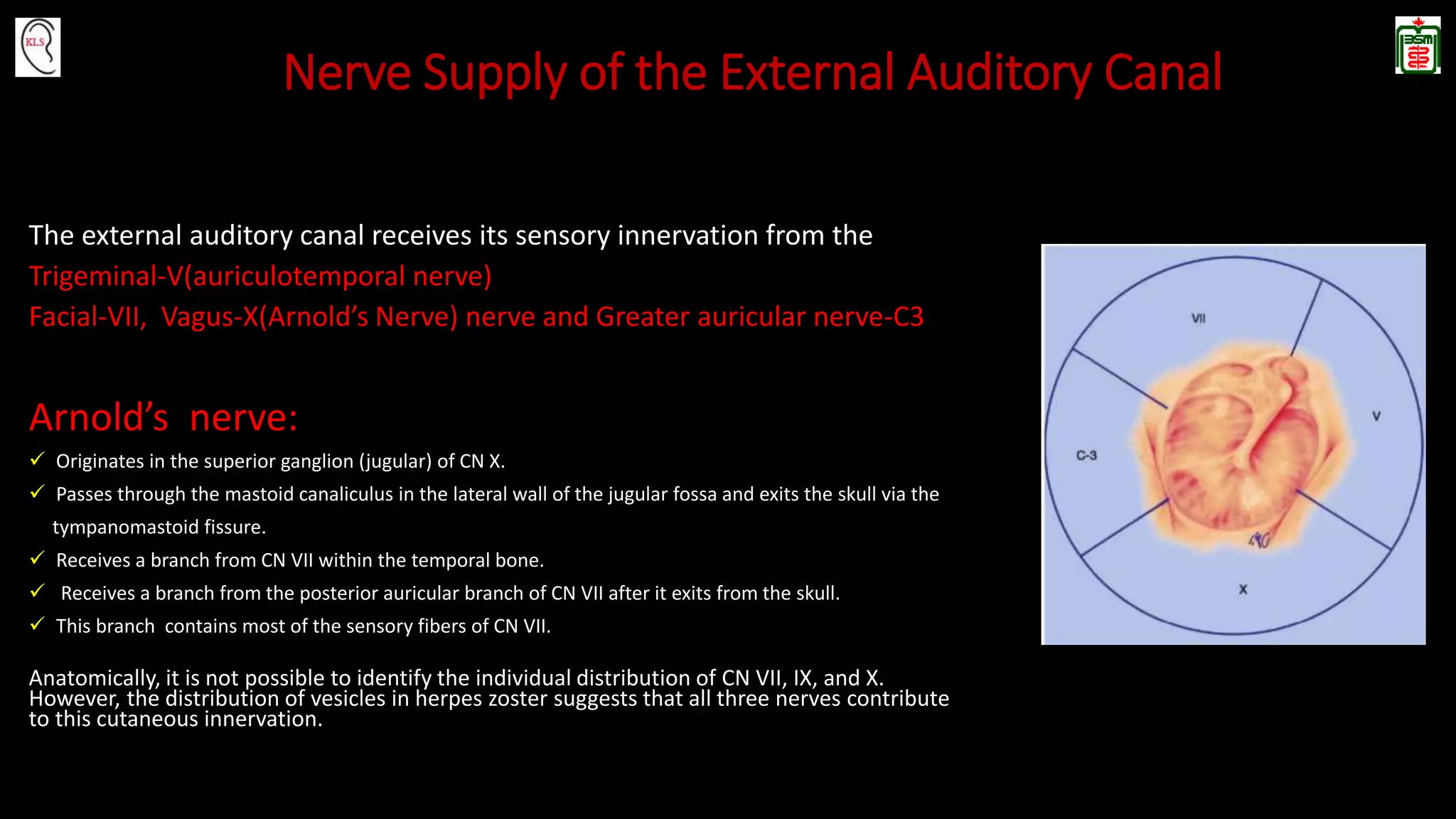
1. Anatomy of External Auditory Canal
Dr Kanu Lal Saha
Associate Professor
Otology Division
Department of Otolaryngology - Head and Neck Surgery
Bangabandhu Sheikh Mujib Medical University, Dhaka
2. The external ear
The external ear comprises
Auricle (pinna)
External auditory canal (meatus)
3. Auricle (Pinna)
Attaches to the side of the head at an angle of about 30 degrees.
Its growth parallels overall body growth until approximately 9 years of age.
Has a single plate of elastic cartilage measuring between 0.5 and 2.0 mm in thickness.
Is continuous with the cartilage of the external meatus.
Skin is thin and firmly attached to the cartilage, particularly on its lateral aspect, which
contains little subcutaneous fat.
Contains many hair-containing sebaceous glands, which are particularly abundant in the
concha and the scaphoid fossa.
Fissures of Santorini, which are not constant in number, extent, or course, are located in
the tragal cartilage and filled by fibrous tissue.
5. Lateral Surface of the Auricle
Helix (or snail)
- Is the prominent rim of the auricle.
-A small tubercle where the helix turns inferiorly is called Darwin's tubercle.
- Antihelix is the curved prominence parallel and anterior to the helix.
Triangular fossa lies between the two crura of the antihelix.
Scaphoid fossa (scoop or shovel) is a narrow, curved depression between the helix
and the antihelix.
Concha (shell) is partially divided by the crus of the helix into two portions:
Upper ( cymba conchae)
Lower (cavum conchae)
Lies at an angle of 90 degrees (± 15 degrees) to the cortex of the mastoid bone.
6. Lateral Surface of the Auricle
Tragus: Is the eminence projecting posteriorly over the external auditory
canal. From the Greek word meaning goat, the tragus is so called
because hair growth on its medial surface is said to resemble a
goat's beard.
Antitragus is located posteriorly, opposite the tragus, and is
separated from it by the intertragic notch.
Lobule Consists of fibrous and fatty tissues and is devoid of cartilage.
A strong fibrous band connects the tragus to the beginning of the
helix,completing the meatus anteriorly.
The endaural Lempert incision passes through this fibrous band.
9. Blood supply of the Auricle
• Posterior auricular artery, a branch of
the external carotid artery.
• Anterior auricular artery, a branch of the
superficial temporal artery.
• A branch from the occipital artery.
• Venous drainage accompanying arteries
10. Innervation of the
ear
1.Auricular branch of CN IX
2. Auricular branch of CN X
3. Greater auricular nerve
4. Lesser occipital nerve
5. Auriculotemporal branch of
CN V
6. Inferior alveolar nerve
7. Lingual n. and chorda tympani
n
8.CN VII (facial nerve)
11. Nerve Supply of Auricle
• A temporal branch of CN VII supplies the anterior auricular muscle
and the superior auricular muscle.
• The posterior auricular branch of CN VII supplies the posterior
auricular muscle and the intrinsic muscles of the cranial aspect of
the pinna.
Sensory nerves of the auricle include
13. THE EXTERNAL ACOUSTIC MEATUS (AUDITORY CANAL)
The external acoustic meatus is a bony-cartilaginous canal extending from
the concha of the auricle to the tympanic membrane
Owing to the obliquity of the tympanic membrane, the posterosuperior wall
of the meatus is slightly shorter than the anteroinferior wall (approximately
25 mm and 31 mm respectively).
Its internal diameter is 7 mm by 9 mm, and it is elliptical and not circular.
The cartilaginous portion is slightly concave towards anterior and the
osseous portion towards posterior. Therefore, the canal has an italic “S”
shape.
Deep to the isthmus, the anteroinferior portion of the canal dips forwards
forming a wedge-shaped anterior recess between the tympanic membrane
and the canal.
The EAC length is approx. 2.4 cm and volume is 15.4 cm3 in the adult.
Two constrictions: one at the junction of the cartilaginous and bony portions
and the other, the isthmus (narrowest), 5mm from the tympanic membrane
where a prominence of the anterior canal wall reduces the diameter.
The resonance of the external auditory canal in the adult is approximately
2.5–3 kilohertz (kHz).
The EAC is covered by skin which is continuous with that of the auricle and
also forms the external layer of the tympanic membrane.
14. THE EXTERNAL ACOUSTIC MEATUS (AUDITORY CANAL)
EAC-Cartilaginous segment
Measures 8 mm long and forms the lateral one-third
of the canal.
It Is continuous with the cartilage of the auricle and
strongly attached to the bony canal. The medial
border of the meatal cartilage is attached to the rim
of the bony canal by fibrous bands.
There are two horizontal fissures (of Santorini) that
lie anteroinferiorly in the cartilaginous portion that
possibly increase flexibility but allow passage of
infection or tumour into the parotid gland.
EAC-Bony Segment
Measures 16mm long and forms the medial two-third
of the canal.
The anterior wall, floor and lower posterior wall
formed by the tympanic portion of the temporal
bone. Its roof and the upper part of the posterior wall
are formed by the squamous portion.
In infants, the bony canal is very short, and the
tympanic membrane is more horizon- tally situated.
Pneumatization of the bone is rare.
Foramen of Huschke of the External Auditory Canal
Is a dehiscence in the anteroinferior wall of the bony
canal up-to 4 years of age . Infections may pass through
it between the parotid gland and the meatus.
15. Skin of the Cartilaginous and Bony canal
Skin of the Cartilaginous Canal:
The skin is tightly bound to the perichondrium
Thickness : 0.5–1 mm
The skin has sebaceous and ceruminous glands as well as
hair follicles in the superior and posterior portion
Skin of the Osseous Canal:
The skin is not firmly bound to the dermis
Continuous with the external (lateral) aspect of the tympanic
membrane
It is thin (0.2 mm)
No hair follicles or sebaceous glands
17. Blood supply of EAC
The arterial supply of the external meatus is derived from branches of the external
carotid.
The auricular branches of the superficial temporal artery supply the roof and anterior
portion of the canal.
The deep auricular branch of the first part of the maxillary artery arises in the parotid
gland behind the temporomandibular joint pierces the cartilage or bone of the
external meatus and supplies the anterior meatal wall skin and the epithelium of the
outer surface of the tympanic membrane.
Finally, auricular branches of the posterior auricular artery pierce the cartilage of the
auricle and supply the posterior portions of the canal.
The veins drain into the external jugular vein, the maxillary veins and the pterygoid
plexus. The lymphatic drainage follows that of the auricle.
18. Nerve Supply of the External Auditory Canal
The external auditory canal receives its sensory innervation from the
Trigeminal-V(auriculotemporal nerve)
Facial-VII, Vagus-X(Arnold’s Nerve) nerve and Greater auricular nerve-C3
Arnold’s nerve:
Originates in the superior ganglion (jugular) of CN X.
Passes through the mastoid canaliculus in the lateral wall of the jugular fossa and exits the skull via the
tympanomastoid fissure.
Receives a branch from CN VII within the temporal bone.
Receives a branch from the posterior auricular branch of CN VII after it exits from the skull.
This branch contains most of the sensory fibers of CN VII.
Anatomically, it is not possible to identify the individual distribution of CN VII, IX, and X.
However, the distribution of vesicles in herpes zoster suggests that all three nerves contribute
to this cutaneous innervation.
Because the cartilage of the external canal is an extension of the auricular cartilage, movement of the auricle is painful in external otitis; this is a useful clinical sign for differentiating it from otitis media.
Auriculitis causes severe pain because of the firm attachment of the soft tissue to the underlying cartilage. The auricle is susceptible to frostbite because of the thinness of the overlying skin. Auricular hematomas frequently develop in the upper lateral aspect of the pinna.
Surgical importance :In lop ear deformity of the external ear, an antihelix is missing, causing protrusion of the auricle.
What is Resonance?
When an object produces a sound, it causes changes in the air that surrounds it. These changes translate into sound waves that travel away from their source. When these sound waves reach an object, this object tends to repel these sounds and to attenuate them. However, certain frequencies are not attenuated and make the object vibrate. These are called the resonant frequencies. All objects have resonant frequencies and this will depend on their size, form, material, etc. Heavy objects resonate with low frequencies; light and small objects with high frequencies and the ear canal with frequencies between 2.5 and 3 kHz.
Hematomas are common in Cartilaginous canal. Due to its thickness it is preferable to inject local anesthetics at this level in a slow fashion“dissecting medially” (“blanching the ear canal. Infections and furuncles occur here as well as tumors of the glands and skin (squamous and basocellular).
Th skin of osseous canal: it can be d epithelialized completely with surgical instruments)
The bony posterior canal wall abuts the mastoid air cells, and the superior wall of the bony canal abuts the epitympanic recess. These air cells are separated from the canal by thin bone.
Mastoiditis may extend into the bony canal through this thin bone, producing a subperiosteal abscess.
Frequently, patients can cough when undergoing microsuction of the canal which is caused by stimulation of the vagus nerve via Arnold’s nerve, its auricular branch.