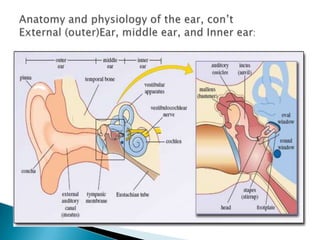
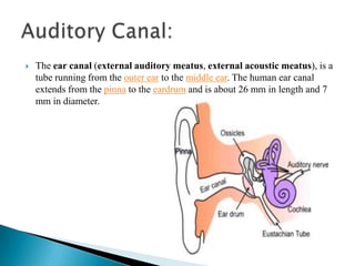
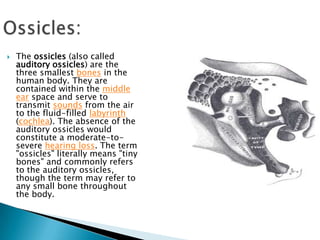
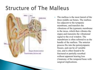
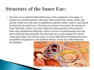
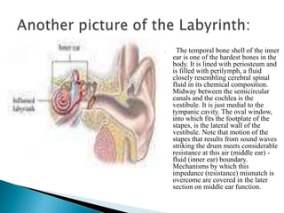
The document summarizes the anatomy and physiology of the human ear. It describes the three main parts of the ear - the outer, middle, and inner ear. The outer ear collects sound waves and directs them through the ear canal to the eardrum. The middle ear contains three small bones that vibrate the eardrum and transmit sound to the inner ear. The inner ear contains the cochlea for hearing and semicircular canals for balance. It detects vibrations and converts them into nerve signals that are sent to the brain.

![The Eustachian tube (or auditory tube or pharyngotympanic tube) is a tube that links the pharynx to the middle ear. In adults the Eustachian tube is approximately 35 mm long. It is named after the sixteenth century anatomist Eustachius.[1] Some modern medical books call this the pharyngotympanic tubeEustachian Tube](https://image.slidesharecdn.com/presentation13-091126140356-phpapp01/85/Presentation-13-9-320.jpg)
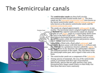
![The Semicircular canalsThe semicircular canals are three half-circular, interconnected tubes located inside each ear. The three canals are the horizontal semicircular canal (also known as the lateral semicircular canal), superior semicircular canal (also known as the anterior semicircular canal), and the posterior semicircular canal.The canals are aligned approximately orthogonally to one another. The horizontal canal is aligned roughly horizontally in the head. The superior and anterior canals are aligned roughly at a 45 degree angle to a vertical plane drawn from the nose to the back of the skull. [1] Thus, the horizontal canal detects horizontal head movements (such as when doing a pirouette), while the superior and posterior canals detect vertical head movements.Each canal is filled with a fluid called endolymph and contains a motion sensor with little hairs (cilia) whose ends are embedded in a gelatinous structure called the cupula. As the skull twists in any direction, the endolymph is thrown into different sections of the canals. The cilia detect when the endolymph rushes past, and a signal is then sent to the brain.The semicircular canals are a component of the Labyrinth.Among species of mammals, the size of the semicircular canals is correlated with their type of locomotion. Specifically, species that are agile and have fast, jerky locomotion have larger canals relative to their body size than those that move more cautiously](https://image.slidesharecdn.com/presentation13-091126140356-phpapp01/85/Presentation-13-12-320.jpg)