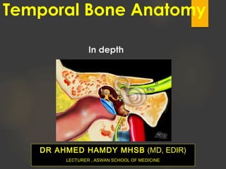
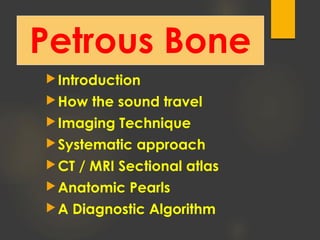
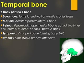
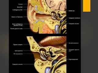
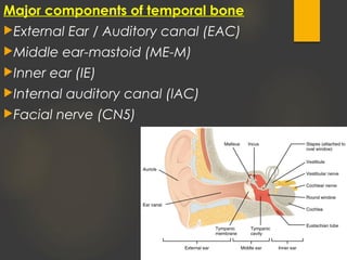
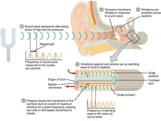
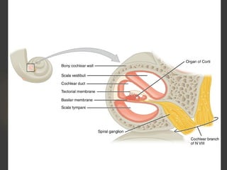
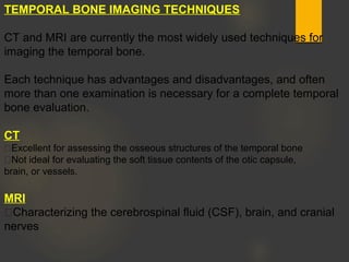
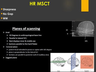
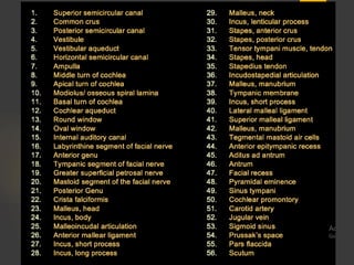
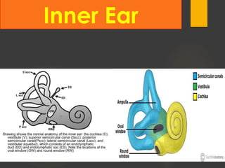
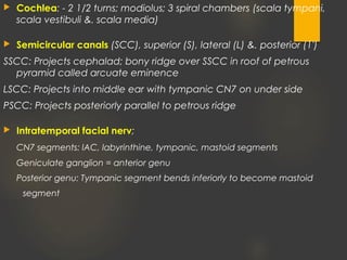
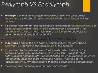
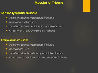
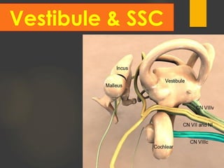
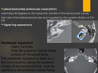
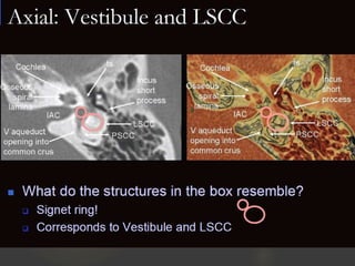
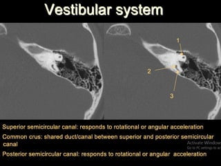
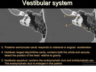
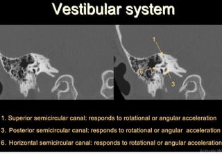
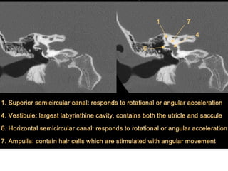
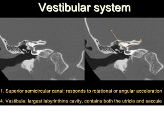
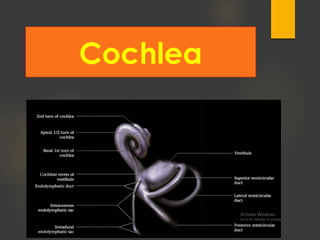
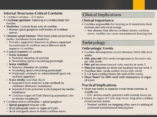
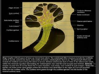
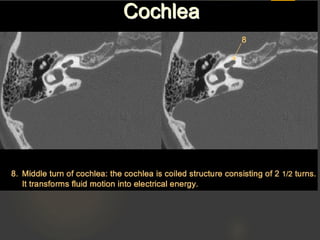
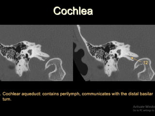
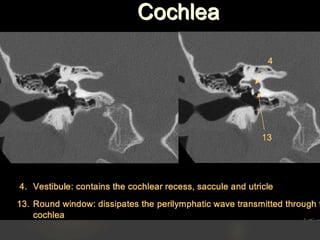
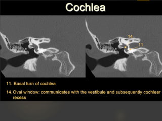
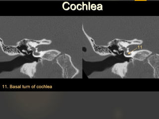
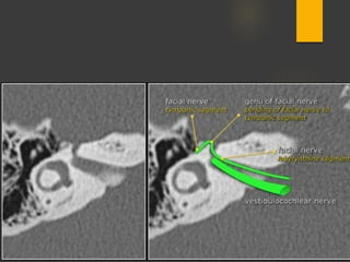
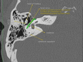
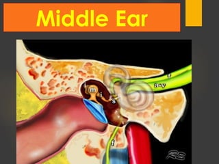
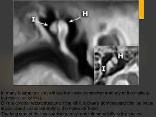
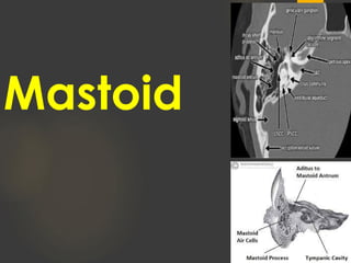
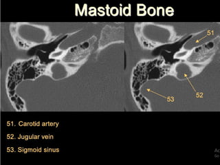
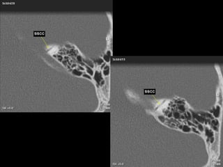
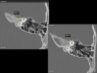
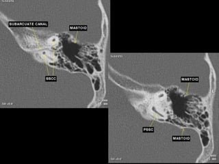
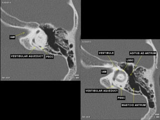
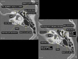
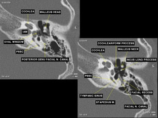
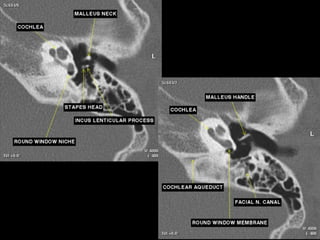
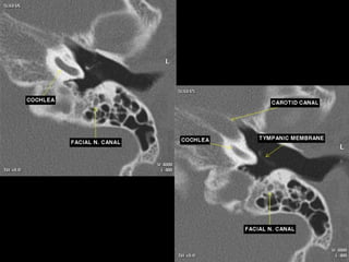
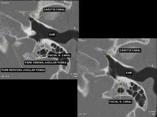
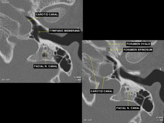
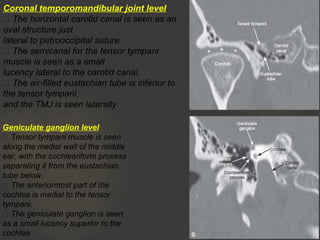
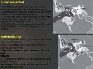
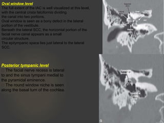
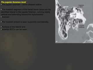
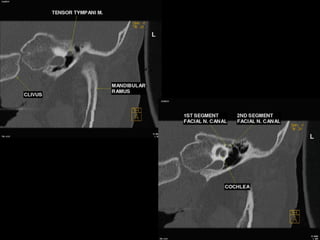
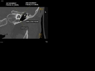
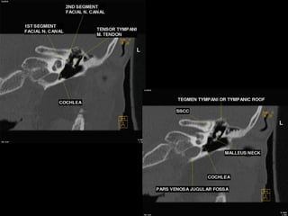
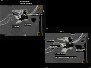
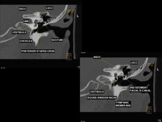
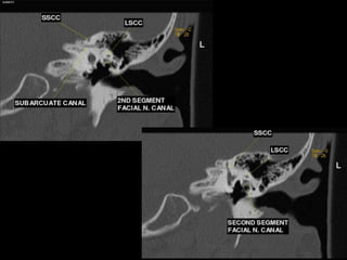
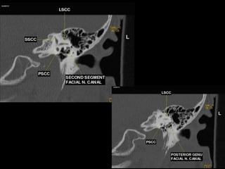
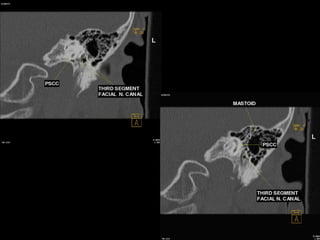
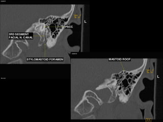
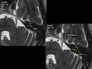
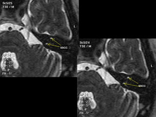
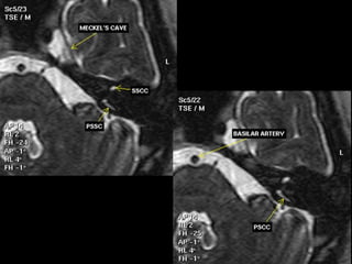
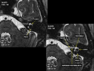
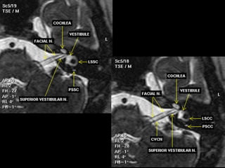
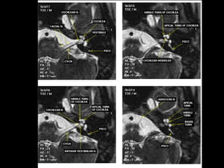
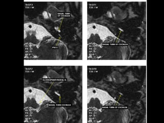
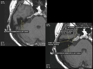
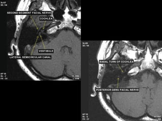
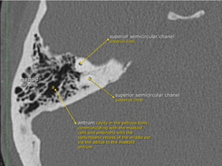
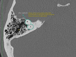
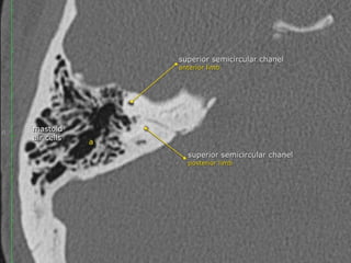
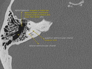
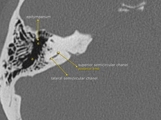
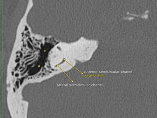
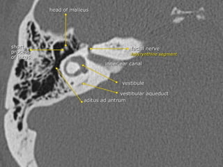
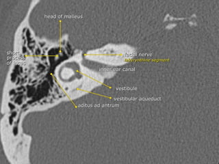
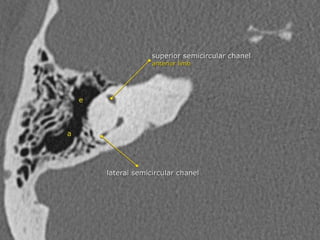
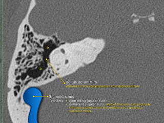
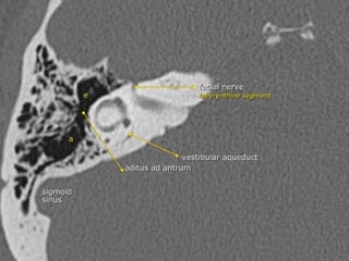
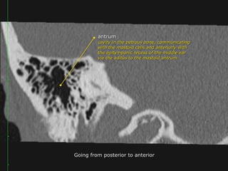
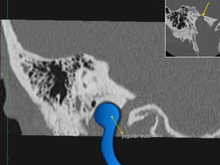
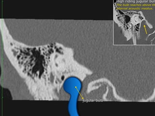
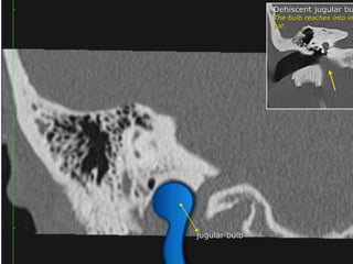
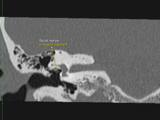
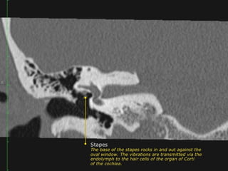
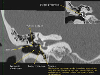
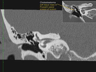
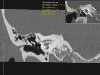
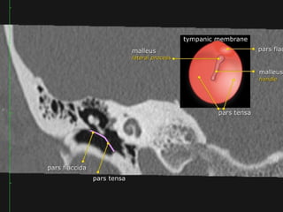
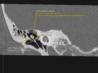
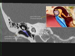
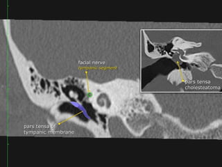
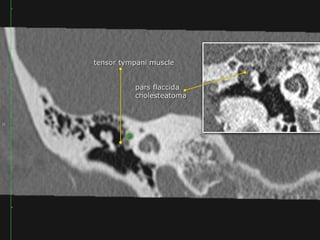
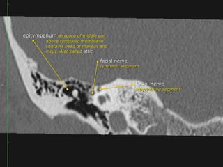
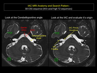
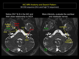
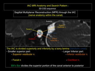
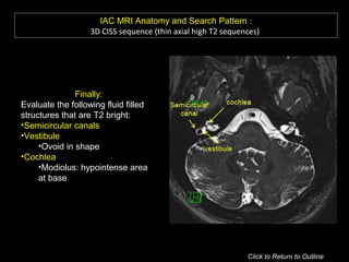
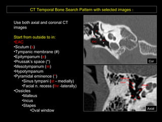
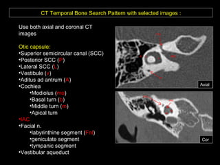
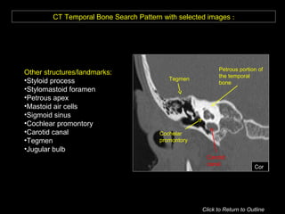
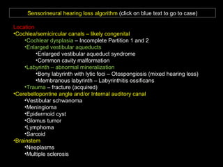
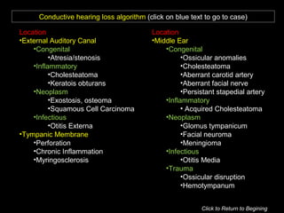
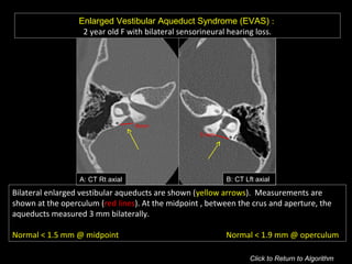
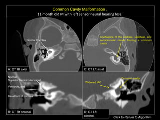
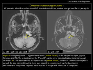
DR AHMED HAMDY MHSB (MD, EDIR)
LECTURER , ASWAN SCHOOL OF MEDICINE
Temporal Bone Anatomy
In depth
Petrous Bone
Introduction
How the sound travel
Imaging Technique
Systematic approach
CT / MRI Sectional atlas
Anatomic Pearls
A Diagnostic Algorithm
Introduction
Temporal bone
5 bony parts to T-bone
Squamous: Forms lateral wall of middle cranial fossa
Mastoid: Aerated posterolateral T-bone
Petrous: Pyramidal shape medial T-bone containing inner
ear, internal auditory canal &. petrous apex
Tympanic: V-shaped bone forming bony EAC
Styloid: Forms styloid process after birth
Major components of temporal bone
External Ear / Auditory canal (EAC)
Middle ear-mastoid (ME-M)
Inner ear (IE)
Internal auditory canal (lAC)
Facial nerve (CN5)
How the sound
travel ?
Imaging
Technique
TEMPORAL BONE IMAGING TECHNIQUES
CT and MRI are currently the most widely used techniques for
imaging the temporal bone.
Each technique has advantages and disadvantages, and often
more than one examination is necessary for a complete temporal
bone evaluation.
CT
Excellent for assessing the osseous structures of the temporal bone
Not ideal for evaluating the soft tissue contents of the otic capsule,
brain, or vessels.
MRI
Characterizing the cerebrospinal fluid (CSF), brain, and cranial
nerves
HR MSCT
Sharpness
No Gap
WW
WL
Systematic
approach
Inner Ear
Inner ear
It refers to the bony labyrinth, the membranous labyrinth and their
contents. It may also be referred to as the vestibulocochlear organ,
supplied by the vestibulocochlear nerve (CN VIII).
Bony labyrinth: Bone confining cochlear, vestibule &. semicircular
canals
Membranous labyrinth; Includes vestibule (utricle &. saccule),
semicircular ducts, scala media of cochlea, endolymphatic duct&. sac
 It is divided into three main parts:
the cochlea housing the cochlear duct for hearing as it connects to :
Stapes → oval window
the vestibule housing the utricle and saccule for static balance
the semicircular canals connect to the vestibule and housing the 
semicircular ducts for kinetic balance
 Cochlea: - 2 1/2 turns; modiolus; 3 spiral chambers (scala tympani,
scala vestibuli &. scala media)
 Semicircular canals (SCC), superior (S), lateral (L) &. posterior (1')
SSCC: Projects cephalad; bony ridge over SSCC in roof of petrous
pyramid called arcuate eminence
LSCC: Projects into middle ear with tympanic CN7 on under side
PSCC: Projects posteriorly parallel to petrous ridge
 Intratemporal facial nerv;
CN7 segments: lAC, labyrinthine, tympanic, mastoid segments
Geniculate ganglion = anterior genu
Posterior genu: Tympanic segment bends inferiorly to become mastoid
segment
Perilymph VS Endolymph
 Perilymph is one of the two types of cochlear fluids, the other being 
endolymph. It is located in the scala vestibuli and scala tympani of the 
cochlea.
 It is a clear fluid with an ionic composition very similar to cerebrospinal fluid as
the scala tympaniand scala tympani communicate directly with the 
subarachnoid space. It has a high level of sodium (Na+) and helps to
generate the endolymphatic potential.
--------------------------------------------------------------------------
 Endolymph is one of the two types of cochlear fluids, the other being 
perilymph. It is located in the scala media of the cochlea.
 It is secreted by the stria vascularis (colloquially called 'battery of the
cochlea') on the outer wall of the scala media. It has a high level of
potassium (K+) and generates a current called the endolymphatic potential,
with positivity inside the scala media and negativity outside its wall
(approximately 80 mV more positive than the perilymphatic compartment).
 The endolymph and perilymph do not communicate.
Muscles of T-bone
Tensor tympani muscle
 Dampens sound; hyperacusis if injured
 Innervation: V3 branch
 Location: Anteromedial wall, mesotympanum
 Attachment: Tendon inserts on malleus
Stapedius muscle
 Dampens sound; hyperacusis if injured
 Innervation: CN?
 Location: Muscle belly in pyramidal eminence
 Attachment: Tendon attaches on head of stapes
Vestibule & SSC
The vestibule is an approximately 4 mm central chamber of the 
bony labyrinth. It is dominated by depressions housing parts of the 
membranous labyrinth:
utricle (elliptical recess)
saccule (spherical recess)
basal end of the cochlear duct (cochlear recess)
It is located medial to the tympanic cavity, posterior to the cochlea
 and anterior to the semicircular canals. In the lateral wall is the 
oval window with the stapes foot plate covering it.
The cribrose areas have perforations through which the nerve bundles
gain access to the inner ear. The endolymphatic duct is housed within
the bony vestibular aqueduct and originates at the posteroinferior
aspect of the vestibule, via the utriculosaccular duct.
The vestibule
-The endolymphatic duct extends from the posterior aspect of the vestibule
toward the posterior cranial fossa, and ends in a blind pouch, the
endolymphatic sac, at the posterior margin of the petrous ridge
-The bony vestibular aqueduct surrounds the endolymphatic duct and normally
measures up to 1 mm at the midpoint and 2 mm at the operculum, according to
the Cincinnati criteria
Large vestibular aqueduct syndrome, also known as large
endolymphatic sac anomaly, refers to the presence of congenital
sensorineural hearing loss with an enlarged vestibular aqueduct due to
enlargement of the endolymphatic duct. It is thought to be one of the most
common congenital causes of sensorineural hearing loss. 
A measurement of the vestibular aqueduct of 1.5 mm is considered the upper
limit of normal. This measurement is ideally made halfway between the crus
and the aperture on an axial view (i.e. AP dimension). Some studies suggest
consideration of upper limit measurements of the midpoint of the aqueduct and
operculum being at 1 mm and 2 mm, respectively 5
.  
The semicircular canals 
 are components of the bony labyrinth within the 
petrous temporal bone (PTB) along with the cochlea and vestibule.
They contain the semicircular ducts, part of the 
membranous labyrinth which are responsible for kinetic balance.
 There are three semicircular canals on each side, each forming two
thirds of a full circle which are orientated perpendicular (at right
angles) to each other. They all open into the vestibule. They have
differing and variable lengths but uniform diameters of approximately
1 mm.
 Each semicircular canal has a dilatation termed ampulla at one end,
the posterior and superior semicircular canals share a common crus
formed by the fusion of the posterior crus of the superior semicircular
canal and the anterior crus of the posterior semicircular canal, which
then opens into the superomedial part of the vestibule, the lateral
semicircular canal, on the other hand, has two separate openings
into the vestibule
 Superior (anterior) semicircular canal (SSCC)
orientated in the vertical plane perpendicular (transverse) to the long axis of the
PTB. it lies under the arcuate eminence on the anterior surface of the PTB. Hair
cells of the superior semicircular duct supplied by the superior division of the 
vestibulocochlear nerve (CN VIII). Seen perpendicular to long axis of
temporal bone
 Posterior semicircular canal (PSCC)
orientated in the vertical plane parallel to the long axis of the PTB
hair cells of the posterior semicircular duct supplied by the inferior division of CN
VIII
Lateral (horizontal) semicircular canal (LSCC)
orientated 30 degrees to the horizontal. shortest of the semicircular canals
hair cells of the lateral semicircular duct supplied by the superior division of CN
VIII.
** Signet ring appearance
Vestibular Aqueduct
Veers Ventrally
Onto the posterior cranial fossa
surface of Temporal bone
The vestibular aqueduct is seen as a
thin bony lucency along the posterior
margin of the temporal bone, near its
opening to the posterior cranial fossa.
Cochlea
 It is part of the inner ear osseous labyrinth found in the petrous temporal bone. It
contains the cochlear duct, part of the membranous labyrinth which senses hearing.
 It is a shell-shaped spiral that turns between two-and-a-half and two-and-three-quarters
times around the modiolus (a central column of porous bone).
 The spiral of the cochlea is separated by the osseous spiral lamina and consists of the 
scala tympani (lower portion), scala media and scala vestibuli (upper portion).
 The scala tympani and scala vestibuli are filled with perilymph and the scala media
by endolymph.
 The organ of Corti, a strip of sensory epithelium located in the scala media, lines the
spiral of the cochlea.
 The round window is an opening (covering by a membrane) in the middle ear that
communicates with the cochlea and enables movement of cochlear lymph.
Communications
 cochlear aperture: opening of the distal internal acoustic canal that transmits
the cochlear nerve with the vestibule posterosuperiorly
 The lateral aspect of the basal turn of the cochlea bulges into the middle ear cavity,
forming the cochlear promontory
Oval & Round Window
The base of the stapes rocks in and out against the membrane in the
oval window.
The vibrations are transmitted from the oval window via the endolymph
to the hair cells of the organ of Corti in the cochlea.
The round window dissipates the pressure generated by the fluid
vibrations within the cochlea and thus serves as a release valve.
The cochlear aqueduct
is a narrow bony channel that surrounds the perilymphatic duct and extends
from the basal turn of the cochlea, anterior to the round window, to the
subarachnoid space adjacent to the pars nervosa
of the jugular foramen, the cochlear aqueduct normally measures up to 0.1-0.2
mm in its midportion and is widest at the medial orifice
** Medial funnelshaped opening seen as a triangular lucency facing the CPA,
progressively enlarging from lateral to medial. The opening of the aqueduct
may be large and mimic the IAC.
IAC
• The internal auditory canal (IAC) is a channel in the petrous bone, the
medial opening of the IAC is termed the porus acousticus, the lateral end of
the IAC is termed the fundus and abuts the labyrinth
• At the fundus, a transverse crest (crista falciformis) divides the IAC into
superior and inferior compartments
• Left and right IAC should not differ >2 mm in diameter
• Contents of IAC :
1-Facial nerve (anterior superior) : CN VII (“7 up”)
2-Cochlear part (anterior inferior) : CN VIII (“Coke down”)
3-Superior vestibular nerve (posterior) with superior and
inferior divisions : CN VIII
4-Inferior vestibular nerve (posterior) : CN VIII
• All structures anterior to it are cochlear
• Posterior to it are vestibular
Facial nerve
 The facial nerve is one of the key cranial nerves with a complex and broad range of
functions.
 Although at first glance it is the motor nerve of facial expression which begins as a trunk
and emerges from the parotid gland as five branches it has taste and parasympathetic
fibres that relay in a complex manner.
 The facial nerve is the only cranial nerve that may show normal post-contrast
enhancement, although this applies only to the labyrinthine segment up to
the stylomastoid foramen. Enhancement of the other segments of the facial nerve and
other cranial nerves is considered pathologic.
Segments
 intracranial (cisternal) segment - zero branches
 meatal (canalicular) segment (internal auditory canal): 8 mm long, zero branches
 labyrinthine segment (IAC to geniculate ganglion): 3-4 mm long, 3 branches
(from geniculate ganglion)
 tympanic segment (from geniculate ganglion to pyramidal eminence): 8-11 mm
long, zero branches
 mastoid segment (from pyramidal eminence to stylomastoid foramen): 8-14 mm long, 3
branches
 extratemporal segment (from stylomastoid foramen to division into major branches): 15-
20 mm, 9 branches
**** https://radiopaedia.org/articles/facial-nerve
Along the posterior wall of the middle ear is the
pyramidal eminence, with the facial nerve recess
lateral and the sinus tympani medial to the eminence.
External Ear
External Ear :
 The external ear includes the auricle and external auditory
 canal (EAC), which extends medially to the tympanic membrane
 The tympanic membrane attaches to the tympanic annulus and
measures approximately 10 mm in diameter
 The normal tympanic membrane can often be faintly discerned on
CT images
 The lateral third of the EAC is fibrocartilagenous, while the
medial two-thirds of the EAC is surrounded by the tympanic portion of
the temporal bone
 The anterior wall of the EAC also forms the posterior aspect of the
glenoid fossa
 The posterior wall of the EAC forms the anterior margin of the mastoid
segment of the temporal bone and is resected during canal wall-
down mastoidectomy
Middle Ear
 Epitympanum (attic):
Middle ear above line from
scutum tip to tympanic
 Mesotympanum: Middle
ear proper
 Hypotympanum: Shallow
trough in floor of ME
Three compartments in coronal plane.
• A line drawn from lower edge of the scutum to the tympanic
portion of the facial nerve -epi/mesotympanum.
• parallel to the floor of the external auditory canal
meso/hypotympanum
a) The Epitympanum :
• Communicates with the mastoid via the aditus ad antrum
• Normally, the mastoid is an air-filled cavity divided into
numerous compartments by mastoid septations
• The mastoid air-cell size and configuration are highly variable
• The mastoid air cells are traversed by the Koerner septum,
which is a thin bony structure formed by the petrosquamous
suture that extends posteriorly from the epitympanum,
separating the mastoid air cells into medial and lateral
compartments
• The medial mastoid air cells are separated from the adjacent
sigmoid sinus by the sigmoid plate
b) The Mesotympanum :
Three ossicles in the mesotympanum transmit
sound waves from the tympanic membrane to
the oval window in the vestibule :
1-Malleus ((head, neck, anterior process, lateral
process, and manubrium)
2-Incus (body, short process, long process, and
lenticular process)
3-Stapes (head/capitellum, anterior crus, posterior
crus, and footplate )
-Prussak space (superior recess), this space is marginated by the pars
flaccida and scutum laterally, the lateral malleal ligament superiorly,
and the neck of the malleus medially
-The manubrium of the malleus is attached to the tympanic membrane, and the
head of the malleus articulates with the body of the incus in the epitympanum
forming the incudomalleal joint, which has a characteristic “ice cream cone”
configuration on axial sections
-The lenticular process of the incus extends at approximately a right angle from
the long process of the incus to articulate with the head of the stapes, forming
the incudostapedial joint.
The footplate of the stapes attaches to the oval window of the vestibule
-There are four suspensory ossicular ligaments: superior
malleal, lateral malleal, posterior malleal, and posterior incudal
-These ligaments are sometimes visible on CT images as thin linear structures
-The lateral malleal ligament is most commonly identifiable among the
suspensory ligaments
-The tensor tympani muscle arises from the superior surface of the
cartilaginous part of the eustachian tube, courses posteriorly in the medial
portion of the middle ear, turns sharply at the terminus of the cochleariform
process, and attaches to the neck of the malleus
c) The Hypotympanum :
Contains the opening to the eustachian tube, with the internal
carotid artery seen along its medial margin
Boundaries of the middle ear :
 tympanic membrane laterally , the tegmen superiorly and the inner ear (ottic
capsule & promontory) medially and the jugular wall (floor) inferiorly
-The tegmen refers to a thin plate of bone that separates the dura of the middle
cranial fossa from the middle ear and the mastoid cavity
-The tegmen tympani is the roof of the middle ear, and the tegmen
mastoideum is the roof of the mastoid
-The posterior wall of the middle ear cavity is irregular and includes the facial
recess (also referred to as the facial nerve recess), pyramidal eminence, sinus
tympani, and round window niche, from lateral to medial
-The pyramidal eminence overlies the stapedius muscle, which inserts onto the
head of the stapes
** The tensor tympani muscle is lateral and parallel to the carotid canal
The tympanic cavity
 Medial wall
contains the oval and round window and the prominence of the
tympanic segment of the facial nerve.
 Lateral wall
is mainly formed by the tympanic membrane. The scutum is the bony
prominence at the upper part or pars flaccidum of the tympanic
membrane.
 Roof
is called the tegmen and separates the upper part of the tympanic
cavity or epitympanum from the middle cranial fossa.
 Posterior wall
forms the entrance to the mastoid and is called the aditus ad
antrum.
Tympanic membrane
 The tympanic membrane or eardrum is a cone-shaped membrane
that separates the external ear from the middle ear.
 The pars flaccida is the upper fragile part that is associated with
eustachian tube dysfunction and cholesteatoma.
 The pars tensa is larger and more rubust and associated with
perforations.
The handle or manubrium of the malleus is connected to the central
part of the tympanic membrane, which is called the umbo.
Ossicles
There are three tiny articulating bones in the middle ear known
as ossicles (from lateral to medial):
malleus
incus
Stapes
** Their role is to mechanically amplify the vibrations of the tympanic
membrane and transmit them to the cochlea where they can be
interpreted as sound. They are located in the middle ear cavityand
articulate with each other via two tiny synovial joints. The stapes also
articulates with the oval window via the stapediovestibular joint, which
is a syndesmosis 3
; this joint transmits the ossicular vibrations to
the endolymph in the vestibule.
** only bones in the body that do not grow after birth
The ice-cream cone sign represents the normal appearance of
the malleus and incus on an axial high-resolution CT scan (HRCT) image
of the temporal bone.
The ball (scoop) of the ice cream is formed by the head of malleus
and the cone is formed by the body of the incus.
** Tensor tympani tendon can be seen
making a 90-degree turn and attaching
to the neck of the malleus.
In many illustrations you will see the incus connecting medially to the malleus,
but this is not correct.
On the coronal reconstruction on the left it is clearly demonstated that the incus
is positioned posterolaterally to the malleolar head.
The long crus of the incus subsequently runs inferomedially to the stapes.
Mastoid
 The antrum is a large aircell superior and posterior
to the tympanic cavity and connected to the
tympanic cavity via the aditus ad antrum.
 It is surrounded by smaller mastoid aircells.
 The mastoid air cells are divided by Koerner’s
septum a thin bony structure formed by the
petrosquamous suture that extends posteriorly
from the epitympanum, into medial and lateral
components.
MSCT
AXIAL CUTS
Overview
MSCT
CORONAL CUTS
Coronal temporomandibular joint level
 The horizontal carotid canal is seen as an
oval structure just
lateral to petrooccipital suture.
 The semicanal for the tensor tympani
muscle is seen as a small
lucency lateral to the carotid canal.
 The air-filled eustachian tube is inferior to
the tensor tympani,
and the TMJ is seen laterally
Geniculate ganglion level
 Tensor tympani muscle is seen
along the medial wall of the middle
ear, with the cochleariform process
separating it from the eustachian
tube below.
 The anteriormost part of the
cochlea is medial to the tensor
tympani.
 The geniculate ganglion is seen
as a small lucency superior to the
cochlea
Anterior tympanic level
The TM may be identified as a thin filamentous
structure extending from the scutum superiorly and
coursing parallel to the plane of the long process of the
malleus to attach to the limbus inferiorly.
The head and neck of the malleus can be seen in the
epitympanic space, with the tendon of the tensor
tympani muscle attaching to neck.
The labyrinthine and tympanic segments of the facial
nerve are seen as two lucencies superior to the
cochlea.
Midtympanic level
This level shows the long process and lenticular
process of the incus
and the incudostapedial articulation as an L-
shaped configuration.
Prussak’s space is seen between the incus
and
scutum.
The tympanic segment of the facial nerve canal
is seen along the
medial wall of the middle ear just superior and
lateral to the cochlea.
Oval window level
The full extent of the IAC is well visualized at this level,
with the central crista falciformis dividing
the canal into two portions.
Oval window is seen as a bony defect in the lateral
portion of the vestibule.
Beneath the lateral SCC, the horizontal portion of the
facial nerve canal appears as a small
circular structure.
The epitympanic space lies just lateral to the lateral
SCC.
Posterior tympanic level
 The facial nerve recess is lateral
to and the sinus tympani medial to
the pyramidal eminence.
 The round window niche is seen
along the basal turn of the cochlea.
The jugular foramen level
•The jugular foramen – dome shaped outline.
•The mastoid segment of the facial nerve canal can be
identified lateral to the jugular foramen, running nearly
vertical and extending toward the stylomastoid
•foramen.
•The mastoid antrum is seen superiorly and laterally.
•Portions of the lateral and
•superior SCCs can be seen.
MRI
AXIAL CUTS
T2
 The images are shown from a left temporal bone,
from
 cranially to caudally. All images have a 0.7 mm
slice
 thickness.
T1
 Axial T1-weighted MR Images
 The images are shown from a right temporal
bone, from
 cranially to caudally. All images have a 3.0 mm
slice
 thickness.
Anatomic Pearls
From Radiology Assistant
Navigating the Temporal Bone and IAC
to Reveal Hearing Loss Pathology:
A DIAGNOSTIC ALGORITHM
IAC MRI Anatomy and Search Pattern :
3D CISS sequence (thin axial high T2 sequences)
Review axial images superior to inferior
Look at CN V and Meckel’s cave Look at Petrous Apex
IAC MRI Anatomy and Search Pattern :
3D CISS sequence (thin axial high T2 sequences)
Look at the Cerebellopontine angle Look at the IAC and evaluate it’s origin
IAC MRI Anatomy and Search Pattern :
3D CISS sequence (thin axial high T2 sequences)
Notice CN 7 & 8 in the IAC and
their close relationship to AICA
More inferiorly, evaluate the cochlear
and vestibular nerves
IAC MRI Anatomy and Search Pattern :
3D CISS sequence
Sagittal Multiplanar Reconstruction (MPR) through the IAC
(nerve anatomy within the canal)
Anterior
Superior
Facial n
Cochlear n
Superior
Vestibular n.
Inferior
Vestibular n.
The IAC is divided superiorly and inferiorly by a bony lamina (falciform crest):
- Smaller superior part: - Larger Inferior part:
- Superior vestibular n. - Inferior vestibular n.
- Facial n - Cochlear n.
Bill’s Bar divides the superior portion of the canal anterior to posterior
Falciform crest
IAC MRI Anatomy and Search Pattern :
3D CISS sequence (thin axial high T2 sequences)
Finally:
Evaluate the following fluid filled
structures that are T2 bright:
•Semicircular canals
•Vestibule
•Ovoid in shape
•Cochlea
•Modiolus: hypointense area
at base
Click to Return to Outline
CT Temporal Bone Search Pattern with selected images :
Use both axial and coronal CT
images
Start from outside to in:
•EAC
•Scutum (s)
•Tympanic membrane (#)
•Epitympanum (e)
•Prussak’s space (*)
•Mesotympanum (m)
•Hypotympanum
•Pyramidal eminence (*)
•Sinus tympani (st - medially)
•Facial n. recess (fnr -laterally)
•Ossicles
•Malleus
•Incus
•Stapes
•Oval window
s
m
*
e
st
fnr
Cor
Axial
*
stapes
incus
malleous
EAC
#
CT Temporal Bone Search Pattern with selected images :
Use both axial and coronal CT
images
Otic capsule:
•Superior semicircular canal (SCC)
•Posterior SCC (P)
•Lateral SCC (L)
•Vestibule (v)
•Aditus ad antrum (A)
•Cochlea
•Modiolus (mo)
•Basal turn (b)
•Middle turn (m)
•Apical turn
•IAC
•Facial n.
•labyrinthine segment (Fnl)
•geniculate segment
•tympanic segment
•Vestibular aqueduct
v
IAC
b
m
mo
Fnl
Cor
Axial
Fnl
L
P
A
CT Temporal Bone Search Pattern with selected images :
Other structures/landmarks:
•Styloid process
•Stylomastoid foramen
•Petrous apex
•Mastoid air cells
•Sigmoid sinus
•Cochlear promontory
•Carotid canal
•Tegmen
•Jugular bulb
Tegmen
Petrous portion of
the temporal
bone
Cochelar
promontory
Carotid
canal
Cor
Click to Return to Outline
Sensorineural hearing loss algorithm (click on blue text to go to case)
Location
•Cochlea/semicircular canals – likely congenital
•Cochlear dysplasia – Incomplete Partition 1 and 2
•Enlarged vestibular aqueducts
•Enlarged vestibular aqueduct syndrome
•Common cavity malformation
•Labyrinth – abnormal mineralization
•Bony labyrinth with lytic foci – Otospongiosis (mixed hearing loss)
•Membranous labyrinth – Labyrinthitis ossificans
•Trauma – fracture (acquired)
•Cerebellopontine angle and/or Internal auditory canal
•Vestibular schwanoma
•Meningioma
•Epidermoid cyst
•Glomus tumor
•Lymphoma
•Sarcoid
•Brainstem
•Neoplasms
•Multiple sclerosis
Conductive hearing loss algorithm (click on blue text to go to case)
Location
•External Auditory Canal
•Congenital
•Atresia/stenosis
•Inflammatory
•Cholesteatoma
•Keratois obturans
•Neoplasm
•Exostosis, osteoma
•Squamous Cell Carcinoma
•Infectious
•Otitis Externa
•Tympanic Membrane
•Perforation
•Chronic Inflammation
•Myringosclerosis
Location
•Middle Ear
•Congenital
•Ossicular anomalies
•Cholesteatoma
•Aberrant carotid artery
•Aberrant facial nerve
•Persistant stapedial artery
•Inflammatory
• Acquired Cholesteatoma
•Neoplasm
•Glomus tympanicum
•Facial neuroma
•Meningioma
•Infectious
•Otitis Media
•Trauma
•Ossicular disruption
•Hemotympanum
Click to Return to Begining
Selected Cases
Enlarged Vestibular Aqueduct Syndrome (EVAS) :
2 year old F with bilateral sensorineural hearing loss.
B: CT Lft axialA: CT Rt axial
Bilateral enlarged vestibular aqueducts are shown (yellow arrows). Measurements are
shown at the operculum (red lines). At the midpoint , between the crus and aperture, the
aqueducts measured 3 mm bilaterally.
Normal < 1.5 mm @ midpoint Normal < 1.9 mm @ operculum
Click to Return to Algorithm
6mm
5 mm
Common Cavity Malformation :
11 month old M with left sensorineural hearing loss.
A: CT Rt axial C: CT Lft axial
B: CT Rt coronal D: CT Lft
coronal
Confluence of the cochlea, vestibule, and
semicirucular canals forming a common
cavity
Common cavity
Widened IAC
Normal Cochlea
Normal:
Superior Semicircular canal,
Vestibule, and
Basal turn of cochlea
Click to Return to Algorithm
A: MR T1WI Pre-Contrast
Figure A:– Large expansile T1 hyperintense lesion (yellow arrows) at the left petrous apex, jugular
foramen and IAC. The lesion involves the 7th
and 8th
cranial nerve complex leading to sensorineural
deafness. B – This lesion exhibits T2 hyperintensity (yellow arrows) and rim of T2 hemosiderin (white
arrow). On post contrast imaging, there was no central enhancement but faint peripheral
enhancement. The patient required trans-mastoid drainage with resolution of symptoms.
Complex cholesterol granuloma :
19 year old M with sudden onset left sensorineural loss, severe vertigo and facial paralysis.
B: MR T2WI
Click to Return to Algorithm
Vestibular Schwanoma :
44 year old M with left sensorineural hearing loss.
A: MR axial T2 FLAIR B: MR axial T1 Post C: MR cor T1 Post
Figure A: Heterogeneous, FLAIR hyperintense 4.8 cm extra-axial mass in the left
cerebellar pontine angle extending into the internal auditory canal with widening and
involvement of the vestibular n. (yellow arrow)
Figures B & C: Heterogeneous enhancement with central cavitation representing
necrosis (yellow arrows). There is mass effect on the pons and left cerebellum with
stenosis of upper 4th
ventricle causing mild hydrocephalus. Click to Return to Algorithm
Epidermoid :
48 year old F with left sensorineural hearing loss.
A: MR T1 axial B: MR DWI axial C: MR DWI axial
Figure A: Lesion at the cerebellopontine angle involving the left internal auditory canal
and nerve complex that is isointense to CSF on T1WI (yellow arrow).
Figures B & C: The lesion is hyperintense on DWI and dark on ADC (not shown) due to
restricted diffusion, without post contrast enhancement (not shown) suggesting the
diagnosis of an Epidermoid tumor over an arachnoid cyst (yellow arrows).
Click to Return to Algorithm
Internal Auditory Canal (IAC) Atresia :
5 year old M with left sensorineural hearing loss.
A: MR axial T2 FLAIR B: MR axial T2
C: MR mip axial IAC
Absent
Lft IAC
Absent
Lft IAC
Normal
Rt IAC
Normal
Rt IAC
Absent
Lft IAC
Click to Return to Algorithm
Otospongiosis :
59 year old F with right mixed hearing loss.
A: CT Rt axial B: CT Lft axial
Figure A: Punctate focus of lucency in the region of the fistula antefenestrum (ie. In front
of the oval window) consistent with fenestral (stapedial) otospongiosis.
The retro-fenestral (cochlear) subtype is less common and involves demineralization of
the cochlear capsule.
Figure B: Normal left side for comparison. Click to Return to Algorithm
No lucency
Otitis Externa :
7 year old F with fever, left ear swelling, tenderness, erythema, discharge and
intermittent left sided conductive hearing loss.
B: CT Lft axialA: CT Rt axial
C: CT Head post contrast axial
Normal Rt
external ear
canal with small
focus of
cerumen.
Soft tissue attenuation
material within the external
auditory canal with
abnormal enhancement
(Fig C). Canal is obliterated
medially with extension into
the middle ear cavity.
Click to Return to Algorithm
Congential Cholesteatoma :
2 year old M with left conductive hearing loss.
A: CT Rt axial C: CT Lft axial
B: CT Rt cor D: CT Lft cor
A 3 mm nodular soft tissue density (yellow arrows) is identified along the left tensor
tympani tendon and inferomedial to the malleus (manubrium). There is no associated
bony erosion differentiating it from acquired cholesteatoma. Normal right side shown
for comparison (Figures A &B)
Click to Return to Algorithm
Acquired Cholesteatoma :
39 year old F with left conductive hearing loss.
Click to Return to Algorithm
A: CT Rt axial
B: CT Rt cor
C: CT Lft axial
D: CT Lft cor
Normal
Scutum
Soft tissue density within the
middle ear cavity. It
obliterates Prussak’s space
and erodes the scutum and
ossicles.
Clear
Middle
ear
cavity
External Auditory Canal (EAC) Atresia :
14 year old M with right conductive hearing loss.
A: CT Rt axial C: CT Lft axial
B: CT Rt coronal D: CT Lft
coronal
Absent EAC
Absent EAC
Small malleus and
middle ear cavity
Normal EAC
Normal size malleus/incus
and middle ear cavity
Click to Return to Algorithm
Squamous Cell Carcinoma involving EAC :
77 year old M with right conductive hearing loss.
A: MR T1 post axial B: MR T1 post cor
A lobulated, heterogeneously enhancing extracranial tumor is shown arising in the right
infratemporal region (yellow arrows). It extends from the right pinna to the inferior
aspect of the parotid gland and obliterates a portion of the external auditory canal.
Click to Return to Algorithm
Trauma :
42 year old M with left conductive hearing loss s/p trauma with blood in left ear.
A: CT Rt axial
B: CT Rt cor
C: CT Lft axial
D: CT Lft cor
Longitudinal fracture of mastoid and petrous portions of the left temporal bone which
also involves the left external auditory canal and middle ear cavity (yellow arrows).
Hemorrhage within the left epitympanum , hypotympanum, left EAC, and mastoid air
cells (red arrows). The left carotid canal is intact (*). Normal Rt side for comparison.
*
Click to Return to Algorithm
Temporal bone Radiologic anatomy.. In depth

Temporal bone Radiologic anatomy.. In depth

  • 1.
    DR AHMED HAMDYMHSB (MD, EDIR) LECTURER , ASWAN SCHOOL OF MEDICINE Temporal Bone Anatomy In depth
  • 2.
    Petrous Bone Introduction How thesound travel Imaging Technique Systematic approach CT / MRI Sectional atlas Anatomic Pearls A Diagnostic Algorithm
  • 3.
  • 4.
    Temporal bone 5 bonyparts to T-bone Squamous: Forms lateral wall of middle cranial fossa Mastoid: Aerated posterolateral T-bone Petrous: Pyramidal shape medial T-bone containing inner ear, internal auditory canal &. petrous apex Tympanic: V-shaped bone forming bony EAC Styloid: Forms styloid process after birth
  • 6.
    Major components oftemporal bone External Ear / Auditory canal (EAC) Middle ear-mastoid (ME-M) Inner ear (IE) Internal auditory canal (lAC) Facial nerve (CN5)
  • 7.
  • 10.
  • 11.
    TEMPORAL BONE IMAGINGTECHNIQUES CT and MRI are currently the most widely used techniques for imaging the temporal bone. Each technique has advantages and disadvantages, and often more than one examination is necessary for a complete temporal bone evaluation. CT Excellent for assessing the osseous structures of the temporal bone Not ideal for evaluating the soft tissue contents of the otic capsule, brain, or vessels. MRI Characterizing the cerebrospinal fluid (CSF), brain, and cranial nerves
  • 12.
  • 13.
  • 15.
  • 16.
    Inner ear It refersto the bony labyrinth, the membranous labyrinth and their contents. It may also be referred to as the vestibulocochlear organ, supplied by the vestibulocochlear nerve (CN VIII). Bony labyrinth: Bone confining cochlear, vestibule &. semicircular canals Membranous labyrinth; Includes vestibule (utricle &. saccule), semicircular ducts, scala media of cochlea, endolymphatic duct&. sac  It is divided into three main parts: the cochlea housing the cochlear duct for hearing as it connects to : Stapes → oval window the vestibule housing the utricle and saccule for static balance the semicircular canals connect to the vestibule and housing the  semicircular ducts for kinetic balance
  • 17.
     Cochlea: -2 1/2 turns; modiolus; 3 spiral chambers (scala tympani, scala vestibuli &. scala media)  Semicircular canals (SCC), superior (S), lateral (L) &. posterior (1') SSCC: Projects cephalad; bony ridge over SSCC in roof of petrous pyramid called arcuate eminence LSCC: Projects into middle ear with tympanic CN7 on under side PSCC: Projects posteriorly parallel to petrous ridge  Intratemporal facial nerv; CN7 segments: lAC, labyrinthine, tympanic, mastoid segments Geniculate ganglion = anterior genu Posterior genu: Tympanic segment bends inferiorly to become mastoid segment
  • 18.
    Perilymph VS Endolymph Perilymph is one of the two types of cochlear fluids, the other being  endolymph. It is located in the scala vestibuli and scala tympani of the  cochlea.  It is a clear fluid with an ionic composition very similar to cerebrospinal fluid as the scala tympaniand scala tympani communicate directly with the  subarachnoid space. It has a high level of sodium (Na+) and helps to generate the endolymphatic potential. --------------------------------------------------------------------------  Endolymph is one of the two types of cochlear fluids, the other being  perilymph. It is located in the scala media of the cochlea.  It is secreted by the stria vascularis (colloquially called 'battery of the cochlea') on the outer wall of the scala media. It has a high level of potassium (K+) and generates a current called the endolymphatic potential, with positivity inside the scala media and negativity outside its wall (approximately 80 mV more positive than the perilymphatic compartment).  The endolymph and perilymph do not communicate.
  • 19.
    Muscles of T-bone Tensortympani muscle  Dampens sound; hyperacusis if injured  Innervation: V3 branch  Location: Anteromedial wall, mesotympanum  Attachment: Tendon inserts on malleus Stapedius muscle  Dampens sound; hyperacusis if injured  Innervation: CN?  Location: Muscle belly in pyramidal eminence  Attachment: Tendon attaches on head of stapes
  • 20.
  • 21.
    The vestibule is an approximately4 mm central chamber of the  bony labyrinth. It is dominated by depressions housing parts of the  membranous labyrinth: utricle (elliptical recess) saccule (spherical recess) basal end of the cochlear duct (cochlear recess) It is located medial to the tympanic cavity, posterior to the cochlea  and anterior to the semicircular canals. In the lateral wall is the  oval window with the stapes foot plate covering it. The cribrose areas have perforations through which the nerve bundles gain access to the inner ear. The endolymphatic duct is housed within the bony vestibular aqueduct and originates at the posteroinferior aspect of the vestibule, via the utriculosaccular duct. The vestibule
  • 22.
    -The endolymphatic ductextends from the posterior aspect of the vestibule toward the posterior cranial fossa, and ends in a blind pouch, the endolymphatic sac, at the posterior margin of the petrous ridge -The bony vestibular aqueduct surrounds the endolymphatic duct and normally measures up to 1 mm at the midpoint and 2 mm at the operculum, according to the Cincinnati criteria Large vestibular aqueduct syndrome, also known as large endolymphatic sac anomaly, refers to the presence of congenital sensorineural hearing loss with an enlarged vestibular aqueduct due to enlargement of the endolymphatic duct. It is thought to be one of the most common congenital causes of sensorineural hearing loss.  A measurement of the vestibular aqueduct of 1.5 mm is considered the upper limit of normal. This measurement is ideally made halfway between the crus and the aperture on an axial view (i.e. AP dimension). Some studies suggest consideration of upper limit measurements of the midpoint of the aqueduct and operculum being at 1 mm and 2 mm, respectively 5 .  
  • 23.
    The semicircular canals   arecomponents of the bony labyrinth within the  petrous temporal bone (PTB) along with the cochlea and vestibule. They contain the semicircular ducts, part of the  membranous labyrinth which are responsible for kinetic balance.  There are three semicircular canals on each side, each forming two thirds of a full circle which are orientated perpendicular (at right angles) to each other. They all open into the vestibule. They have differing and variable lengths but uniform diameters of approximately 1 mm.  Each semicircular canal has a dilatation termed ampulla at one end, the posterior and superior semicircular canals share a common crus formed by the fusion of the posterior crus of the superior semicircular canal and the anterior crus of the posterior semicircular canal, which then opens into the superomedial part of the vestibule, the lateral semicircular canal, on the other hand, has two separate openings into the vestibule
  • 24.
     Superior (anterior)semicircular canal (SSCC) orientated in the vertical plane perpendicular (transverse) to the long axis of the PTB. it lies under the arcuate eminence on the anterior surface of the PTB. Hair cells of the superior semicircular duct supplied by the superior division of the  vestibulocochlear nerve (CN VIII). Seen perpendicular to long axis of temporal bone  Posterior semicircular canal (PSCC) orientated in the vertical plane parallel to the long axis of the PTB hair cells of the posterior semicircular duct supplied by the inferior division of CN VIII
  • 25.
    Lateral (horizontal) semicircular canal(LSCC) orientated 30 degrees to the horizontal. shortest of the semicircular canals hair cells of the lateral semicircular duct supplied by the superior division of CN VIII. ** Signet ring appearance Vestibular Aqueduct Veers Ventrally Onto the posterior cranial fossa surface of Temporal bone The vestibular aqueduct is seen as a thin bony lucency along the posterior margin of the temporal bone, near its opening to the posterior cranial fossa.
  • 36.
  • 37.
     It ispart of the inner ear osseous labyrinth found in the petrous temporal bone. It contains the cochlear duct, part of the membranous labyrinth which senses hearing.  It is a shell-shaped spiral that turns between two-and-a-half and two-and-three-quarters times around the modiolus (a central column of porous bone).  The spiral of the cochlea is separated by the osseous spiral lamina and consists of the  scala tympani (lower portion), scala media and scala vestibuli (upper portion).  The scala tympani and scala vestibuli are filled with perilymph and the scala media by endolymph.  The organ of Corti, a strip of sensory epithelium located in the scala media, lines the spiral of the cochlea.  The round window is an opening (covering by a membrane) in the middle ear that communicates with the cochlea and enables movement of cochlear lymph. Communications  cochlear aperture: opening of the distal internal acoustic canal that transmits the cochlear nerve with the vestibule posterosuperiorly  The lateral aspect of the basal turn of the cochlea bulges into the middle ear cavity, forming the cochlear promontory
  • 38.
    Oval & RoundWindow The base of the stapes rocks in and out against the membrane in the oval window. The vibrations are transmitted from the oval window via the endolymph to the hair cells of the organ of Corti in the cochlea. The round window dissipates the pressure generated by the fluid vibrations within the cochlea and thus serves as a release valve.
  • 39.
    The cochlear aqueduct isa narrow bony channel that surrounds the perilymphatic duct and extends from the basal turn of the cochlea, anterior to the round window, to the subarachnoid space adjacent to the pars nervosa of the jugular foramen, the cochlear aqueduct normally measures up to 0.1-0.2 mm in its midportion and is widest at the medial orifice ** Medial funnelshaped opening seen as a triangular lucency facing the CPA, progressively enlarging from lateral to medial. The opening of the aqueduct may be large and mimic the IAC.
  • 55.
  • 56.
    • The internalauditory canal (IAC) is a channel in the petrous bone, the medial opening of the IAC is termed the porus acousticus, the lateral end of the IAC is termed the fundus and abuts the labyrinth • At the fundus, a transverse crest (crista falciformis) divides the IAC into superior and inferior compartments • Left and right IAC should not differ >2 mm in diameter • Contents of IAC : 1-Facial nerve (anterior superior) : CN VII (“7 up”) 2-Cochlear part (anterior inferior) : CN VIII (“Coke down”) 3-Superior vestibular nerve (posterior) with superior and inferior divisions : CN VIII 4-Inferior vestibular nerve (posterior) : CN VIII • All structures anterior to it are cochlear • Posterior to it are vestibular
  • 57.
  • 58.
     The facial nerve isone of the key cranial nerves with a complex and broad range of functions.  Although at first glance it is the motor nerve of facial expression which begins as a trunk and emerges from the parotid gland as five branches it has taste and parasympathetic fibres that relay in a complex manner.  The facial nerve is the only cranial nerve that may show normal post-contrast enhancement, although this applies only to the labyrinthine segment up to the stylomastoid foramen. Enhancement of the other segments of the facial nerve and other cranial nerves is considered pathologic. Segments  intracranial (cisternal) segment - zero branches  meatal (canalicular) segment (internal auditory canal): 8 mm long, zero branches  labyrinthine segment (IAC to geniculate ganglion): 3-4 mm long, 3 branches (from geniculate ganglion)  tympanic segment (from geniculate ganglion to pyramidal eminence): 8-11 mm long, zero branches  mastoid segment (from pyramidal eminence to stylomastoid foramen): 8-14 mm long, 3 branches  extratemporal segment (from stylomastoid foramen to division into major branches): 15- 20 mm, 9 branches **** https://radiopaedia.org/articles/facial-nerve
  • 64.
    Along the posteriorwall of the middle ear is the pyramidal eminence, with the facial nerve recess lateral and the sinus tympani medial to the eminence.
  • 76.
  • 77.
    External Ear : The external ear includes the auricle and external auditory  canal (EAC), which extends medially to the tympanic membrane  The tympanic membrane attaches to the tympanic annulus and measures approximately 10 mm in diameter  The normal tympanic membrane can often be faintly discerned on CT images  The lateral third of the EAC is fibrocartilagenous, while the medial two-thirds of the EAC is surrounded by the tympanic portion of the temporal bone  The anterior wall of the EAC also forms the posterior aspect of the glenoid fossa  The posterior wall of the EAC forms the anterior margin of the mastoid segment of the temporal bone and is resected during canal wall- down mastoidectomy
  • 78.
  • 79.
     Epitympanum (attic): Middleear above line from scutum tip to tympanic  Mesotympanum: Middle ear proper  Hypotympanum: Shallow trough in floor of ME Three compartments in coronal plane. • A line drawn from lower edge of the scutum to the tympanic portion of the facial nerve -epi/mesotympanum. • parallel to the floor of the external auditory canal meso/hypotympanum
  • 80.
    a) The Epitympanum: • Communicates with the mastoid via the aditus ad antrum • Normally, the mastoid is an air-filled cavity divided into numerous compartments by mastoid septations • The mastoid air-cell size and configuration are highly variable • The mastoid air cells are traversed by the Koerner septum, which is a thin bony structure formed by the petrosquamous suture that extends posteriorly from the epitympanum, separating the mastoid air cells into medial and lateral compartments • The medial mastoid air cells are separated from the adjacent sigmoid sinus by the sigmoid plate
  • 81.
    b) The Mesotympanum: Three ossicles in the mesotympanum transmit sound waves from the tympanic membrane to the oval window in the vestibule : 1-Malleus ((head, neck, anterior process, lateral process, and manubrium) 2-Incus (body, short process, long process, and lenticular process) 3-Stapes (head/capitellum, anterior crus, posterior crus, and footplate ) -Prussak space (superior recess), this space is marginated by the pars flaccida and scutum laterally, the lateral malleal ligament superiorly, and the neck of the malleus medially
  • 82.
    -The manubrium ofthe malleus is attached to the tympanic membrane, and the head of the malleus articulates with the body of the incus in the epitympanum forming the incudomalleal joint, which has a characteristic “ice cream cone” configuration on axial sections -The lenticular process of the incus extends at approximately a right angle from the long process of the incus to articulate with the head of the stapes, forming the incudostapedial joint. The footplate of the stapes attaches to the oval window of the vestibule -There are four suspensory ossicular ligaments: superior malleal, lateral malleal, posterior malleal, and posterior incudal -These ligaments are sometimes visible on CT images as thin linear structures -The lateral malleal ligament is most commonly identifiable among the suspensory ligaments -The tensor tympani muscle arises from the superior surface of the cartilaginous part of the eustachian tube, courses posteriorly in the medial portion of the middle ear, turns sharply at the terminus of the cochleariform process, and attaches to the neck of the malleus
  • 83.
    c) The Hypotympanum: Contains the opening to the eustachian tube, with the internal carotid artery seen along its medial margin
  • 84.
    Boundaries of themiddle ear :  tympanic membrane laterally , the tegmen superiorly and the inner ear (ottic capsule & promontory) medially and the jugular wall (floor) inferiorly -The tegmen refers to a thin plate of bone that separates the dura of the middle cranial fossa from the middle ear and the mastoid cavity -The tegmen tympani is the roof of the middle ear, and the tegmen mastoideum is the roof of the mastoid -The posterior wall of the middle ear cavity is irregular and includes the facial recess (also referred to as the facial nerve recess), pyramidal eminence, sinus tympani, and round window niche, from lateral to medial -The pyramidal eminence overlies the stapedius muscle, which inserts onto the head of the stapes ** The tensor tympani muscle is lateral and parallel to the carotid canal
  • 85.
    The tympanic cavity Medial wall contains the oval and round window and the prominence of the tympanic segment of the facial nerve.  Lateral wall is mainly formed by the tympanic membrane. The scutum is the bony prominence at the upper part or pars flaccidum of the tympanic membrane.  Roof is called the tegmen and separates the upper part of the tympanic cavity or epitympanum from the middle cranial fossa.  Posterior wall forms the entrance to the mastoid and is called the aditus ad antrum.
  • 86.
    Tympanic membrane  Thetympanic membrane or eardrum is a cone-shaped membrane that separates the external ear from the middle ear.  The pars flaccida is the upper fragile part that is associated with eustachian tube dysfunction and cholesteatoma.  The pars tensa is larger and more rubust and associated with perforations. The handle or manubrium of the malleus is connected to the central part of the tympanic membrane, which is called the umbo.
  • 87.
  • 88.
    There are threetiny articulating bones in the middle ear known as ossicles (from lateral to medial): malleus incus Stapes ** Their role is to mechanically amplify the vibrations of the tympanic membrane and transmit them to the cochlea where they can be interpreted as sound. They are located in the middle ear cavityand articulate with each other via two tiny synovial joints. The stapes also articulates with the oval window via the stapediovestibular joint, which is a syndesmosis 3 ; this joint transmits the ossicular vibrations to the endolymph in the vestibule. ** only bones in the body that do not grow after birth The ice-cream cone sign represents the normal appearance of the malleus and incus on an axial high-resolution CT scan (HRCT) image of the temporal bone. The ball (scoop) of the ice cream is formed by the head of malleus and the cone is formed by the body of the incus.
  • 102.
    ** Tensor tympanitendon can be seen making a 90-degree turn and attaching to the neck of the malleus.
  • 103.
    In many illustrationsyou will see the incus connecting medially to the malleus, but this is not correct. On the coronal reconstruction on the left it is clearly demonstated that the incus is positioned posterolaterally to the malleolar head. The long crus of the incus subsequently runs inferomedially to the stapes.
  • 104.
  • 105.
     The antrumis a large aircell superior and posterior to the tympanic cavity and connected to the tympanic cavity via the aditus ad antrum.  It is surrounded by smaller mastoid aircells.  The mastoid air cells are divided by Koerner’s septum a thin bony structure formed by the petrosquamous suture that extends posteriorly from the epitympanum, into medial and lateral components.
  • 113.
  • 124.
  • 125.
    Coronal temporomandibular jointlevel  The horizontal carotid canal is seen as an oval structure just lateral to petrooccipital suture.  The semicanal for the tensor tympani muscle is seen as a small lucency lateral to the carotid canal.  The air-filled eustachian tube is inferior to the tensor tympani, and the TMJ is seen laterally Geniculate ganglion level  Tensor tympani muscle is seen along the medial wall of the middle ear, with the cochleariform process separating it from the eustachian tube below.  The anteriormost part of the cochlea is medial to the tensor tympani.  The geniculate ganglion is seen as a small lucency superior to the cochlea
  • 126.
    Anterior tympanic level TheTM may be identified as a thin filamentous structure extending from the scutum superiorly and coursing parallel to the plane of the long process of the malleus to attach to the limbus inferiorly. The head and neck of the malleus can be seen in the epitympanic space, with the tendon of the tensor tympani muscle attaching to neck. The labyrinthine and tympanic segments of the facial nerve are seen as two lucencies superior to the cochlea. Midtympanic level This level shows the long process and lenticular process of the incus and the incudostapedial articulation as an L- shaped configuration. Prussak’s space is seen between the incus and scutum. The tympanic segment of the facial nerve canal is seen along the medial wall of the middle ear just superior and lateral to the cochlea.
  • 127.
    Oval window level Thefull extent of the IAC is well visualized at this level, with the central crista falciformis dividing the canal into two portions. Oval window is seen as a bony defect in the lateral portion of the vestibule. Beneath the lateral SCC, the horizontal portion of the facial nerve canal appears as a small circular structure. The epitympanic space lies just lateral to the lateral SCC. Posterior tympanic level  The facial nerve recess is lateral to and the sinus tympani medial to the pyramidal eminence.  The round window niche is seen along the basal turn of the cochlea.
  • 128.
    The jugular foramenlevel •The jugular foramen – dome shaped outline. •The mastoid segment of the facial nerve canal can be identified lateral to the jugular foramen, running nearly vertical and extending toward the stylomastoid •foramen. •The mastoid antrum is seen superiorly and laterally. •Portions of the lateral and •superior SCCs can be seen.
  • 138.
  • 139.
    T2  The imagesare shown from a left temporal bone, from  cranially to caudally. All images have a 0.7 mm slice  thickness.
  • 147.
    T1  Axial T1-weightedMR Images  The images are shown from a right temporal bone, from  cranially to caudally. All images have a 3.0 mm slice  thickness.
  • 151.
  • 180.
    Navigating the TemporalBone and IAC to Reveal Hearing Loss Pathology: A DIAGNOSTIC ALGORITHM
  • 181.
    IAC MRI Anatomyand Search Pattern : 3D CISS sequence (thin axial high T2 sequences) Review axial images superior to inferior Look at CN V and Meckel’s cave Look at Petrous Apex
  • 182.
    IAC MRI Anatomyand Search Pattern : 3D CISS sequence (thin axial high T2 sequences) Look at the Cerebellopontine angle Look at the IAC and evaluate it’s origin
  • 183.
    IAC MRI Anatomyand Search Pattern : 3D CISS sequence (thin axial high T2 sequences) Notice CN 7 & 8 in the IAC and their close relationship to AICA More inferiorly, evaluate the cochlear and vestibular nerves
  • 184.
    IAC MRI Anatomyand Search Pattern : 3D CISS sequence Sagittal Multiplanar Reconstruction (MPR) through the IAC (nerve anatomy within the canal) Anterior Superior Facial n Cochlear n Superior Vestibular n. Inferior Vestibular n. The IAC is divided superiorly and inferiorly by a bony lamina (falciform crest): - Smaller superior part: - Larger Inferior part: - Superior vestibular n. - Inferior vestibular n. - Facial n - Cochlear n. Bill’s Bar divides the superior portion of the canal anterior to posterior Falciform crest
  • 185.
    IAC MRI Anatomyand Search Pattern : 3D CISS sequence (thin axial high T2 sequences) Finally: Evaluate the following fluid filled structures that are T2 bright: •Semicircular canals •Vestibule •Ovoid in shape •Cochlea •Modiolus: hypointense area at base Click to Return to Outline
  • 186.
    CT Temporal BoneSearch Pattern with selected images : Use both axial and coronal CT images Start from outside to in: •EAC •Scutum (s) •Tympanic membrane (#) •Epitympanum (e) •Prussak’s space (*) •Mesotympanum (m) •Hypotympanum •Pyramidal eminence (*) •Sinus tympani (st - medially) •Facial n. recess (fnr -laterally) •Ossicles •Malleus •Incus •Stapes •Oval window s m * e st fnr Cor Axial * stapes incus malleous EAC #
  • 187.
    CT Temporal BoneSearch Pattern with selected images : Use both axial and coronal CT images Otic capsule: •Superior semicircular canal (SCC) •Posterior SCC (P) •Lateral SCC (L) •Vestibule (v) •Aditus ad antrum (A) •Cochlea •Modiolus (mo) •Basal turn (b) •Middle turn (m) •Apical turn •IAC •Facial n. •labyrinthine segment (Fnl) •geniculate segment •tympanic segment •Vestibular aqueduct v IAC b m mo Fnl Cor Axial Fnl L P A
  • 188.
    CT Temporal BoneSearch Pattern with selected images : Other structures/landmarks: •Styloid process •Stylomastoid foramen •Petrous apex •Mastoid air cells •Sigmoid sinus •Cochlear promontory •Carotid canal •Tegmen •Jugular bulb Tegmen Petrous portion of the temporal bone Cochelar promontory Carotid canal Cor Click to Return to Outline
  • 189.
    Sensorineural hearing lossalgorithm (click on blue text to go to case) Location •Cochlea/semicircular canals – likely congenital •Cochlear dysplasia – Incomplete Partition 1 and 2 •Enlarged vestibular aqueducts •Enlarged vestibular aqueduct syndrome •Common cavity malformation •Labyrinth – abnormal mineralization •Bony labyrinth with lytic foci – Otospongiosis (mixed hearing loss) •Membranous labyrinth – Labyrinthitis ossificans •Trauma – fracture (acquired) •Cerebellopontine angle and/or Internal auditory canal •Vestibular schwanoma •Meningioma •Epidermoid cyst •Glomus tumor •Lymphoma •Sarcoid •Brainstem •Neoplasms •Multiple sclerosis
  • 190.
    Conductive hearing lossalgorithm (click on blue text to go to case) Location •External Auditory Canal •Congenital •Atresia/stenosis •Inflammatory •Cholesteatoma •Keratois obturans •Neoplasm •Exostosis, osteoma •Squamous Cell Carcinoma •Infectious •Otitis Externa •Tympanic Membrane •Perforation •Chronic Inflammation •Myringosclerosis Location •Middle Ear •Congenital •Ossicular anomalies •Cholesteatoma •Aberrant carotid artery •Aberrant facial nerve •Persistant stapedial artery •Inflammatory • Acquired Cholesteatoma •Neoplasm •Glomus tympanicum •Facial neuroma •Meningioma •Infectious •Otitis Media •Trauma •Ossicular disruption •Hemotympanum Click to Return to Begining
  • 191.
  • 192.
    Enlarged Vestibular AqueductSyndrome (EVAS) : 2 year old F with bilateral sensorineural hearing loss. B: CT Lft axialA: CT Rt axial Bilateral enlarged vestibular aqueducts are shown (yellow arrows). Measurements are shown at the operculum (red lines). At the midpoint , between the crus and aperture, the aqueducts measured 3 mm bilaterally. Normal < 1.5 mm @ midpoint Normal < 1.9 mm @ operculum Click to Return to Algorithm 6mm 5 mm
  • 193.
    Common Cavity Malformation: 11 month old M with left sensorineural hearing loss. A: CT Rt axial C: CT Lft axial B: CT Rt coronal D: CT Lft coronal Confluence of the cochlea, vestibule, and semicirucular canals forming a common cavity Common cavity Widened IAC Normal Cochlea Normal: Superior Semicircular canal, Vestibule, and Basal turn of cochlea Click to Return to Algorithm
  • 194.
    A: MR T1WIPre-Contrast Figure A:– Large expansile T1 hyperintense lesion (yellow arrows) at the left petrous apex, jugular foramen and IAC. The lesion involves the 7th and 8th cranial nerve complex leading to sensorineural deafness. B – This lesion exhibits T2 hyperintensity (yellow arrows) and rim of T2 hemosiderin (white arrow). On post contrast imaging, there was no central enhancement but faint peripheral enhancement. The patient required trans-mastoid drainage with resolution of symptoms. Complex cholesterol granuloma : 19 year old M with sudden onset left sensorineural loss, severe vertigo and facial paralysis. B: MR T2WI Click to Return to Algorithm
  • 195.
    Vestibular Schwanoma : 44year old M with left sensorineural hearing loss. A: MR axial T2 FLAIR B: MR axial T1 Post C: MR cor T1 Post Figure A: Heterogeneous, FLAIR hyperintense 4.8 cm extra-axial mass in the left cerebellar pontine angle extending into the internal auditory canal with widening and involvement of the vestibular n. (yellow arrow) Figures B & C: Heterogeneous enhancement with central cavitation representing necrosis (yellow arrows). There is mass effect on the pons and left cerebellum with stenosis of upper 4th ventricle causing mild hydrocephalus. Click to Return to Algorithm
  • 196.
    Epidermoid : 48 yearold F with left sensorineural hearing loss. A: MR T1 axial B: MR DWI axial C: MR DWI axial Figure A: Lesion at the cerebellopontine angle involving the left internal auditory canal and nerve complex that is isointense to CSF on T1WI (yellow arrow). Figures B & C: The lesion is hyperintense on DWI and dark on ADC (not shown) due to restricted diffusion, without post contrast enhancement (not shown) suggesting the diagnosis of an Epidermoid tumor over an arachnoid cyst (yellow arrows). Click to Return to Algorithm
  • 197.
    Internal Auditory Canal(IAC) Atresia : 5 year old M with left sensorineural hearing loss. A: MR axial T2 FLAIR B: MR axial T2 C: MR mip axial IAC Absent Lft IAC Absent Lft IAC Normal Rt IAC Normal Rt IAC Absent Lft IAC Click to Return to Algorithm
  • 198.
    Otospongiosis : 59 yearold F with right mixed hearing loss. A: CT Rt axial B: CT Lft axial Figure A: Punctate focus of lucency in the region of the fistula antefenestrum (ie. In front of the oval window) consistent with fenestral (stapedial) otospongiosis. The retro-fenestral (cochlear) subtype is less common and involves demineralization of the cochlear capsule. Figure B: Normal left side for comparison. Click to Return to Algorithm No lucency
  • 199.
    Otitis Externa : 7year old F with fever, left ear swelling, tenderness, erythema, discharge and intermittent left sided conductive hearing loss. B: CT Lft axialA: CT Rt axial C: CT Head post contrast axial Normal Rt external ear canal with small focus of cerumen. Soft tissue attenuation material within the external auditory canal with abnormal enhancement (Fig C). Canal is obliterated medially with extension into the middle ear cavity. Click to Return to Algorithm
  • 200.
    Congential Cholesteatoma : 2year old M with left conductive hearing loss. A: CT Rt axial C: CT Lft axial B: CT Rt cor D: CT Lft cor A 3 mm nodular soft tissue density (yellow arrows) is identified along the left tensor tympani tendon and inferomedial to the malleus (manubrium). There is no associated bony erosion differentiating it from acquired cholesteatoma. Normal right side shown for comparison (Figures A &B) Click to Return to Algorithm
  • 201.
    Acquired Cholesteatoma : 39year old F with left conductive hearing loss. Click to Return to Algorithm A: CT Rt axial B: CT Rt cor C: CT Lft axial D: CT Lft cor Normal Scutum Soft tissue density within the middle ear cavity. It obliterates Prussak’s space and erodes the scutum and ossicles. Clear Middle ear cavity
  • 202.
    External Auditory Canal(EAC) Atresia : 14 year old M with right conductive hearing loss. A: CT Rt axial C: CT Lft axial B: CT Rt coronal D: CT Lft coronal Absent EAC Absent EAC Small malleus and middle ear cavity Normal EAC Normal size malleus/incus and middle ear cavity Click to Return to Algorithm
  • 203.
    Squamous Cell Carcinomainvolving EAC : 77 year old M with right conductive hearing loss. A: MR T1 post axial B: MR T1 post cor A lobulated, heterogeneously enhancing extracranial tumor is shown arising in the right infratemporal region (yellow arrows). It extends from the right pinna to the inferior aspect of the parotid gland and obliterates a portion of the external auditory canal. Click to Return to Algorithm
  • 204.
    Trauma : 42 yearold M with left conductive hearing loss s/p trauma with blood in left ear. A: CT Rt axial B: CT Rt cor C: CT Lft axial D: CT Lft cor Longitudinal fracture of mastoid and petrous portions of the left temporal bone which also involves the left external auditory canal and middle ear cavity (yellow arrows). Hemorrhage within the left epitympanum , hypotympanum, left EAC, and mastoid air cells (red arrows). The left carotid canal is intact (*). Normal Rt side for comparison. * Click to Return to Algorithm