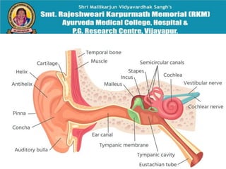
ANATOMY OF
EAR
By :
Dr Manjula Vastrad
Asst Prof
Dept of Rachana Shareera
SMVVS RKM AMC
VIJAYAPUR
INTRODUCTION
The ear is the organ of hearing and balance.
the ear is having three parts—
the outer ear,
the middle ear and
the inner ear.
OUTER EAR
The outer ear is the external portion of the ear and includes the
fleshy visible pinna (also called the auricle), the ear canal, and the
outer layer of the eardrum (also called the tympanic membrane).
The pinna consists of the curving outer rim called the helix, the
inner curved rim called the antihelix, and opens into the ear canal.
The tragus protrudes and partially obscures the ear canal, as
does the facing antitragus. The hollow region in front of the ear
canal is called the concha. The ear canal stretches for about
1 inch. The first part of the canal is surrounded by cartilage, while
the second part near the eardrum is surrounded by bone. This
bony part is known as the auditory bulla and is formed by
the tympanic part of the temporal bone. The skin surrounding the
ear canal contains ceruminous and sebaceous glands that
produce protective ear wax. The ear canal ends at the external
surface of the eardrum.
MIDDLE EAR :
The middle ear lies between the outer ear and the inner ear. It
consists of an air-filled cavity called the tympanic cavity and
includes the three ossicles and their attaching ligaments;
the auditory tube; and the round and oval windows. The
ossicles are three small bones that function together to
receive, amplify, and transmit the sound from the eardrum to
the inner ear. The ossicles are
the malleus (hammer), incus (anvil), and the stapes (stirrup).
The stapes is the smallest named bone in the body. The
middle ear also connects to the upper throat at
the nasopharynx via the pharyngeal opening of the
Eustachian tube.
The three ossicles transmit sound from the outer ear
to the inner ear. The malleus receives vibrations
from sound pressure on the eardrum, where it is
connected at its longest part (the manubrium or
handle) by a ligament. It transmits vibrations to the
incus, which in turn transmits the vibrations to the
small stapes bone. The wide base of the stapes
rests on the oval window. As the stapes vibrates,
vibrations are transmitted through the oval window,
causing movement of fluid within the cochlea.
INTERNAL EAR :
The inner ear sits within the temporal bone in a
complex cavity called the bony labyrinth. A central
area known as the vestibule contains two small fluid-
filled recesses, the utricle and saccule. These
connect to the semicircular canals and the cochlea.
There are three semicircular canals angled at right
angles to each other which are responsible for
dynamic balance. The cochlea is a spiral shell-
shaped organ responsible for the sense of hearing.
These structures together create the membranous
labyrinth.
The bony labyrinth refers to the bony compartment which
contains the membranous labyrinth, contained within the
temporal bone. The inner ear structurally begins at the oval
window, which receives vibrations from the incus of the
middle ear. Vibrations are transmitted into the inner ear into a
fluid called endolymph, which fills the membranous labyrinth.
The endolymph is situated in two vestibules,
the utricle and saccule, and eventually transmits to the
cochlea, a spiral-shaped structure. The cochlea consists of
three fluid-filled spaces: the vestibular duct, the cochlear duct,
and the tympanic duct. Hair cells responsible
for transduction—changing mechanical changes into
electrical stimuli are present in the organ of Corti in the
cochlea.
Blood supply
The blood supply of the ear differs according to each part of the ear.
The posterior auricular artery
The anterior auricular arteries
the superficial temporal artery.
The occipital artery
 the maxillary artery
the middle meningeal artery,
 ascending pharyngeal artery,
internal carotid artery, and
 the artery of the pterygoid canal.
the labyrinthine artery,
 the anterior inferior cerebellar artery or the basilar artery.
Venous Drainage :
pterygoid plexus,
external jugular and
maxillary veins.
Innervation
temporal branch of the facial nerve
the posterior auricular branch of the facial nerve
the great auricular nerve
 auriculotemporal nerve
auriculotemporal branch of the mandibular nerve
the auricular branch of the vagus nerve
the auricular branch of the vagus nerve
Clinical significance :
•Hearing loss
•Congenital abnormalities
Vertigo
•Tinnitus
Ear functions

Ear functions

  • 1.
    ANATOMY OF EAR By : DrManjula Vastrad Asst Prof Dept of Rachana Shareera SMVVS RKM AMC VIJAYAPUR
  • 4.
    INTRODUCTION The ear isthe organ of hearing and balance. the ear is having three parts— the outer ear, the middle ear and the inner ear.
  • 6.
    OUTER EAR The outerear is the external portion of the ear and includes the fleshy visible pinna (also called the auricle), the ear canal, and the outer layer of the eardrum (also called the tympanic membrane). The pinna consists of the curving outer rim called the helix, the inner curved rim called the antihelix, and opens into the ear canal. The tragus protrudes and partially obscures the ear canal, as does the facing antitragus. The hollow region in front of the ear canal is called the concha. The ear canal stretches for about 1 inch. The first part of the canal is surrounded by cartilage, while the second part near the eardrum is surrounded by bone. This bony part is known as the auditory bulla and is formed by the tympanic part of the temporal bone. The skin surrounding the ear canal contains ceruminous and sebaceous glands that produce protective ear wax. The ear canal ends at the external surface of the eardrum.
  • 8.
    MIDDLE EAR : Themiddle ear lies between the outer ear and the inner ear. It consists of an air-filled cavity called the tympanic cavity and includes the three ossicles and their attaching ligaments; the auditory tube; and the round and oval windows. The ossicles are three small bones that function together to receive, amplify, and transmit the sound from the eardrum to the inner ear. The ossicles are the malleus (hammer), incus (anvil), and the stapes (stirrup). The stapes is the smallest named bone in the body. The middle ear also connects to the upper throat at the nasopharynx via the pharyngeal opening of the Eustachian tube.
  • 9.
    The three ossiclestransmit sound from the outer ear to the inner ear. The malleus receives vibrations from sound pressure on the eardrum, where it is connected at its longest part (the manubrium or handle) by a ligament. It transmits vibrations to the incus, which in turn transmits the vibrations to the small stapes bone. The wide base of the stapes rests on the oval window. As the stapes vibrates, vibrations are transmitted through the oval window, causing movement of fluid within the cochlea.
  • 11.
    INTERNAL EAR : Theinner ear sits within the temporal bone in a complex cavity called the bony labyrinth. A central area known as the vestibule contains two small fluid- filled recesses, the utricle and saccule. These connect to the semicircular canals and the cochlea. There are three semicircular canals angled at right angles to each other which are responsible for dynamic balance. The cochlea is a spiral shell- shaped organ responsible for the sense of hearing. These structures together create the membranous labyrinth.
  • 12.
    The bony labyrinthrefers to the bony compartment which contains the membranous labyrinth, contained within the temporal bone. The inner ear structurally begins at the oval window, which receives vibrations from the incus of the middle ear. Vibrations are transmitted into the inner ear into a fluid called endolymph, which fills the membranous labyrinth. The endolymph is situated in two vestibules, the utricle and saccule, and eventually transmits to the cochlea, a spiral-shaped structure. The cochlea consists of three fluid-filled spaces: the vestibular duct, the cochlear duct, and the tympanic duct. Hair cells responsible for transduction—changing mechanical changes into electrical stimuli are present in the organ of Corti in the cochlea.
  • 13.
    Blood supply The bloodsupply of the ear differs according to each part of the ear. The posterior auricular artery The anterior auricular arteries the superficial temporal artery. The occipital artery  the maxillary artery the middle meningeal artery,  ascending pharyngeal artery, internal carotid artery, and  the artery of the pterygoid canal. the labyrinthine artery,  the anterior inferior cerebellar artery or the basilar artery.
  • 14.
    Venous Drainage : pterygoidplexus, external jugular and maxillary veins.
  • 15.
    Innervation temporal branch ofthe facial nerve the posterior auricular branch of the facial nerve the great auricular nerve  auriculotemporal nerve auriculotemporal branch of the mandibular nerve the auricular branch of the vagus nerve the auricular branch of the vagus nerve
  • 16.
    Clinical significance : •Hearingloss •Congenital abnormalities Vertigo •Tinnitus