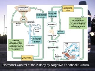
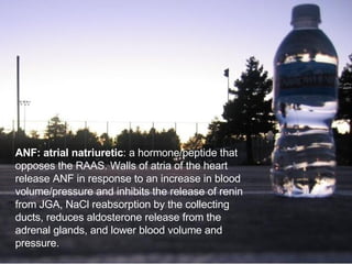
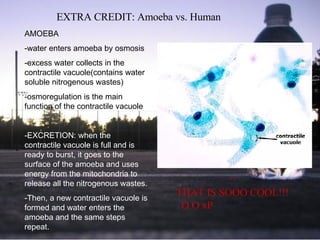
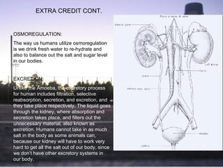
Osmoregulation &  Excretion Kevin Kwan Jeffery Khou Jessica Dai Jason Yiu
Introduction  to Osmoregularity and Excretion The physiological systems of animals operate within a fluid environment   The relative concentration of water and solutes must be maintained Metabolism presents organisms with the problem of waste disposal thus animals use two key homeostatic processes: - Osmoregulation – how animals regulate  solute concentrations and balance the gain  and loss of water - Excretion – how animals get rid of nitrogen- containing waste products of metabolism Homeostasis – steady state of physiological condition of the body
Osmosis and -osmotic All animals face the central problem of osmoregularity. The rates of the uptake and loss of water must balance. This is done with the help of osmosis. -Osmosis: diffusion or movement of water  across  a selectively permeable  membrane   Osmolarity happens when two solutions, separated by a membrane, differ in osmolarity. - Osmolarity: total solute concentration  expressed as molarity, or moles of solute  per liter of solution
Osmosis and –osmotic cont. Different terms relating to osmolarity: -Isoosmotic: refers to two solutions of the same  molarity that are separated by a selectively  permeable membrane.  There is no net  movement of water between the solutions,  although water molecules are continually  crossing the membrane at equal rates in both  directions   -Hyperosmotic: refers to the solution that has  the greater concentration of solutes   -Hypoosmotic: refers to the solution that has  the more dilute solution   Thus, when the osmolarities of solutions differ, water flows by osmosis from a hypoosmotic solution to a hyperosmotic solution
Osmotic Challenges There are two basic solutions to the problem of balancing water gain with water loss. One is to be an osmoconformer. - Osmoconformer:  an animal that does not actively  adjust its internal osmolarity. The internal osmolarity  is the same as that of its environment. There is no  tendency to lose or gain water, and they often live in  water with stable composition, so as to keep a  constant internal osmolarity.   Another is to be an osmoregulator. -Osmoregulator:  an animal that must control its own  osmolarity because its own body fluids are not  isoosmotic with its outside environment.   If the environment is hypoosmotic, an osmoregulator must discharge extra water.   If the environment is hyperosmotic, an osmoregulator must take in water to offset osmotic loss.   Nonetheless, osmoregulation costs energy depending on the difference between its osmolarity and its surroundings.
Osmotic Challenges cont. Most animals, though, are stenohaline animals: -Stenohaline:  animals (osmoconformers/  osmoregulators) that cannot tolerate  substantial changes in external  osmolarity.   In contrast, some are: -Euryhaline:  animals (certain  osmoconformers/ certain  osmoregulators) that can survive large  fluctuations in external osmolarity .
Osmoregulation and adaptation and water balance  Marine Animals, Freshwater Animals, Animals of Temp Waters, Land animals Most marine invertebrates are osmoconformers. But, the concentration of its specific solutes differ from each other. Thus, even an osmoconformer regulates its internal composition of solutes. The ocean is a strongly dehydrating environment because it is much saltier, thus: Marine animals: drink large amount of  seawater; gills dispose sodium chloride; kidney disposes excess calcium, magnesium, and sulfate ions or uses the rectal gland and feces to excrete salt and water slowly enters the body by osmosis and food.   Freshwater animals constantly gain water and lose salts, thus: Freshwater animals: Excrete large amount of dilute urine, salts are replenished by food and by uptake across the gills.
Osmoregulation and adaptation and water balance cont.  Animals that live in temporary waters can withstand huge fluctuations of moisture in their environments.  (Anhydrobiosis: survival in a dormant state when one habitat dries up) Their habitats may dry up, thus: Animals that live in temporary water (and hydrobiotic animals): utilize large amounts of sugar (trehalose)  Land animals face the threat  of desiccation as a major regulatory problem, thus Land animals: utilize adaptations that reduce water loss such as body coverings, some are nocturnal, drinking and eating moist food and by using metabolic water, and simple anatomical features
Countercurrent   heat exchange   Four processes that account for heat exchange: conduction (direct transfer of heat), convection (transfer of heat by movement of liquid or air against the body), radiation (the emission of electromagnetic waves produced by something containing heat), evaporation  (loss of heat from the surface of a liquid that is losing some of its molecules as gas). Circulation aids in heat exchange: it alters the amount of blood flowing to the skin; increased blood flow usually results in vasodilation (increase in diameter of superficial blood vessels) causing more blood flow, thus heat transfer. Vasoconstriction reduces the blood flow and heat loss by decreasing the diameter of the blood vessels.
Countercurrent heat exchange cont. • Countercurrent heat exchange involves a special arrangement of arteries and veins • The countercurrent heat exchange conserves heat by arteries that carry warm blood which circulate through limbs, which come into contact with veins that convey blood in the opposite direction. This arrangement  facilitates heat transfer from arteries to veins along the entire length of the blood vessel.  • Examples of how an ectotherm maintains higher than expected temperature: behavioral adaptations (seek ideal environments), physiological adaptations (vasoconstriction).
Countercurrent heat exchange  cont. How feedback mechanism regulates temperature (and diagram): nerve cells that control thermoregulation, concentrated in the hypothalamus, contain a thermostat that responds to change in body temperature above/below a set point by activating mechanisms that promote heat loss/gain (located in the skin).   Others are cold receptors and respond by inhibiting heat loss mechanisms and activate heat-saving ones (vasoconstriction, erection of fur, heat-generating mechanisms).  In response to elevated temperatures: shuts down heat-saving mechanisms, promotes cooling (vasodilation, sweating, panting)
 
Transport Epithelia The ultimate function of osmoregulation is to maintain composition of cellular cytoplasm, by managing the composition of the internal body fluid that bathes the cells (insects and other animals with an open circulatory system: hemolymph; vertebrates with closed circulatory system: interstitial fluid)   Transport epithelium: layer or layers of specialized epithelial cells that regulates solute movements, essential components of osmotic regulation/ metabolic waste disposal   Seagulls and high salt diet : The nasal glands secrete a fluid much saltier than that of ocean water so even if the bird drinks ocean water, the net gain is water. Salt glands empty it via a duct into the nostrils and the salty solution either drips off the tips of the beak or is exhaled in a fine mist.
Nitrogenous waste   Metabolic waste in general: must be dissolved in water, type and quantity have a large impact on water balanc e Different types of waste: - Ammonia: soluble but tolerated  at very low concentrations  -Urea: low toxicity, combination of  ammonia and carbon dioxide and  high concentrations  -Uric Acid: relatively non toxic,  insoluble in water, semi-solid  paste with very little water loss
Nitrogenous waste cont.  The kinds of nitrogenous waste depend on the animal’s evolutionary history and habitat (influence).  If there is minimal water, uric acid rather than urea may be the favored waste products because it needs less water to be produced. Also, different waste products are a result with different forms of reproductions.   For some animals such as tortoises, if temperature changes and goes up, or if water becomes less available, uric acid will be produced, for example, in place of urea. Evolution determines the limits of physiological organisms  and organisms make physiological adjustments   within these evolutionary constraints. It is also dependent on the energy budget, which is affected by the food in the habitat.
Excretory Process First, body fluid is collected, involving filtration through selectively permeable membranes (filtrate)-> selective reabsorption-> secretion-> excretion. -Filtration: blood or other body fluids  are exposed to filtering device of  selectively permeable membranes of  transport epithelia -Selective reabsorption: excretory systems  use active transport to reabsorb valuable  solutes (glucose, certain salts, amino acids) -Secretion; solutes are removed from  animal body fluids and added to the  filtrate -Excretion: disposal of nitrogen- containing waste products of  metabolism
Excretion cont. Different excretory systems: Protonephridia: Flame-Bulb System Flatworms utilize  protonephidium: a network  of dead-end tubules lacking  internal opening .  Metanephridia:  It is another type of tubular  excretory system; has  internal openings that collect  body fluids. Malphighian tubules:  A type of tubular excretory  system in which organs in  insects and other terrestrial  arthropods remove  nitrogenous wastes and also  function in osmoregulation.
Excretion cont. Metanephridia of  Earthworm
Malpighian Tubules of insects Excretion cont.
Nephron and Associated Structures Structure and Function of the Nephron:   -nephron: functional unit of the vertebrate kidney -renal cortex: outer part of a mammalian kidney -renal medulla: inner part of a mammalian kidney -glomerulus: single long tubule and a ball of  capillaries - Bowman’s capsule: blind end of a tubule that forms a cup-shaped swelling surrounding the glomerulus. 80% of the nephrons in the human kidney are cortical nephrons and they have reduced loops of Henle. Juxtamedullary nephrons are about 20% and have well-developed loops that extend deeply into the renal medulla.  
The relationship of the kidney and the circulatory system: The kidneys produce urine and regulate the composition of the blood. The urine is conveyed to the urinary bladder via the ureter and  to the outside via the   urethra. Branches of the aorta, retinal arteries, convey blood to the kidneys; renal veins drain blood from the kidneys into the posterior vena cava.   Excretion cont. Posterior Vena cava Kidney Renal artery and vein Aorta Ureter Urinary Bladder Urethra Bowman’s capsule Glomerulus Afferent arteriole from renal artery Efferent arteriole from glomerulus Branch of renal vein Proximal tubule Peritubular capillaries Distal tubule Collecting duct Loop of Henle Descending limb Ascending limb Vasa recta Renal pelvis Ureter Renal Medulla Renal cortex Juxtamedullarnephron Cortical nephron Renal cortex Renal medulla
Filtration of the Blood: It occurs as blood pressure forces fluid from the blood in the glomerulus into the lumen of the Bowman’s capsule. It contains a variety of substances including nitrogenous wastes.  Pathway of the filtrate: (Blood filtrate to Urine) proximal tubule-> Descending limb of the loop of Henle-> Ascending limb of the loop of Henle-> Distal tubule-> Collecting duct-> renal pelvis-> drained by ureter Nephron and Associated Structures
Blood Vessels Associated with the Nephrons Afferent arteriole: supplies blood to each Nephron Efferent arteriole: formed by the capillaries that converge as they leave the glomerulus Peritubular capillaries: formed by vessels subdividing Vasa recta: capillaries that serve the loop for Henle
Solute Gradients and Water Conservation The cooperative action and precise arrangement of the loops of Henle and the collecting ducts are responsible for the concentration of urine The nephrons, especially the loops of Henle, consume energy to produce a region of high osmolarity in the kidney, which extract water from the filtrate .
How the human kidney concentrates urine
Regulation of Kidney Function One of the most important aspects of the mammalian kidney is its ability to adjust both the volume and osmolarity of urine depending on the animals’ water and salt balance and rate of urea production. Filtrates starts at the Bowman's capsule which then leads to the proximal tubule, the filtrate starts at an osmolarity of 300 mosm/L. In the proximal tubule, a large amount of water and salt is reabsorbed, which decreases the volume of the filtrate, because both water and NaCl is lost, osmolarity remains the same. As the filtrate flows from the cortex to the outer medulla, in the descending limb of the loop of Henle, water leaves the tubule by osmosis. Osmolarity increases, and the highest osmolarity of NaCl occurs here, at 1,200 mosm/L. As it rounds the curve and starts to ascend the next limb, which is only permeable to salt, not to water. This loss of water and NaCl maintains the osmolarity of the interstitial fluid in the kidney.
Hormonal Control of the Kidney by Negative Feedback Circuits
One of the most important aspects of the mammalian kidney is its ability to adjust both the volume and osmolarity of urine, depending on the animal’s water and salt balance and the rate of urea production. This versatility in osemoregulatory function is managed with a combination of nervous and hormonal controls. Regulation of Kidney Functions
ADH: antidiuretic hormone  - Produced in the hypothalamus, stored in and released from the posterior pituitary gland, osmoreceptor cells monitor the osmolarity of the blood. If it rises about a set point of 300 mosm/L, more ADH is released into the bloodstream/reaches kidney. The main targets are the distal tubules and collecting ducts where it increases the permeability  of the epithelium to water, amplify water reabsorption, reduce urine volume and prevent further increase of blood osmolarity above the set point. If negative feedback- less ADH. Only gain of additional water in food/drink can bring osmolarity back. Large intake of water-> little ADH is released thus decreases permeability of the distal tubules and collecting ducts (more urine).
·  JGA: juxtaglomerular apparatus : locate near the afferent arteriole that supplies blood to the glomulus. When pressure/blood volume drops, enzyme rennin initiates chemical reactions that convert a plasma protein called angiotensinogen to a peptide called angiotensin II-> raises blood pressure by constricting arterioles, decreasing blood  flow to many capillaries, stimulates   proximal tubules to nephrons to reabsorb more NaCl and water (raises blood volume/pressure), stimulation of adrenal glands to release a hormone called aldosterone (causes reabsorption of sodium and water, increasing blood volume/pressure).
·          RAAS: renin-angiotensin-aldosterone system : part of a complex feedback circuit that functions in homeostasis, drop in blood pressure/blood volume triggers renin release from JGA. In turn, rise in blood pressure/volume from various actions of angiotensin II/aldsterone reduce the release of renin.  ·          ADH vs RAAS : ADH is a response to an increase in osmolarity of the blood and lowers blood sodium ion concentration by stimulating water reabsorption in the kidney. RAAS responds to a fall in blood volume/pressure by increasing water and sodium ion reabsorption.
ANF: atrial natriuretic : a hormone/peptide that opposes the RAAS. Walls of atria of the heart release ANF in response to an increase in blood volume/pressure and inhibits the release of renin from JGA, NaCl reabsorption by the collecting ducts, reduces aldosterone release from the adrenal glands, and lower blood volume and pressure.
All animals have different habitats, functions of osmoregulations, physiological machines (organs) to maintain solute and water balance and excreting nitrogenous wastes.
AMOEBA -water enters amoeba by osmosis -excess water collects in the contractile vacuole(contains water soluble nitrogenous wastes) -osmoregulation is the main function of the contractile vacuole -EXCRETION: when the contractile vacuole is full and is ready to burst, it goes to the surface of the amoeba and uses energy from the mitochondria to release all the nitrogenous wastes.  -Then, a new contractile vacuole is formed and water enters the amoeba and the same steps repeat.  EXTRA CREDIT: Amoeba vs. Human ^   THAT IS SOOO COOL!!!    O.O xP
OSMOREGULATION: The way us humans utilize osmoregulation is we drink fresh water to re-hydrate and also to balance out the salt and sugar level in our bodies. EXCRETION: Unlike the Amoeba, the excretory process for human includes filtration, selective reabsorption, secretion, and excretion, and they take place respectively. The liquid goes through the kidney, where absorption and secretion takes place, and filters out the unnecessary material, also known as excretion. Humans cannot take in as much salt in the body as some animals can, because our kidney will have to work very hard to get all the salt out of our body, since we don’t have other excretory systems in our body.   EXTRA CREDIT CONT.

Presentation1

  • 1.
    Osmoregulation & Excretion Kevin Kwan Jeffery Khou Jessica Dai Jason Yiu
  • 2.
    Introduction toOsmoregularity and Excretion The physiological systems of animals operate within a fluid environment The relative concentration of water and solutes must be maintained Metabolism presents organisms with the problem of waste disposal thus animals use two key homeostatic processes: - Osmoregulation – how animals regulate solute concentrations and balance the gain and loss of water - Excretion – how animals get rid of nitrogen- containing waste products of metabolism Homeostasis – steady state of physiological condition of the body
  • 3.
    Osmosis and -osmoticAll animals face the central problem of osmoregularity. The rates of the uptake and loss of water must balance. This is done with the help of osmosis. -Osmosis: diffusion or movement of water across a selectively permeable membrane Osmolarity happens when two solutions, separated by a membrane, differ in osmolarity. - Osmolarity: total solute concentration expressed as molarity, or moles of solute per liter of solution
  • 4.
    Osmosis and –osmoticcont. Different terms relating to osmolarity: -Isoosmotic: refers to two solutions of the same molarity that are separated by a selectively permeable membrane. There is no net movement of water between the solutions, although water molecules are continually crossing the membrane at equal rates in both directions -Hyperosmotic: refers to the solution that has the greater concentration of solutes -Hypoosmotic: refers to the solution that has the more dilute solution Thus, when the osmolarities of solutions differ, water flows by osmosis from a hypoosmotic solution to a hyperosmotic solution
  • 5.
    Osmotic Challenges Thereare two basic solutions to the problem of balancing water gain with water loss. One is to be an osmoconformer. - Osmoconformer: an animal that does not actively adjust its internal osmolarity. The internal osmolarity is the same as that of its environment. There is no tendency to lose or gain water, and they often live in water with stable composition, so as to keep a constant internal osmolarity. Another is to be an osmoregulator. -Osmoregulator: an animal that must control its own osmolarity because its own body fluids are not isoosmotic with its outside environment. If the environment is hypoosmotic, an osmoregulator must discharge extra water. If the environment is hyperosmotic, an osmoregulator must take in water to offset osmotic loss. Nonetheless, osmoregulation costs energy depending on the difference between its osmolarity and its surroundings.
  • 6.
    Osmotic Challenges cont.Most animals, though, are stenohaline animals: -Stenohaline: animals (osmoconformers/ osmoregulators) that cannot tolerate substantial changes in external osmolarity. In contrast, some are: -Euryhaline: animals (certain osmoconformers/ certain osmoregulators) that can survive large fluctuations in external osmolarity .
  • 7.
    Osmoregulation and adaptationand water balance Marine Animals, Freshwater Animals, Animals of Temp Waters, Land animals Most marine invertebrates are osmoconformers. But, the concentration of its specific solutes differ from each other. Thus, even an osmoconformer regulates its internal composition of solutes. The ocean is a strongly dehydrating environment because it is much saltier, thus: Marine animals: drink large amount of seawater; gills dispose sodium chloride; kidney disposes excess calcium, magnesium, and sulfate ions or uses the rectal gland and feces to excrete salt and water slowly enters the body by osmosis and food. Freshwater animals constantly gain water and lose salts, thus: Freshwater animals: Excrete large amount of dilute urine, salts are replenished by food and by uptake across the gills.
  • 8.
    Osmoregulation and adaptationand water balance cont. Animals that live in temporary waters can withstand huge fluctuations of moisture in their environments. (Anhydrobiosis: survival in a dormant state when one habitat dries up) Their habitats may dry up, thus: Animals that live in temporary water (and hydrobiotic animals): utilize large amounts of sugar (trehalose) Land animals face the threat of desiccation as a major regulatory problem, thus Land animals: utilize adaptations that reduce water loss such as body coverings, some are nocturnal, drinking and eating moist food and by using metabolic water, and simple anatomical features
  • 9.
    Countercurrent heat exchange Four processes that account for heat exchange: conduction (direct transfer of heat), convection (transfer of heat by movement of liquid or air against the body), radiation (the emission of electromagnetic waves produced by something containing heat), evaporation (loss of heat from the surface of a liquid that is losing some of its molecules as gas). Circulation aids in heat exchange: it alters the amount of blood flowing to the skin; increased blood flow usually results in vasodilation (increase in diameter of superficial blood vessels) causing more blood flow, thus heat transfer. Vasoconstriction reduces the blood flow and heat loss by decreasing the diameter of the blood vessels.
  • 10.
    Countercurrent heat exchangecont. • Countercurrent heat exchange involves a special arrangement of arteries and veins • The countercurrent heat exchange conserves heat by arteries that carry warm blood which circulate through limbs, which come into contact with veins that convey blood in the opposite direction. This arrangement facilitates heat transfer from arteries to veins along the entire length of the blood vessel. • Examples of how an ectotherm maintains higher than expected temperature: behavioral adaptations (seek ideal environments), physiological adaptations (vasoconstriction).
  • 11.
    Countercurrent heat exchange cont. How feedback mechanism regulates temperature (and diagram): nerve cells that control thermoregulation, concentrated in the hypothalamus, contain a thermostat that responds to change in body temperature above/below a set point by activating mechanisms that promote heat loss/gain (located in the skin). Others are cold receptors and respond by inhibiting heat loss mechanisms and activate heat-saving ones (vasoconstriction, erection of fur, heat-generating mechanisms). In response to elevated temperatures: shuts down heat-saving mechanisms, promotes cooling (vasodilation, sweating, panting)
  • 12.
  • 13.
    Transport Epithelia Theultimate function of osmoregulation is to maintain composition of cellular cytoplasm, by managing the composition of the internal body fluid that bathes the cells (insects and other animals with an open circulatory system: hemolymph; vertebrates with closed circulatory system: interstitial fluid) Transport epithelium: layer or layers of specialized epithelial cells that regulates solute movements, essential components of osmotic regulation/ metabolic waste disposal Seagulls and high salt diet : The nasal glands secrete a fluid much saltier than that of ocean water so even if the bird drinks ocean water, the net gain is water. Salt glands empty it via a duct into the nostrils and the salty solution either drips off the tips of the beak or is exhaled in a fine mist.
  • 14.
    Nitrogenous waste Metabolic waste in general: must be dissolved in water, type and quantity have a large impact on water balanc e Different types of waste: - Ammonia: soluble but tolerated at very low concentrations -Urea: low toxicity, combination of ammonia and carbon dioxide and high concentrations -Uric Acid: relatively non toxic, insoluble in water, semi-solid paste with very little water loss
  • 15.
    Nitrogenous waste cont. The kinds of nitrogenous waste depend on the animal’s evolutionary history and habitat (influence). If there is minimal water, uric acid rather than urea may be the favored waste products because it needs less water to be produced. Also, different waste products are a result with different forms of reproductions. For some animals such as tortoises, if temperature changes and goes up, or if water becomes less available, uric acid will be produced, for example, in place of urea. Evolution determines the limits of physiological organisms and organisms make physiological adjustments within these evolutionary constraints. It is also dependent on the energy budget, which is affected by the food in the habitat.
  • 16.
    Excretory Process First,body fluid is collected, involving filtration through selectively permeable membranes (filtrate)-> selective reabsorption-> secretion-> excretion. -Filtration: blood or other body fluids are exposed to filtering device of selectively permeable membranes of transport epithelia -Selective reabsorption: excretory systems use active transport to reabsorb valuable solutes (glucose, certain salts, amino acids) -Secretion; solutes are removed from animal body fluids and added to the filtrate -Excretion: disposal of nitrogen- containing waste products of metabolism
  • 17.
    Excretion cont. Differentexcretory systems: Protonephridia: Flame-Bulb System Flatworms utilize protonephidium: a network of dead-end tubules lacking internal opening . Metanephridia: It is another type of tubular excretory system; has internal openings that collect body fluids. Malphighian tubules: A type of tubular excretory system in which organs in insects and other terrestrial arthropods remove nitrogenous wastes and also function in osmoregulation.
  • 18.
  • 19.
    Malpighian Tubules ofinsects Excretion cont.
  • 20.
    Nephron and AssociatedStructures Structure and Function of the Nephron: -nephron: functional unit of the vertebrate kidney -renal cortex: outer part of a mammalian kidney -renal medulla: inner part of a mammalian kidney -glomerulus: single long tubule and a ball of capillaries - Bowman’s capsule: blind end of a tubule that forms a cup-shaped swelling surrounding the glomerulus. 80% of the nephrons in the human kidney are cortical nephrons and they have reduced loops of Henle. Juxtamedullary nephrons are about 20% and have well-developed loops that extend deeply into the renal medulla.  
  • 21.
    The relationship ofthe kidney and the circulatory system: The kidneys produce urine and regulate the composition of the blood. The urine is conveyed to the urinary bladder via the ureter and to the outside via the urethra. Branches of the aorta, retinal arteries, convey blood to the kidneys; renal veins drain blood from the kidneys into the posterior vena cava. Excretion cont. Posterior Vena cava Kidney Renal artery and vein Aorta Ureter Urinary Bladder Urethra Bowman’s capsule Glomerulus Afferent arteriole from renal artery Efferent arteriole from glomerulus Branch of renal vein Proximal tubule Peritubular capillaries Distal tubule Collecting duct Loop of Henle Descending limb Ascending limb Vasa recta Renal pelvis Ureter Renal Medulla Renal cortex Juxtamedullarnephron Cortical nephron Renal cortex Renal medulla
  • 22.
    Filtration of theBlood: It occurs as blood pressure forces fluid from the blood in the glomerulus into the lumen of the Bowman’s capsule. It contains a variety of substances including nitrogenous wastes. Pathway of the filtrate: (Blood filtrate to Urine) proximal tubule-> Descending limb of the loop of Henle-> Ascending limb of the loop of Henle-> Distal tubule-> Collecting duct-> renal pelvis-> drained by ureter Nephron and Associated Structures
  • 23.
    Blood Vessels Associatedwith the Nephrons Afferent arteriole: supplies blood to each Nephron Efferent arteriole: formed by the capillaries that converge as they leave the glomerulus Peritubular capillaries: formed by vessels subdividing Vasa recta: capillaries that serve the loop for Henle
  • 24.
    Solute Gradients andWater Conservation The cooperative action and precise arrangement of the loops of Henle and the collecting ducts are responsible for the concentration of urine The nephrons, especially the loops of Henle, consume energy to produce a region of high osmolarity in the kidney, which extract water from the filtrate .
  • 25.
    How the humankidney concentrates urine
  • 26.
    Regulation of KidneyFunction One of the most important aspects of the mammalian kidney is its ability to adjust both the volume and osmolarity of urine depending on the animals’ water and salt balance and rate of urea production. Filtrates starts at the Bowman's capsule which then leads to the proximal tubule, the filtrate starts at an osmolarity of 300 mosm/L. In the proximal tubule, a large amount of water and salt is reabsorbed, which decreases the volume of the filtrate, because both water and NaCl is lost, osmolarity remains the same. As the filtrate flows from the cortex to the outer medulla, in the descending limb of the loop of Henle, water leaves the tubule by osmosis. Osmolarity increases, and the highest osmolarity of NaCl occurs here, at 1,200 mosm/L. As it rounds the curve and starts to ascend the next limb, which is only permeable to salt, not to water. This loss of water and NaCl maintains the osmolarity of the interstitial fluid in the kidney.
  • 27.
    Hormonal Control ofthe Kidney by Negative Feedback Circuits
  • 28.
    One of themost important aspects of the mammalian kidney is its ability to adjust both the volume and osmolarity of urine, depending on the animal’s water and salt balance and the rate of urea production. This versatility in osemoregulatory function is managed with a combination of nervous and hormonal controls. Regulation of Kidney Functions
  • 29.
    ADH: antidiuretic hormone - Produced in the hypothalamus, stored in and released from the posterior pituitary gland, osmoreceptor cells monitor the osmolarity of the blood. If it rises about a set point of 300 mosm/L, more ADH is released into the bloodstream/reaches kidney. The main targets are the distal tubules and collecting ducts where it increases the permeability of the epithelium to water, amplify water reabsorption, reduce urine volume and prevent further increase of blood osmolarity above the set point. If negative feedback- less ADH. Only gain of additional water in food/drink can bring osmolarity back. Large intake of water-> little ADH is released thus decreases permeability of the distal tubules and collecting ducts (more urine).
  • 30.
    ·  JGA: juxtaglomerularapparatus : locate near the afferent arteriole that supplies blood to the glomulus. When pressure/blood volume drops, enzyme rennin initiates chemical reactions that convert a plasma protein called angiotensinogen to a peptide called angiotensin II-> raises blood pressure by constricting arterioles, decreasing blood flow to many capillaries, stimulates proximal tubules to nephrons to reabsorb more NaCl and water (raises blood volume/pressure), stimulation of adrenal glands to release a hormone called aldosterone (causes reabsorption of sodium and water, increasing blood volume/pressure).
  • 31.
    ·         RAAS:renin-angiotensin-aldosterone system : part of a complex feedback circuit that functions in homeostasis, drop in blood pressure/blood volume triggers renin release from JGA. In turn, rise in blood pressure/volume from various actions of angiotensin II/aldsterone reduce the release of renin. ·         ADH vs RAAS : ADH is a response to an increase in osmolarity of the blood and lowers blood sodium ion concentration by stimulating water reabsorption in the kidney. RAAS responds to a fall in blood volume/pressure by increasing water and sodium ion reabsorption.
  • 32.
    ANF: atrial natriuretic: a hormone/peptide that opposes the RAAS. Walls of atria of the heart release ANF in response to an increase in blood volume/pressure and inhibits the release of renin from JGA, NaCl reabsorption by the collecting ducts, reduces aldosterone release from the adrenal glands, and lower blood volume and pressure.
  • 33.
    All animals havedifferent habitats, functions of osmoregulations, physiological machines (organs) to maintain solute and water balance and excreting nitrogenous wastes.
  • 34.
    AMOEBA -water entersamoeba by osmosis -excess water collects in the contractile vacuole(contains water soluble nitrogenous wastes) -osmoregulation is the main function of the contractile vacuole -EXCRETION: when the contractile vacuole is full and is ready to burst, it goes to the surface of the amoeba and uses energy from the mitochondria to release all the nitrogenous wastes. -Then, a new contractile vacuole is formed and water enters the amoeba and the same steps repeat. EXTRA CREDIT: Amoeba vs. Human ^ THAT IS SOOO COOL!!! O.O xP
  • 35.
    OSMOREGULATION: The wayus humans utilize osmoregulation is we drink fresh water to re-hydrate and also to balance out the salt and sugar level in our bodies. EXCRETION: Unlike the Amoeba, the excretory process for human includes filtration, selective reabsorption, secretion, and excretion, and they take place respectively. The liquid goes through the kidney, where absorption and secretion takes place, and filters out the unnecessary material, also known as excretion. Humans cannot take in as much salt in the body as some animals can, because our kidney will have to work very hard to get all the salt out of our body, since we don’t have other excretory systems in our body. EXTRA CREDIT CONT.