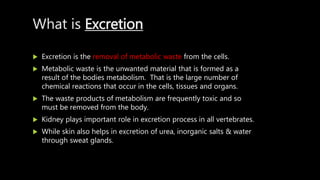
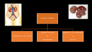
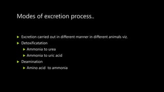
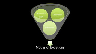
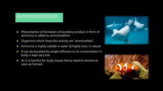
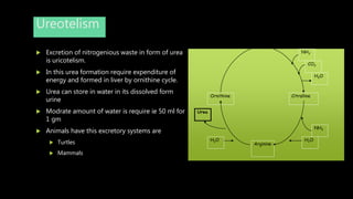
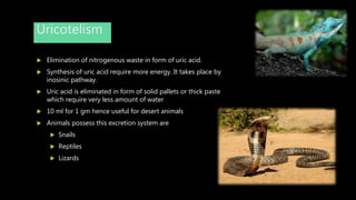
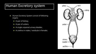
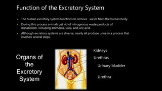
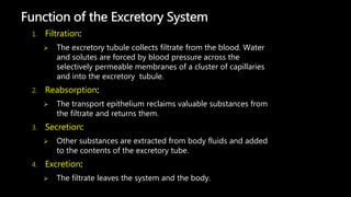
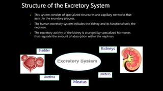
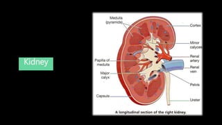
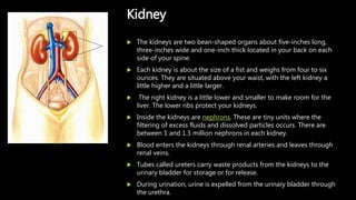
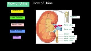
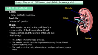
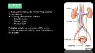
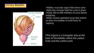
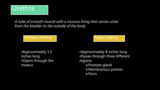
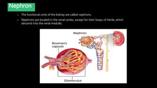
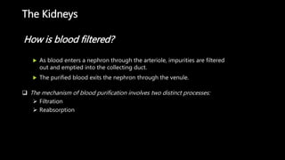
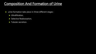
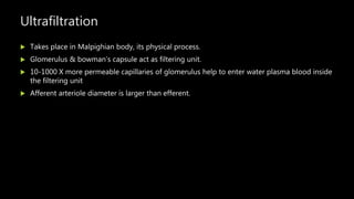
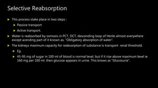
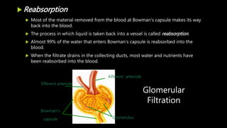
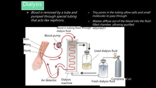
Excretion &
Osmoregulation
CHETAN K.
What is Excretion
 Excretion is the removal of metabolic waste from the cells.
 Metabolic waste is the unwanted material that is formed as a
result of the bodies metabolism. That is the large number of
chemical reactions that occur in the cells, tissues and organs.
 The waste products of metabolism are frequently toxic and so
must be removed from the body.
 Kidney plays important role in excretion process in all vertebrates.
 While skin also helps in excretion of urea, inorganic salts & water
through sweat glands.
Functions of Kidney
Elimination on nitro. waste
Maintain H2O level
ie.
osmoregulation
Maintain H+ ion concentration
ie
iso-tonic
Modes of excretion process..
 Excretion carried out in different manner in different animals viz.
 Detoxificatation
 Ammonia to urea
 Ammonia to uric acid
 Deamination
 Amino acid to ammonia
Modes of Excretions
Uricotelism
Ammonot
elism
ureotelism
Ammonotelism
 Phenomenon of formation of excretory product in form of
ammonia is called as ammonotelism.
 Organisms which show this activity are “ammonotelic”.
 Ammonia is highly soluble in water & highly toxic in nature
 It can be excreted by simple diffusion so its concentration in
body is kept very low.
 As it is harmful for body tissues hence need to remove as
soon as formed.
Ammonotelic animals
 For elimination of ammonia large quantity of
water is required.
 Due to this reason ammonotelism is seen in
aquatic animals, bony fish, tadpole larva.
 Such animals ammonia diffuse through the skin
and gills and kidney.
Ureotelism
 Excretion of nitrogenious waste in form of urea
is uricotelism.
 In this urea formation require expenditure of
energy and formed in liver by ornithine cycle.
 Urea can store in water in its dissolved form
urine
 Modrate amount of water is require ie 50 ml for
1 gm
 Animals have this excretory systems are
 Turtles
 Mammals
Ornithine Citralline
Arginine
NH3
CO2
H2O
NH3
H2O
Urea
H2O
Uricotelism
 Elimination of nitrogenous waste in form of uric acid.
 Synthesis of uric acid require more energy. It takes place by
inosinic pathway.
 Uric acid is eliminated in form of solid pallets or thick paste
which require very less amount of water
 10 ml for 1 gm hence useful for desert animals
 Animals possess this excretion system are
 Snails
 Reptiles
 Lizards
Human Excretory system
 Human Excretory System consist of following
organs
 A pair of kidney,
 A pair of ureters,
 A single unpaired urinary bladder,
 A urethra in males / vestibule in females.
Function of the Excretory System
 The human excretory system functions to remove waste from the human body.
 During this process animals get rid of nitrogenous waste products of
metabolism, including ammonia, urea, and uric acid.
 Although excretory systems are diverse, nearly all produce urine in a process that
involves several steps.
Kidneys
Urethras
Urinary bladder
Urethra
Organs of
the
Excretory
System
1. Filtration:
 The excretory tubule collects filtrate from the blood. Water
and solutes are forced by blood pressure across the
selectively permeable membranes of a cluster of capillaries
and into the excretory tubule.
2. Reabsorption:
 The transport epithelium reclaims valuable substances from
the filtrate and returns them.
3. Secretion:
 Other substances are extracted from body fluids and added
to the contents of the excretory tube.
4. Excretion:
 The filtrate leaves the system and the body.
Function of the Excretory System
Structure of the Excretory System
 This system consists of specialized structures and capillary networks that
assist in the excretory process.
 The human excretory system includes the kidney and its functional unit, the
nephron.
 The excretory activity of the kidney is changed by specialized hormones
that regulate the amount of absorption within the nephron.
Bladder Kidneys
Urethra
Ureters
Meatus
Kidney
 The kidneys are two bean-shaped organs about five-inches long,
three-inches wide and one-inch thick located in your back on each
side of your spine.
 Each kidney is about the size of a fist and weighs from four to six
ounces. They are situated above your waist, with the left kidney a
little higher and a little larger.
 The right kidney is a little lower and smaller to make room for the
liver. The lower ribs protect your kidneys.
 Inside the kidneys are nephrons. These are tiny units where the
filtering of excess fluids and dissolved particles occurs. There are
between 1 and 1.3 million nephrons in each kidney.
 Blood enters the kidneys through renal arteries and leaves through
renal veins.
 Tubes called ureters carry waste products from the kidneys to the
urinary bladder for storage or for release.
 During urination, urine is expelled from the urinary bladder through
the urethra.
Kidney
Flow of UrineFlow of Urine
Glomerulus
Renal Tubules
Renal Pelvis
Renal Calices
Ureters
Renal cortex
Renal capsule
Renal medulla
Ureter
Renal vein
Renal artery
Nephron
Kidneys filter about 1700 liters of blood daily in the average adult.
Parts of the kidneys
• Cortex
-outer protective portion
• Medulla
-inner soft portion
• Hilum
-a depression located in the middle of the
concave side of the kidney where blood
vessels, nerves, and the ureters enter and exit
the kidneys
Cortex
Medulla
Hilum
 The cortex is where the blood is filtered.
 The medulla contains the collecting ducts which carry filtrate (filtered
substances) to the pelvis.
 The pelvis is a hollow cavity where urine accumulates and drains into the
ureter.
Ureters
Peristalsis, a rhythmic contraction of the ureter
smooth muscle which helps to move the urine into
the bladder.
A tube approximately 6 to 7 inches long attached
to each kidney.
 Made up of three layers of tissue
 Smooth muscle
 Fibrous tissue
 Mucous layer
Urinary Bladder
•Hollow, muscular organ that stores urine
•Sphincter muscles hold the urine in place
•Holds 300 to 400 milliliters of urine before
emptying
•Walls contain epithelial tissue that stretch
to allow the bladder to hold twice its
capacity
Urethra
Prostate
gland
Ureter
Urinary bladder
•The trigone is a triangular area at the
base of the bladder where the ureters
enter and the urethra exits
Urethra
Female Urethra Male Urethra
•Approximately 1.5
inches long
•Opens through the
meatus
•Approximately 8 inches long
•Passes through three different
regions:
Prostate gland
Membranous portion
Penis
A tube of smooth muscle with a mucous lining that carries urine
from the bladder to the outside of the body.
 The functional units of the kidney are called nephrons.
 Nephrons are located in the renal cortex, except for their loops of Henle, which
descend into the renal medulla.
Nephron
Two parts
a. Renal Corpuscle
1. Bowman Capsule
2. Glomerulus
a. Fenestrae - pore in endothelial walls of glomerulus.
b. Podocytes - specialized cells.
c. Filteration slits
b. Renal Tubule - Series of single layer tubules
1. Proximal Convoluted tubule
2. Loop of Henle
3. Distal Convoluted tubule
Nephron
Kidneys are made up of nephrons. Blood enters the nephron, where impurities are
filtered out and emptied into the collecting duct. The purified blood leaves the
nephron through the renal vein.
Nephron
 The glomerulus is a mass of thin-walled capillaries.
 The Bowman’s capsule is a double-walled, cup-shaped structure.
 The proximal tubule leads from the Bowman’s capsule to the Loop of Henle.
 The loop of Henle is a long loop which extends into the medulla.
 The distal tubule connects the loop of Henle to the collecting duct.
 Each nephron has its own blood supply:
 An arteriole
 A venule
 A network of capillaries connecting them
Each nephron releases fluids to a collecting duct, which leads
to the ureter.
Each kidney contains more than 1 million nephrons.
 Blood Flow through the Kidneys
Blood enters through the renal artery Arterioles
Each arteriole leads to a nephron Renal corpuscle
The glomerulus filters fluid from the blood, and is the first place where
urine is formed in the kidneys.
Blood flows through the glomerulus at a constant rate.
Each glomerulus is surrounded by a capsule known as Bowman’s capsule.
Blood then passes into the renal tubules where some substances are
reabsorbed and the remaining become urine.
The Kidneys
 As blood enters a nephron through the arteriole, impurities are filtered
out and emptied into the collecting duct.
 The purified blood exits the nephron through the venule.
How is blood filtered?
 The mechanism of blood purification involves two distinct processes:
 Filtration
 Reabsorption
Filtration
 Passing a liquid or gas through a filter to remove wastes is called filtration.
 The filtration of blood mainly takes place in the glomerulus.
 The glomerulus is a small network of capillaries encased in the top of the nephron
by a hollow, cup-shaped structure called Bowman's capsule.
 Fluid from the blood flows into Bowman’s capsule.
 The materials filtered from the blood include water, urea, glucose, salts, amino acids,
and some vitamins.
 Plasma proteins, cells, and platelets remain in the blood because they are too large
to pass through the capillary walls.
Composition And Formation of Urine
 urine formation take place in three different stages :
 Ultrafiltration,
 Selective Reabsorption,
 Tubular secretion.
Ultrafiltration
 Takes place in Malpighian body, its physical process.
 Glomerulus & bowman’s capsule act as filtering unit.
 10-1000 X more permeable capillaries of glomerulus help to enter water plasma blood inside
the filtering unit
 Afferent arteriole diameter is larger than efferent.
Selective Reabsorption
 This process stake place in two steps :
 Passive transport
 Active transport.
 Water is reabsorbed by osmosis in PCT, DCT, descending loop of Henle almost everywhere
except acending part of it known as, “Obligatory absorption of water”.
 The kidneys maximum capacity for reabsorption of substance is transport renal threshold.
 Eg.
 45-95 mg of sugar in 100 ml of blood is normal level, but if it rise above maximum level ie
160 mg per 100 ml. then glucose appears in urine. This knows as “Glucosuria”.
 Reabsorption
 Most of the material removed from the blood at Bowman's capsule makes its way
back into the blood.
 The process in which liquid is taken back into a vessel is called reabsorption.
 Almost 99% of the water that enters Bowman’s capsule is reabsorbed into the
blood.
 When the filtrate drains in the collecting ducts, most water and nutrients have
been reabsorbed into the blood.
Glomerulus
Afferent arteriole
Efferent arteriole
Bowman’s
capsule
Glomerular
Filtration
 Remaining material, called urine, is emptied into a collecting duct.
 Urine is primarily concentrated in the loop of Henle.
 The loop of Henle is a section of the nephron tubule in which water is conserved
and the volume of urine minimized.
 As the kidney works, purified blood is returned to circulation while urine is
collected in the urinary bladder.
 Urine is stored here until it is released from the body through a tube called the
urethra.
Tubular secretion
 It does not synthesize or add any material in the urine.
 Substance which generally secreted are creatinineand potassium and hydrogen ion.
 Secretion of ion in in DCT and collecting tubule is important to regulate the acidity of blood.
 Some abnormal substance in blood are also secreted in the urine.
 Anti-biotics like penicillin iodine compound, iodopyrace are secreted in urine
Composition of Urine
 Depends on food that individual eat consume on daily basis.
 On an average 1.2-1.5 ltr of urine produced per day.
 Characteristic yellow colour is due to presence of pigment “urochrome”.
 Contains :
 95 % of water
 Rest of urea, uric acid, creatinine
 Tiny pores in the tubing allow salts and small
molecules to pass through.
 Wastes diffuse out of the blood into the fluid-
filled chamber, allowing purified blood to be
returned to the body.
Dialysis
Air detector Dialysis
machine
Fresh dialysis fluid
Compressed air
Vein
Artery
Shunt
Blood pump
Blood in tubing flows through
dialysis fluid
Used dialysis fluid
 Blood is removed by a tube and
pumped through special tubing
that acts like nephrons.

Excretion & osmoregulation.

  • 1.
  • 2.
    What is Excretion Excretion is the removal of metabolic waste from the cells.  Metabolic waste is the unwanted material that is formed as a result of the bodies metabolism. That is the large number of chemical reactions that occur in the cells, tissues and organs.  The waste products of metabolism are frequently toxic and so must be removed from the body.  Kidney plays important role in excretion process in all vertebrates.  While skin also helps in excretion of urea, inorganic salts & water through sweat glands.
  • 3.
    Functions of Kidney Eliminationon nitro. waste Maintain H2O level ie. osmoregulation Maintain H+ ion concentration ie iso-tonic
  • 4.
    Modes of excretionprocess..  Excretion carried out in different manner in different animals viz.  Detoxificatation  Ammonia to urea  Ammonia to uric acid  Deamination  Amino acid to ammonia
  • 5.
  • 6.
    Ammonotelism  Phenomenon offormation of excretory product in form of ammonia is called as ammonotelism.  Organisms which show this activity are “ammonotelic”.  Ammonia is highly soluble in water & highly toxic in nature  It can be excreted by simple diffusion so its concentration in body is kept very low.  As it is harmful for body tissues hence need to remove as soon as formed.
  • 7.
    Ammonotelic animals  Forelimination of ammonia large quantity of water is required.  Due to this reason ammonotelism is seen in aquatic animals, bony fish, tadpole larva.  Such animals ammonia diffuse through the skin and gills and kidney.
  • 8.
    Ureotelism  Excretion ofnitrogenious waste in form of urea is uricotelism.  In this urea formation require expenditure of energy and formed in liver by ornithine cycle.  Urea can store in water in its dissolved form urine  Modrate amount of water is require ie 50 ml for 1 gm  Animals have this excretory systems are  Turtles  Mammals Ornithine Citralline Arginine NH3 CO2 H2O NH3 H2O Urea H2O
  • 9.
    Uricotelism  Elimination ofnitrogenous waste in form of uric acid.  Synthesis of uric acid require more energy. It takes place by inosinic pathway.  Uric acid is eliminated in form of solid pallets or thick paste which require very less amount of water  10 ml for 1 gm hence useful for desert animals  Animals possess this excretion system are  Snails  Reptiles  Lizards
  • 10.
    Human Excretory system Human Excretory System consist of following organs  A pair of kidney,  A pair of ureters,  A single unpaired urinary bladder,  A urethra in males / vestibule in females.
  • 11.
    Function of theExcretory System  The human excretory system functions to remove waste from the human body.  During this process animals get rid of nitrogenous waste products of metabolism, including ammonia, urea, and uric acid.  Although excretory systems are diverse, nearly all produce urine in a process that involves several steps. Kidneys Urethras Urinary bladder Urethra Organs of the Excretory System
  • 12.
    1. Filtration:  Theexcretory tubule collects filtrate from the blood. Water and solutes are forced by blood pressure across the selectively permeable membranes of a cluster of capillaries and into the excretory tubule. 2. Reabsorption:  The transport epithelium reclaims valuable substances from the filtrate and returns them. 3. Secretion:  Other substances are extracted from body fluids and added to the contents of the excretory tube. 4. Excretion:  The filtrate leaves the system and the body. Function of the Excretory System
  • 13.
    Structure of theExcretory System  This system consists of specialized structures and capillary networks that assist in the excretory process.  The human excretory system includes the kidney and its functional unit, the nephron.  The excretory activity of the kidney is changed by specialized hormones that regulate the amount of absorption within the nephron. Bladder Kidneys Urethra Ureters Meatus
  • 14.
  • 15.
     The kidneysare two bean-shaped organs about five-inches long, three-inches wide and one-inch thick located in your back on each side of your spine.  Each kidney is about the size of a fist and weighs from four to six ounces. They are situated above your waist, with the left kidney a little higher and a little larger.  The right kidney is a little lower and smaller to make room for the liver. The lower ribs protect your kidneys.  Inside the kidneys are nephrons. These are tiny units where the filtering of excess fluids and dissolved particles occurs. There are between 1 and 1.3 million nephrons in each kidney.  Blood enters the kidneys through renal arteries and leaves through renal veins.  Tubes called ureters carry waste products from the kidneys to the urinary bladder for storage or for release.  During urination, urine is expelled from the urinary bladder through the urethra. Kidney
  • 16.
    Flow of UrineFlowof Urine Glomerulus Renal Tubules Renal Pelvis Renal Calices Ureters Renal cortex Renal capsule Renal medulla Ureter Renal vein Renal artery Nephron
  • 17.
    Kidneys filter about1700 liters of blood daily in the average adult. Parts of the kidneys • Cortex -outer protective portion • Medulla -inner soft portion • Hilum -a depression located in the middle of the concave side of the kidney where blood vessels, nerves, and the ureters enter and exit the kidneys Cortex Medulla Hilum  The cortex is where the blood is filtered.  The medulla contains the collecting ducts which carry filtrate (filtered substances) to the pelvis.  The pelvis is a hollow cavity where urine accumulates and drains into the ureter.
  • 18.
    Ureters Peristalsis, a rhythmiccontraction of the ureter smooth muscle which helps to move the urine into the bladder. A tube approximately 6 to 7 inches long attached to each kidney.  Made up of three layers of tissue  Smooth muscle  Fibrous tissue  Mucous layer
  • 19.
    Urinary Bladder •Hollow, muscularorgan that stores urine •Sphincter muscles hold the urine in place •Holds 300 to 400 milliliters of urine before emptying •Walls contain epithelial tissue that stretch to allow the bladder to hold twice its capacity Urethra Prostate gland Ureter Urinary bladder •The trigone is a triangular area at the base of the bladder where the ureters enter and the urethra exits
  • 20.
    Urethra Female Urethra MaleUrethra •Approximately 1.5 inches long •Opens through the meatus •Approximately 8 inches long •Passes through three different regions: Prostate gland Membranous portion Penis A tube of smooth muscle with a mucous lining that carries urine from the bladder to the outside of the body.
  • 21.
     The functionalunits of the kidney are called nephrons.  Nephrons are located in the renal cortex, except for their loops of Henle, which descend into the renal medulla. Nephron
  • 22.
    Two parts a. RenalCorpuscle 1. Bowman Capsule 2. Glomerulus a. Fenestrae - pore in endothelial walls of glomerulus. b. Podocytes - specialized cells. c. Filteration slits b. Renal Tubule - Series of single layer tubules 1. Proximal Convoluted tubule 2. Loop of Henle 3. Distal Convoluted tubule Nephron Kidneys are made up of nephrons. Blood enters the nephron, where impurities are filtered out and emptied into the collecting duct. The purified blood leaves the nephron through the renal vein.
  • 23.
  • 24.
     The glomerulusis a mass of thin-walled capillaries.  The Bowman’s capsule is a double-walled, cup-shaped structure.  The proximal tubule leads from the Bowman’s capsule to the Loop of Henle.  The loop of Henle is a long loop which extends into the medulla.  The distal tubule connects the loop of Henle to the collecting duct.  Each nephron has its own blood supply:  An arteriole  A venule  A network of capillaries connecting them Each nephron releases fluids to a collecting duct, which leads to the ureter.
  • 25.
    Each kidney containsmore than 1 million nephrons.  Blood Flow through the Kidneys Blood enters through the renal artery Arterioles Each arteriole leads to a nephron Renal corpuscle The glomerulus filters fluid from the blood, and is the first place where urine is formed in the kidneys. Blood flows through the glomerulus at a constant rate. Each glomerulus is surrounded by a capsule known as Bowman’s capsule. Blood then passes into the renal tubules where some substances are reabsorbed and the remaining become urine.
  • 26.
    The Kidneys  Asblood enters a nephron through the arteriole, impurities are filtered out and emptied into the collecting duct.  The purified blood exits the nephron through the venule. How is blood filtered?  The mechanism of blood purification involves two distinct processes:  Filtration  Reabsorption
  • 27.
    Filtration  Passing aliquid or gas through a filter to remove wastes is called filtration.  The filtration of blood mainly takes place in the glomerulus.  The glomerulus is a small network of capillaries encased in the top of the nephron by a hollow, cup-shaped structure called Bowman's capsule.  Fluid from the blood flows into Bowman’s capsule.  The materials filtered from the blood include water, urea, glucose, salts, amino acids, and some vitamins.  Plasma proteins, cells, and platelets remain in the blood because they are too large to pass through the capillary walls.
  • 28.
    Composition And Formationof Urine  urine formation take place in three different stages :  Ultrafiltration,  Selective Reabsorption,  Tubular secretion.
  • 29.
    Ultrafiltration  Takes placein Malpighian body, its physical process.  Glomerulus & bowman’s capsule act as filtering unit.  10-1000 X more permeable capillaries of glomerulus help to enter water plasma blood inside the filtering unit  Afferent arteriole diameter is larger than efferent.
  • 30.
    Selective Reabsorption  Thisprocess stake place in two steps :  Passive transport  Active transport.  Water is reabsorbed by osmosis in PCT, DCT, descending loop of Henle almost everywhere except acending part of it known as, “Obligatory absorption of water”.  The kidneys maximum capacity for reabsorption of substance is transport renal threshold.  Eg.  45-95 mg of sugar in 100 ml of blood is normal level, but if it rise above maximum level ie 160 mg per 100 ml. then glucose appears in urine. This knows as “Glucosuria”.
  • 31.
     Reabsorption  Mostof the material removed from the blood at Bowman's capsule makes its way back into the blood.  The process in which liquid is taken back into a vessel is called reabsorption.  Almost 99% of the water that enters Bowman’s capsule is reabsorbed into the blood.  When the filtrate drains in the collecting ducts, most water and nutrients have been reabsorbed into the blood. Glomerulus Afferent arteriole Efferent arteriole Bowman’s capsule Glomerular Filtration
  • 32.
     Remaining material,called urine, is emptied into a collecting duct.  Urine is primarily concentrated in the loop of Henle.  The loop of Henle is a section of the nephron tubule in which water is conserved and the volume of urine minimized.  As the kidney works, purified blood is returned to circulation while urine is collected in the urinary bladder.  Urine is stored here until it is released from the body through a tube called the urethra.
  • 33.
    Tubular secretion  Itdoes not synthesize or add any material in the urine.  Substance which generally secreted are creatinineand potassium and hydrogen ion.  Secretion of ion in in DCT and collecting tubule is important to regulate the acidity of blood.  Some abnormal substance in blood are also secreted in the urine.  Anti-biotics like penicillin iodine compound, iodopyrace are secreted in urine
  • 34.
    Composition of Urine Depends on food that individual eat consume on daily basis.  On an average 1.2-1.5 ltr of urine produced per day.  Characteristic yellow colour is due to presence of pigment “urochrome”.  Contains :  95 % of water  Rest of urea, uric acid, creatinine
  • 35.
     Tiny poresin the tubing allow salts and small molecules to pass through.  Wastes diffuse out of the blood into the fluid- filled chamber, allowing purified blood to be returned to the body. Dialysis Air detector Dialysis machine Fresh dialysis fluid Compressed air Vein Artery Shunt Blood pump Blood in tubing flows through dialysis fluid Used dialysis fluid  Blood is removed by a tube and pumped through special tubing that acts like nephrons.