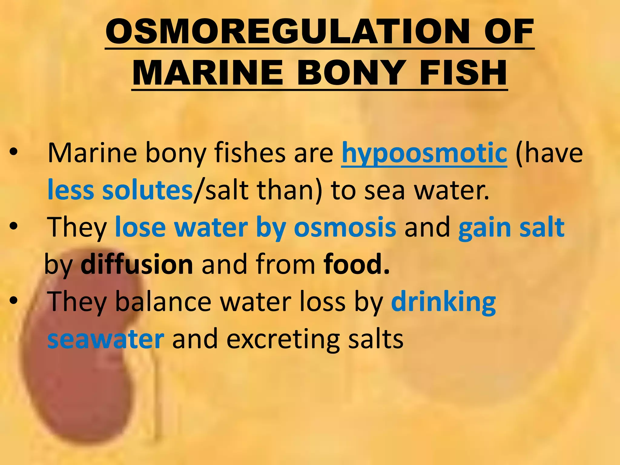
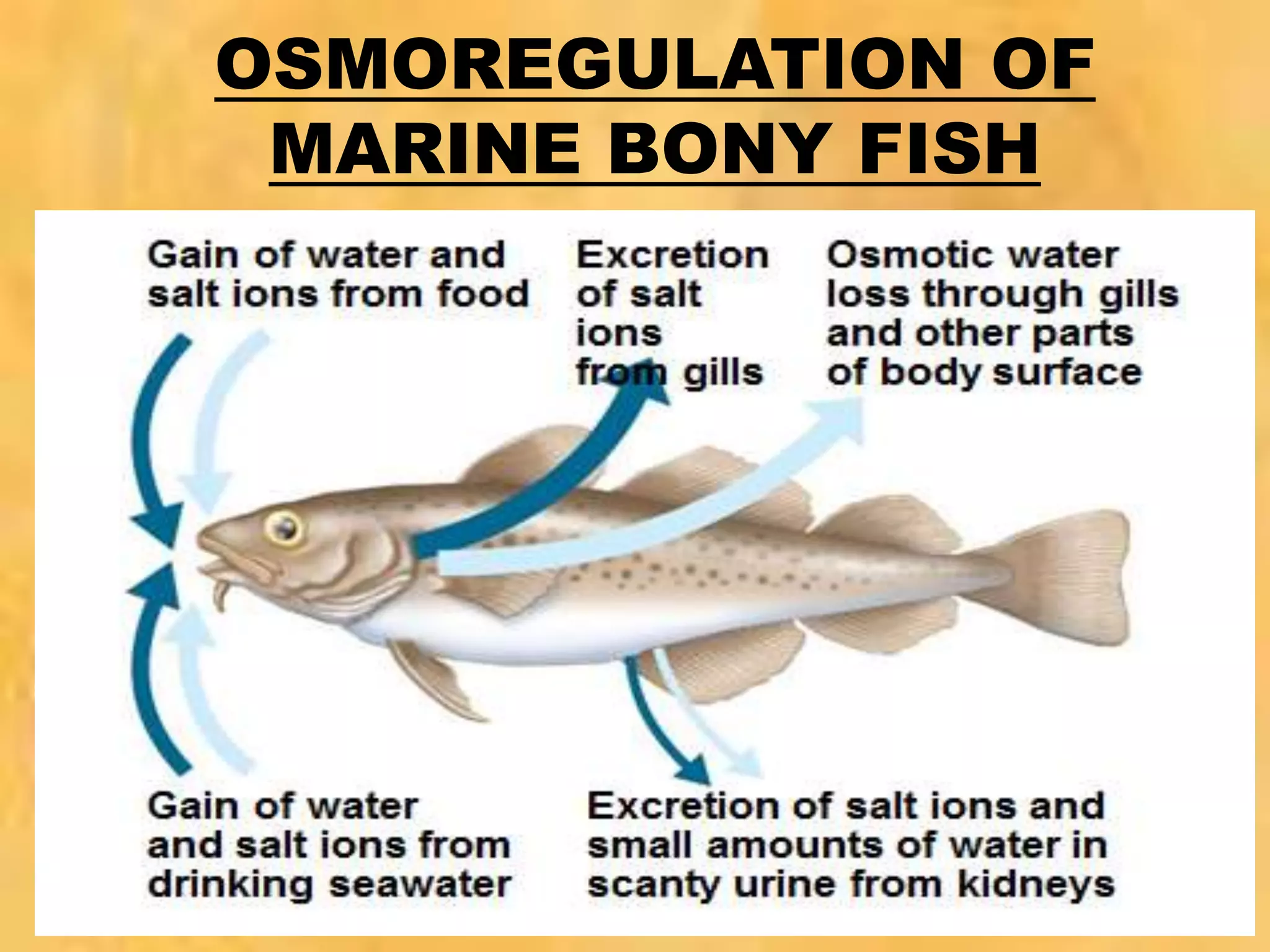
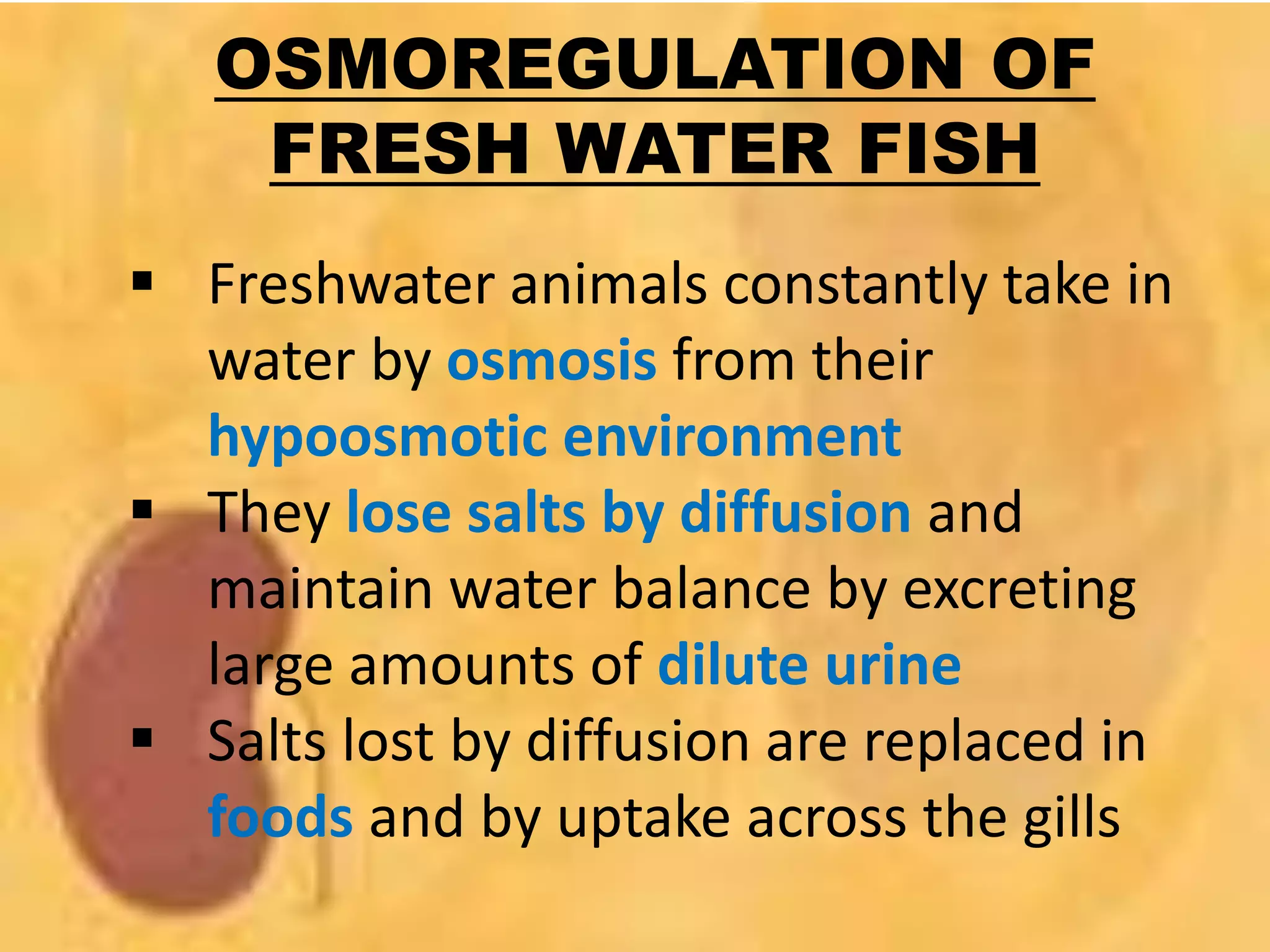
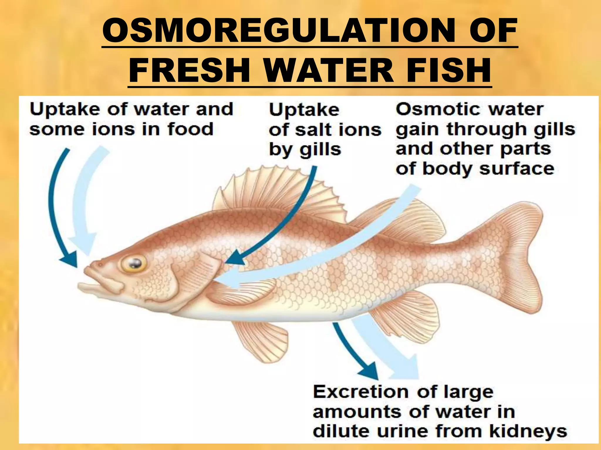
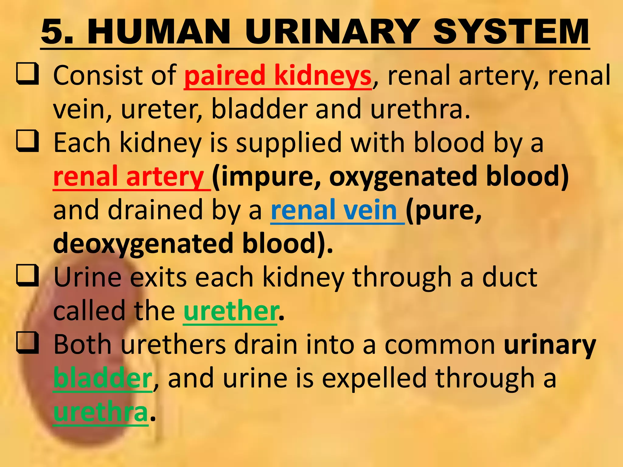
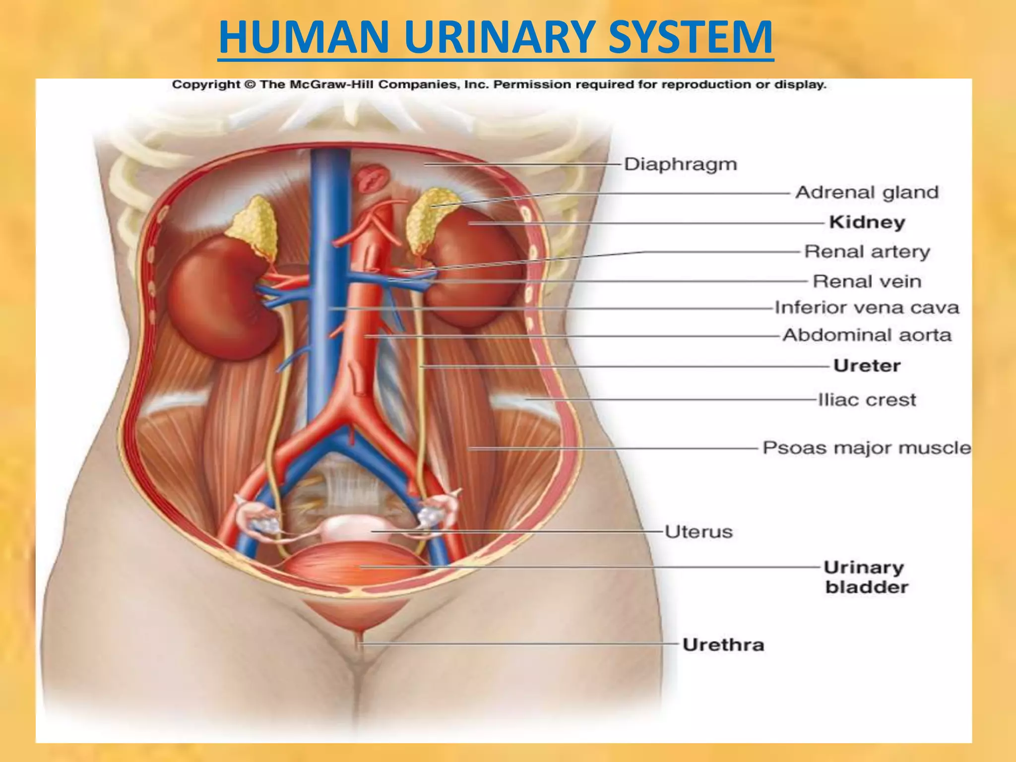
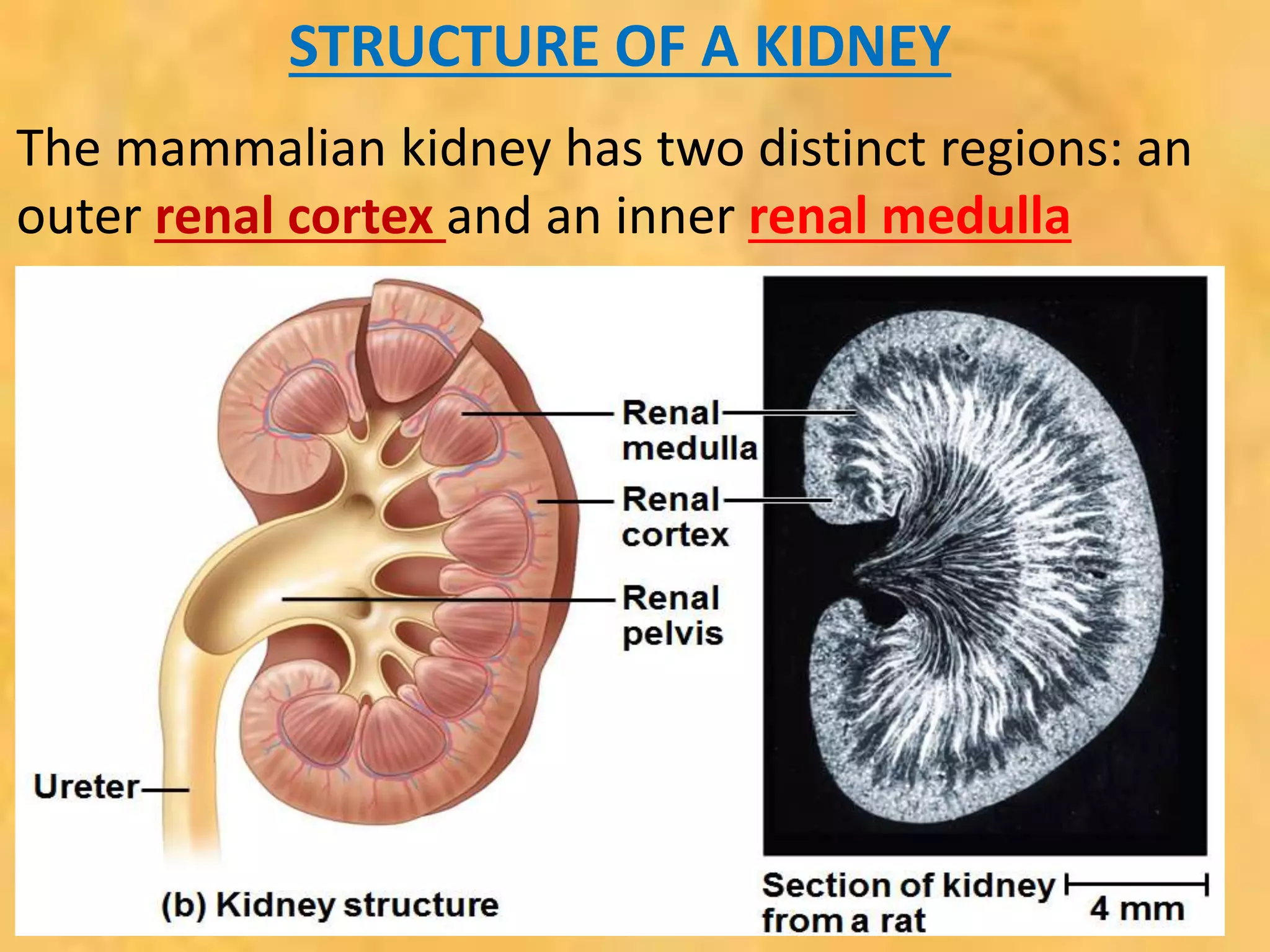
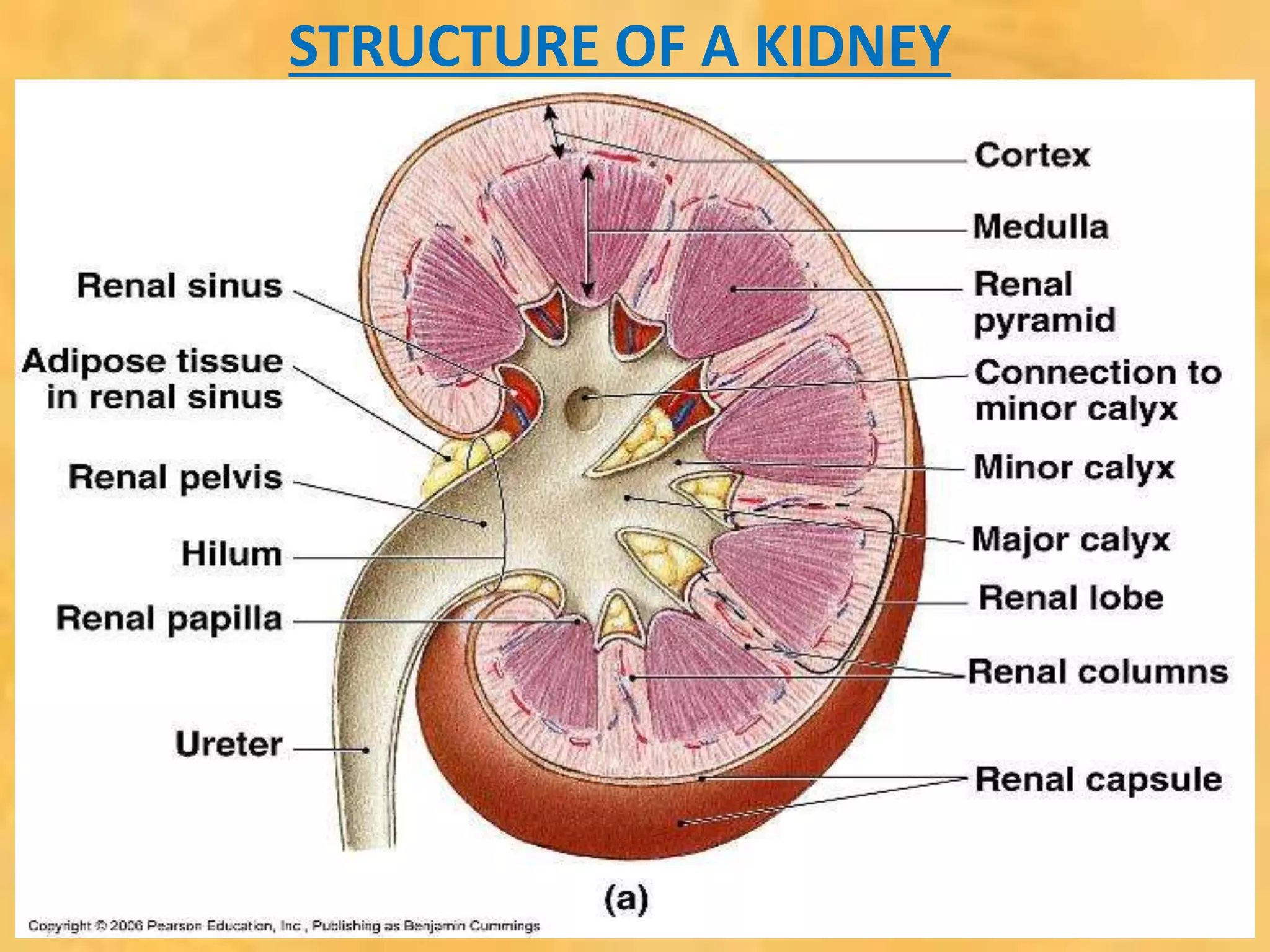
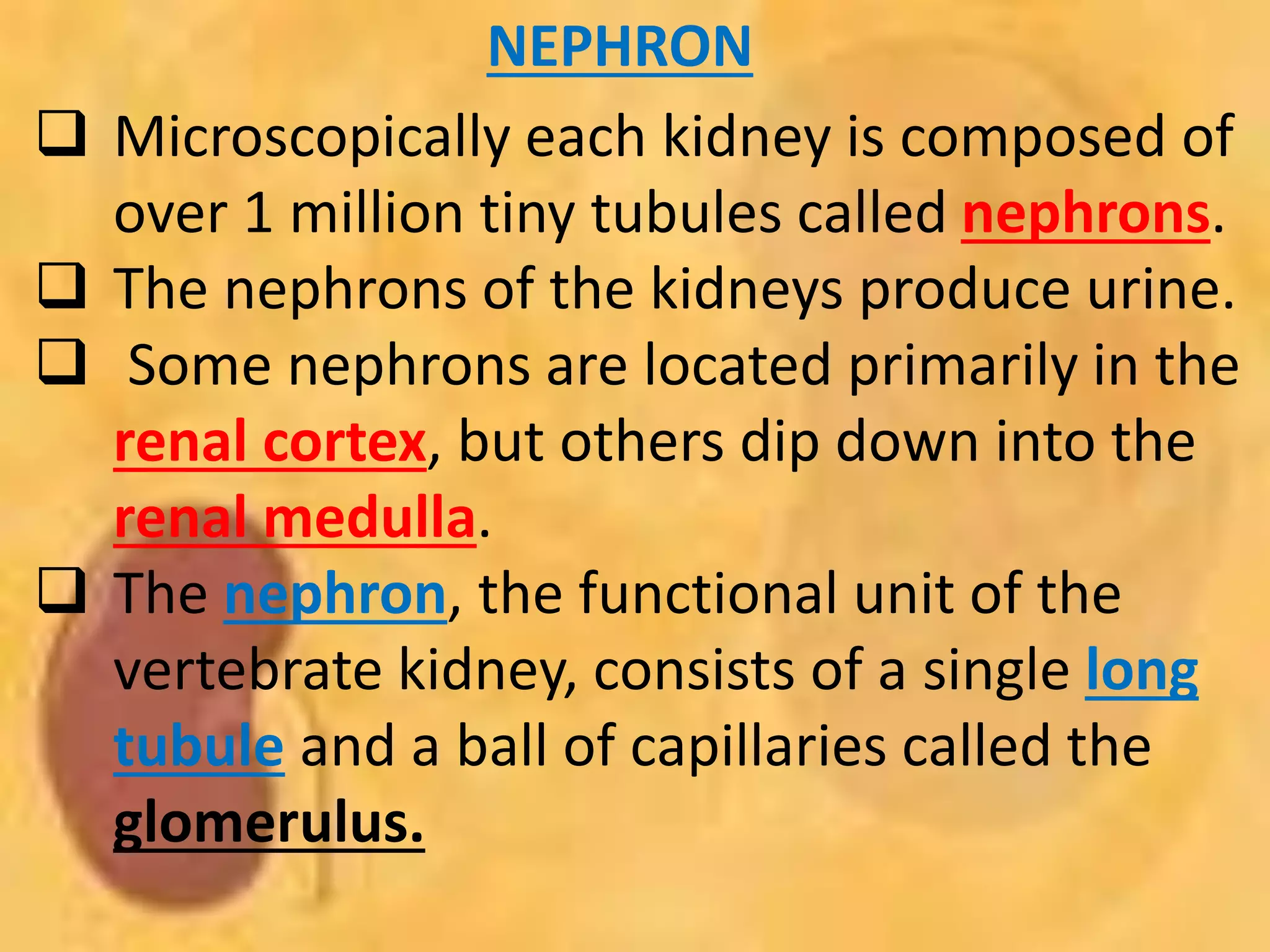
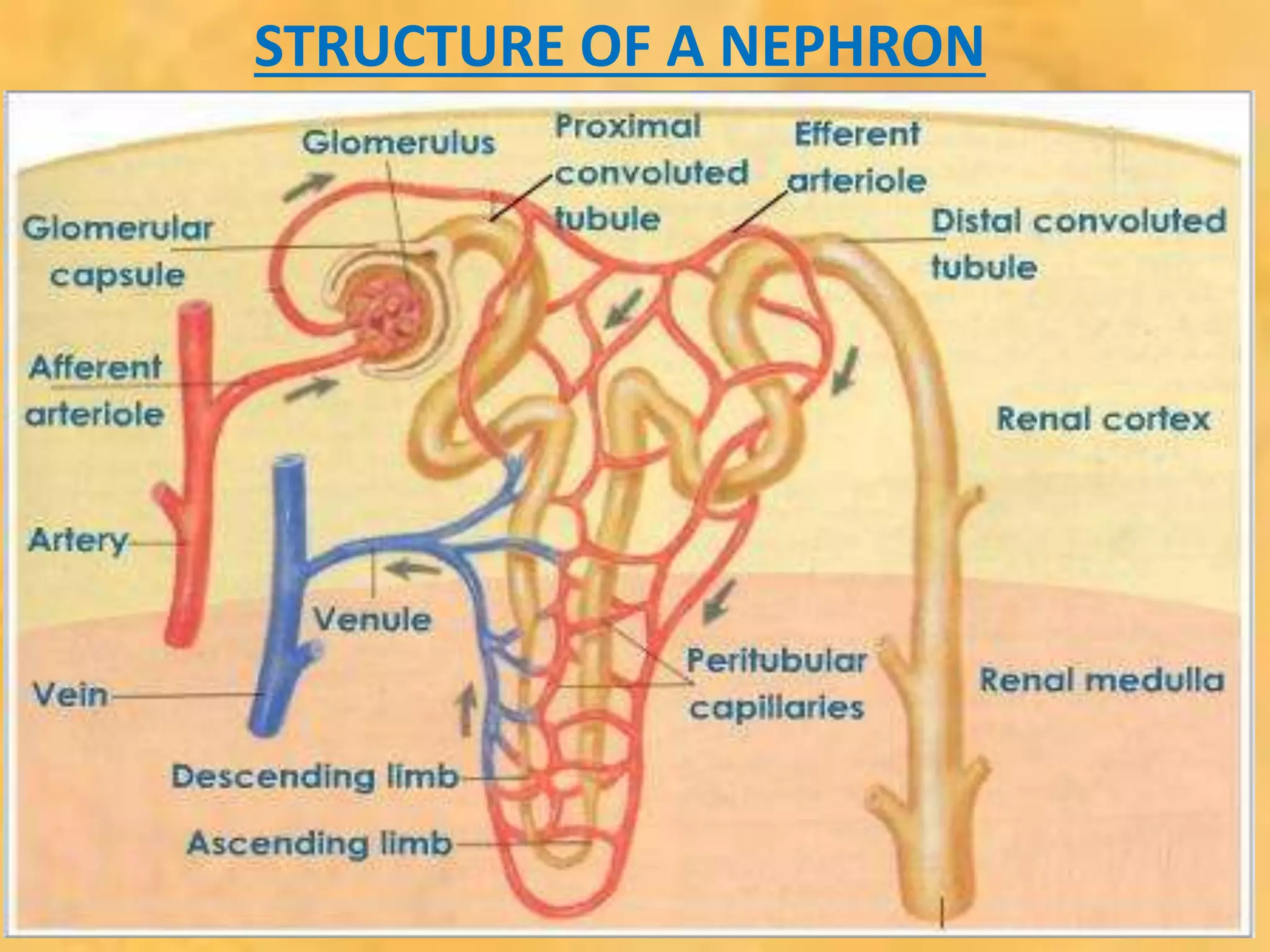
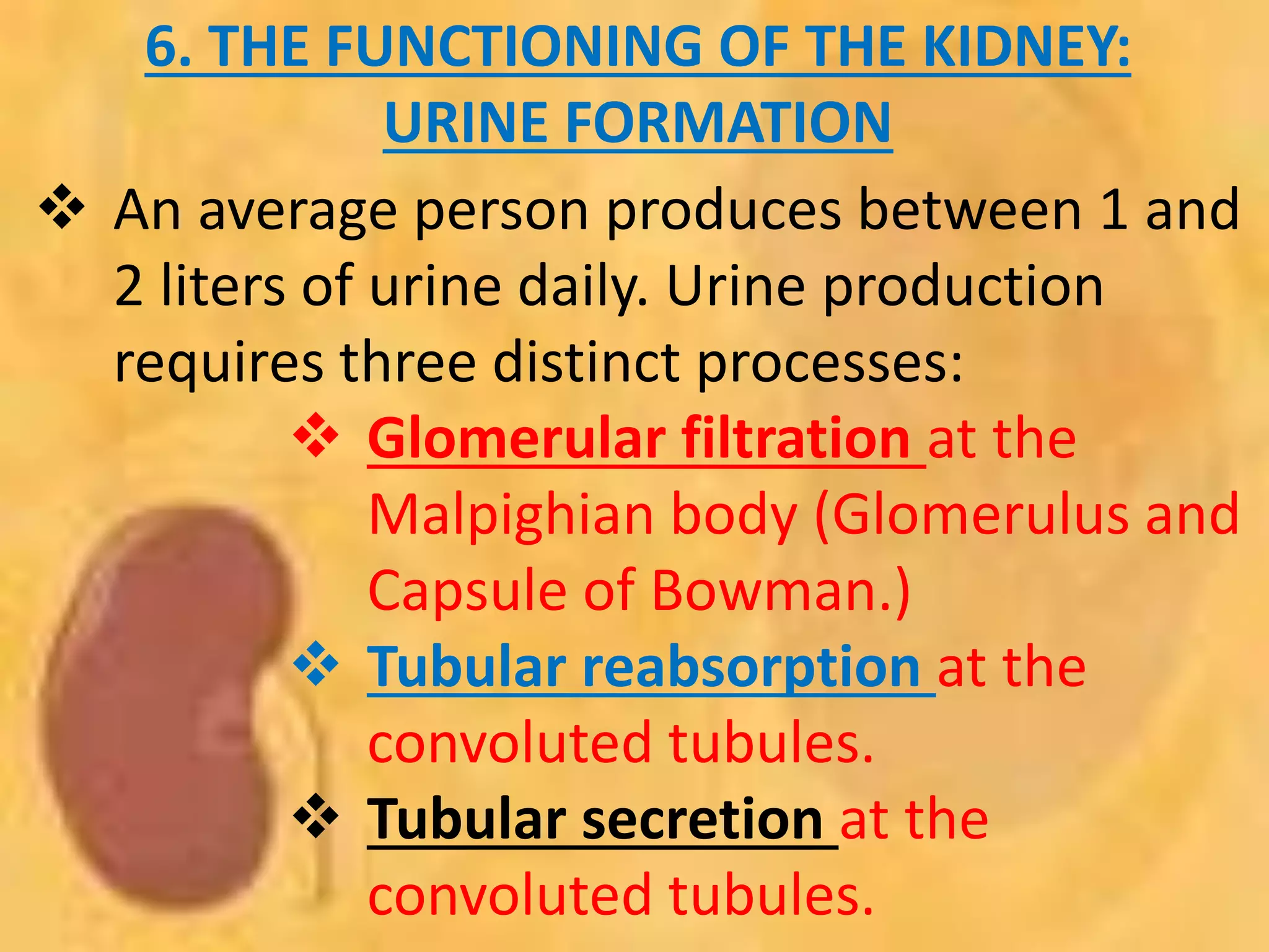
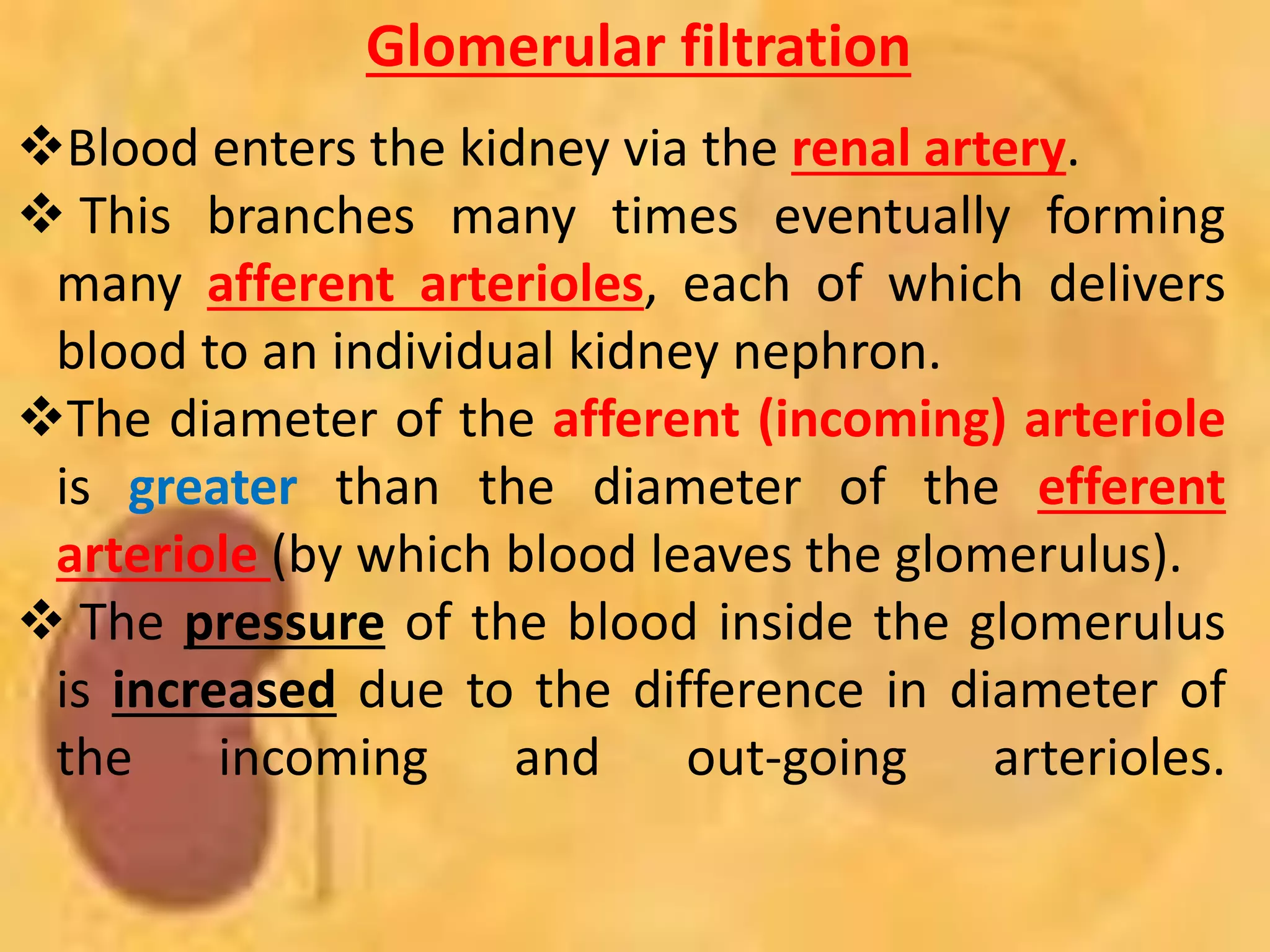
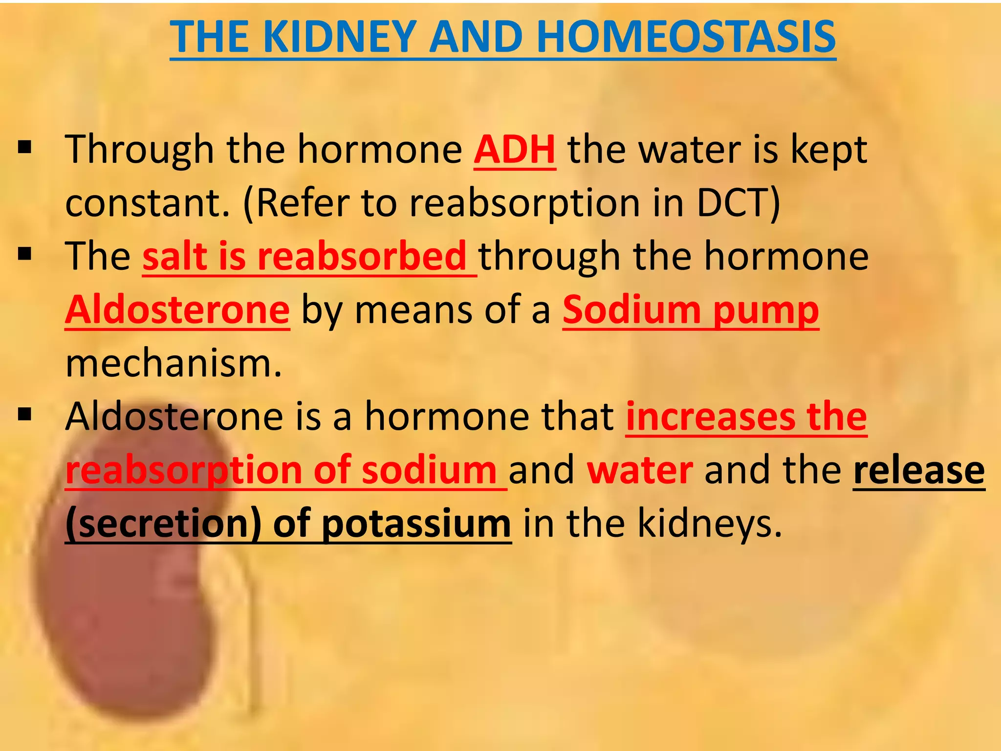
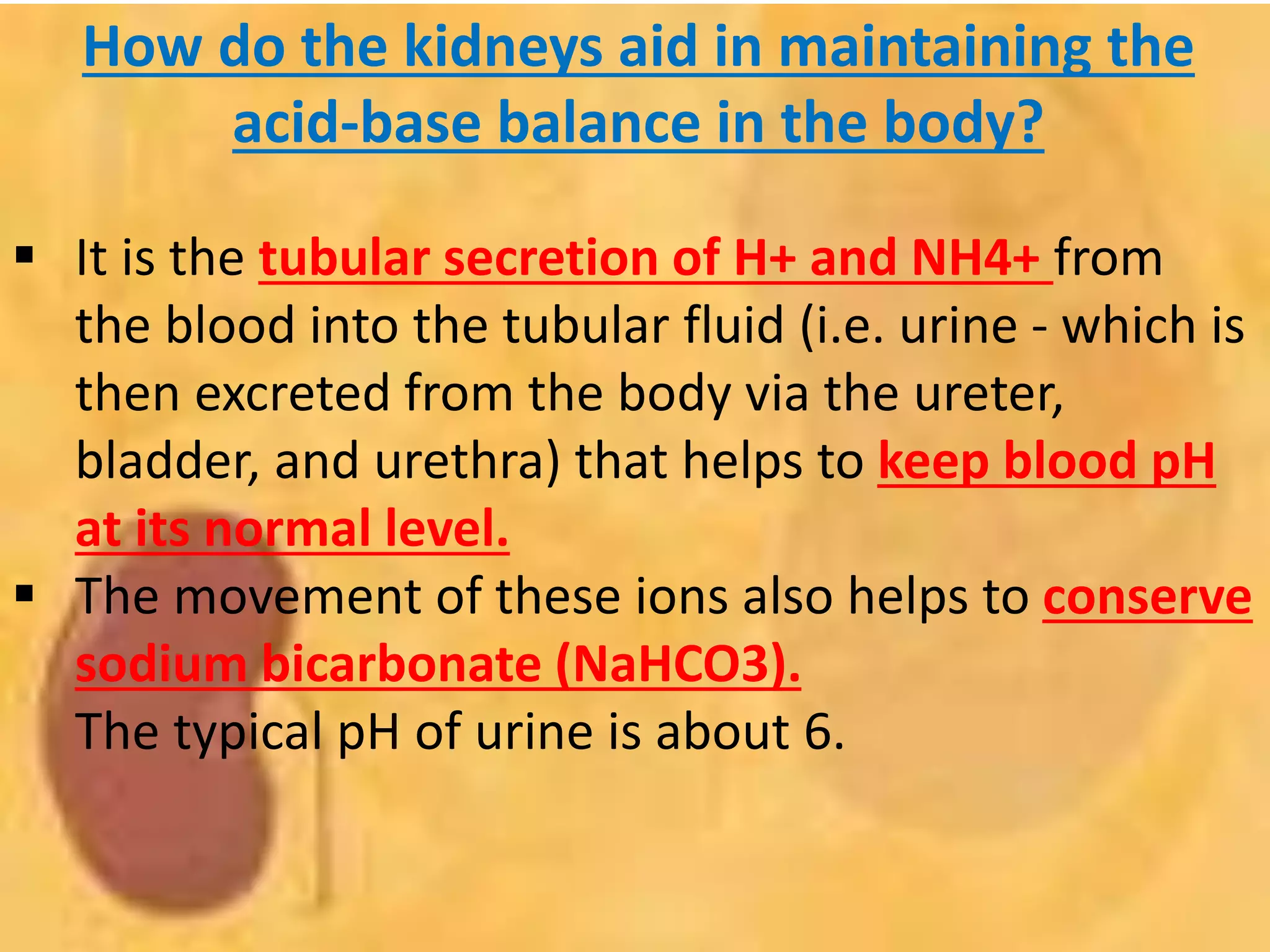
UNIT 2:
EXCRETION
AND
OSMOREGULATION
CAMPBELL & REECE (2010)
CHAPTER 44
1. OSMOREGULATION
AND EXCRETION
• Relative concentrations of water and solutes
must be maintained within fairly narrow
limits
• Osmoregulation regulates solute
concentrations and balances the gain and
loss of water.
• Freshwater animals show adaptations that
reduce water uptake and conserve solutes
OSMOREGULATION AND
EXCRETION
• Desert and marine animals face desiccating
environments that can quickly deplete body
water.
• Excretion gets rid of nitrogenous
metabolites and other waste products.
OSMOREGULATION OF
MARINE BONY FISH
• Marine bony fishes are hypoosmotic (have
less solutes/salt than) to sea water.
• They lose water by osmosis and gain salt
by diffusion and from food.
• They balance water loss by drinking
seawater and excreting salts
OSMOREGULATION OF
MARINE BONY FISH
OSMOREGULATION OF
FRESH WATER FISH
 Freshwater animals constantly take in
water by osmosis from their
hypoosmotic environment
 They lose salts by diffusion and
maintain water balance by excreting
large amounts of dilute urine
 Salts lost by diffusion are replaced in
foods and by uptake across the gills
OSMOREGULATION OF
FRESH WATER FISH
2. WASTE PRODUCTS
 UREA
 AMMONIA
 URIC ACID
 The kinds of nitrogenous wastes
excreted depend on an animal’s
evolutionary history and habitat
 The amount of nitrogenous waste is
coupled to the animal’s energy budget
AMMONIA
 Form when an amino group (NH2) adds a
third hydrogen ion. = NH3.
 Ammonia is rather toxic.
 If enough water is available to wash it from
the body it can be a nitrogenous excretory
product.
 Do not require a lot of energy for
production.
 Ammonia is excreted by most fish whose
gills and skin are in direct contact with the
water of the environment.
UREA
 Requires a lot of energy for production of Urea.
 Carrier molecules take up carbon dioxide and 2
molecules of ammonia to form and excrete urea.
 Urea is less toxic than ammonia.
 Can be excreted as a concentrated solution. –
Important allows terrestrial animals to conserve
water.
 Sharks, frogs and mammals excrete urea as their
main nitrogenous waste.
URIC ACID
 Requires an extreme amount of ATP (energy) for
the production of Uric acid.
 Uric acid is not very toxic and do not dissolve in
water readily, therefore is excreted very
concentrated – to conserve water in the body.
 Uric acid is routinely excreted by insects, reptiles
and birds.
3. EXCRETORY ORGANS
AMONG INVERTEBRATES
 Most animals have tubular excretory organs that
regulate the water-salt balance of the body and
excrete metabolic wastes into the environment.
 3 examples amongst invertebrates:
Planarians – use Flame cells.
Earthworms – use Nephridia.
Insects – use Malpighian tubules.
 On occasion, excretion also involves other organs,
such as the rectum in the earthworm and the gills
in crayfish.
PLANARIANS
(FLATWORMS)with flame cells
 Flatworms use flame cells (bud-like
outgrowths in a branched tubular excretion
system) with cilia to filter out waste and get
rid of excess water.
Earthworm with nephridia
Earthworms have nephridia with a ciliated
opening, called the nephridiostome, that leads
to a coiled tubule surrounded by blood
capillaries. Waste move through the tubules
and is excreted through the nephridiopore.
Insects with Malpighian
tubules
Insects use thin tubules called Malpighian
tubules attached to the gut for excretion
++
4. HUMAN EXCRETORY
ORGANS
 Humans use several excretory organs:
 Lungs – Carbon dioxide (waste product from
cellular respiration)
 Kidneys and bladder - Ammonia, uric acid,
excess water, excess salt and urea (Waste
products in deamination and food)
 Liver - Colorants, alcohol, poisons(Waste
products from food and drink)
 Alimentary canal - Feaces (undigested
food), cellulose.
 Skin - Excess water and salt, ammonia.
5. HUMAN URINARY SYSTEM
 Consist of paired kidneys, renal artery, renal
vein, ureter, bladder and urethra.
 Each kidney is supplied with blood by a
renal artery (impure, oxygenated blood)
and drained by a renal vein (pure,
deoxygenated blood).
 Urine exits each kidney through a duct
called the urether.
 Both urethers drain into a common urinary
bladder, and urine is expelled through a
urethra.
HUMAN URINARY SYSTEM
STRUCTURE OF A KIDNEY
The mammalian kidney has two distinct regions: an
outer renal cortex and an inner renal medulla
STRUCTURE OF A KIDNEY
NEPHRON
 Microscopically each kidney is composed of
over 1 million tiny tubules called nephrons.
 The nephrons of the kidneys produce urine.
 Some nephrons are located primarily in the
renal cortex, but others dip down into the
renal medulla.
 The nephron, the functional unit of the
vertebrate kidney, consists of a single long
tubule and a ball of capillaries called the
glomerulus.
STRUCTURE OF A NEPHRON
6. THE FUNCTIONING OF THE KIDNEY:
URINE FORMATION
 An average person produces between 1 and
2 liters of urine daily. Urine production
requires three distinct processes:
 Glomerular filtration at the
Malpighian body (Glomerulus and
Capsule of Bowman.)
 Tubular reabsorption at the
convoluted tubules.
 Tubular secretion at the
convoluted tubules.
Glomerular filtration
Blood enters the kidney via the renal artery.
 This branches many times eventually forming
many afferent arterioles, each of which delivers
blood to an individual kidney nephron.
The diameter of the afferent (incoming) arteriole
is greater than the diameter of the efferent
arteriole (by which blood leaves the glomerulus).
 The pressure of the blood inside the glomerulus
is increased due to the difference in diameter of
the incoming and out-going arterioles.
+
Glomerular filtration
This increased blood pressure helps to force
the following components of the blood out
of the glomerular capillaries into the cavity
of the capsule of Bowman:
Most of the water;
Most/all of the salts;
Most/all of the glucose;
Most/all of the urea.
Glomerular filtration
• These substances are filtered in preference to
other components of blood based on particle size.
• Blood cells and plasma proteins are not filtered
through the Glomerular capillaries because they
are relatively larger in physical size.
• The water and salts that have been forced out of
the glomerular capillaries pass into the Bowman's
Capsule and are called the glomerular filtrate.
• It contains all the materials present in the blood
except blood cells and most proteins - which are
too large to cross the basement membrane of the
Glomerular.
036+
Tubular reabsorption
Only about 1% of the glomerular filtrate
actually leaves the body because the rest
(the other 99% - useful substances) is
reabsorbed into the blood while it passes
through the renal tubules and ducts.
This is called Tubular reabsorption and
occurs via three mechanisms. They are:
Osmosis
Diffusion
Active Transport
Tubular reabsorption
Reabsorption varies according to the body's
needs, enabling the body to retain most of
its nutrients.
The processes of tubular reabsorption occur
in the following order:
Proximal convoluted tubule (PCT).
Loop of Henle
Distal convoluted tubule (DCT).
Tubular reabsorption: Proximal convoluted
tubule
Most of the volume of the filtrate solution is
reabsorbed in the proximal convoluted tubule.
This includes some water and most/all of the
glucose.
Solutes are selectively moved from the glomerular
filtrate to the plasma by active transport.
However, almost all glucose and amino acids, and
high amounts of ions, are reabsorbed again later.
Following the movement of solutes (including
Na+ ), water is then also reabsorbed by osmosis.
Tubular reabsorption:In the loop of henle
 The water, urea, and salts contained within the
ascending limb of Henle eventually pass into the
distal convoluted tubule (DCT).
 The DCT reacts to the amount of anti-diuretic
hormone (ADH) in the blood:
 The more ADH is present in the blood, the more
water is re-absorbed into it. This happens because
the presence of ADH in the blood causes the cells
of the DCT to become more permeable to water,
therefore they allow more water to pass from the
tubular fluid back into the blood. This results in
more concentrated urine.
Tubular reabsorption:In the loop of henle
 If the level of ADH in the blood is reduced then the
cells in the DCT becomes less permeable to water
therefore less water is able to pass from the
tubular fluid back into the blood - which results in
less concentrated urine.
Tubular secretion
• Involves substances being added to the tubular fluid.
• This removes excessive quantities of certain dissolved
substances from the body.
• Also maintains the blood at a normal healthy pH (pH 7.35
to pH 7.45)
• The substances that are secreted into the tubular fluid
(for removal from the body) include:
• Potassium ions (K+),
• Hydrogen ions (H+),
• Ammonium ions (NH4
+),
• creatinine,
• urea,
• some hormones, and
• some drugs (e.g. penicillin).
7. THE KIDNEY AND HOMEOSTASIS
 The kidneys are organs of homeostasis for four
main reasons:
1. They excrete metabolic waste such as urea, which
is the primary metabolic waste of humans.
2. They maintain the water salt balance in the body.
3. They maintain the acid-base (pH) balance in the
body.
4. They secrete hormones.
THE KIDNEY AND HOMEOSTASIS
 Through the hormone ADH the water is kept
constant. (Refer to reabsorption in DCT)
 The salt is reabsorbed through the hormone
Aldosterone by means of a Sodium pump
mechanism.
 Aldosterone is a hormone that increases the
reabsorption of sodium and water and the release
(secretion) of potassium in the kidneys.
How do the kidneys aid in maintaining the
acid-base balance in the body?
 It is the tubular secretion of H+ and NH4+ from
the blood into the tubular fluid (i.e. urine - which is
then excreted from the body via the ureter,
bladder, and urethra) that helps to keep blood pH
at its normal level.
 The movement of these ions also helps to conserve
sodium bicarbonate (NaHCO3).
The typical pH of urine is about 6.

Unit 2 excretion and osmoregulation

  • 1.
  • 2.
    1. OSMOREGULATION AND EXCRETION •Relative concentrations of water and solutes must be maintained within fairly narrow limits • Osmoregulation regulates solute concentrations and balances the gain and loss of water. • Freshwater animals show adaptations that reduce water uptake and conserve solutes
  • 3.
    OSMOREGULATION AND EXCRETION • Desertand marine animals face desiccating environments that can quickly deplete body water. • Excretion gets rid of nitrogenous metabolites and other waste products.
  • 4.
    OSMOREGULATION OF MARINE BONYFISH • Marine bony fishes are hypoosmotic (have less solutes/salt than) to sea water. • They lose water by osmosis and gain salt by diffusion and from food. • They balance water loss by drinking seawater and excreting salts
  • 5.
  • 6.
    OSMOREGULATION OF FRESH WATERFISH  Freshwater animals constantly take in water by osmosis from their hypoosmotic environment  They lose salts by diffusion and maintain water balance by excreting large amounts of dilute urine  Salts lost by diffusion are replaced in foods and by uptake across the gills
  • 7.
  • 8.
    2. WASTE PRODUCTS UREA  AMMONIA  URIC ACID  The kinds of nitrogenous wastes excreted depend on an animal’s evolutionary history and habitat  The amount of nitrogenous waste is coupled to the animal’s energy budget
  • 9.
    AMMONIA  Form whenan amino group (NH2) adds a third hydrogen ion. = NH3.  Ammonia is rather toxic.  If enough water is available to wash it from the body it can be a nitrogenous excretory product.  Do not require a lot of energy for production.  Ammonia is excreted by most fish whose gills and skin are in direct contact with the water of the environment.
  • 10.
    UREA  Requires alot of energy for production of Urea.  Carrier molecules take up carbon dioxide and 2 molecules of ammonia to form and excrete urea.  Urea is less toxic than ammonia.  Can be excreted as a concentrated solution. – Important allows terrestrial animals to conserve water.  Sharks, frogs and mammals excrete urea as their main nitrogenous waste.
  • 11.
    URIC ACID  Requiresan extreme amount of ATP (energy) for the production of Uric acid.  Uric acid is not very toxic and do not dissolve in water readily, therefore is excreted very concentrated – to conserve water in the body.  Uric acid is routinely excreted by insects, reptiles and birds.
  • 12.
    3. EXCRETORY ORGANS AMONGINVERTEBRATES  Most animals have tubular excretory organs that regulate the water-salt balance of the body and excrete metabolic wastes into the environment.  3 examples amongst invertebrates: Planarians – use Flame cells. Earthworms – use Nephridia. Insects – use Malpighian tubules.  On occasion, excretion also involves other organs, such as the rectum in the earthworm and the gills in crayfish.
  • 13.
    PLANARIANS (FLATWORMS)with flame cells Flatworms use flame cells (bud-like outgrowths in a branched tubular excretion system) with cilia to filter out waste and get rid of excess water.
  • 14.
    Earthworm with nephridia Earthwormshave nephridia with a ciliated opening, called the nephridiostome, that leads to a coiled tubule surrounded by blood capillaries. Waste move through the tubules and is excreted through the nephridiopore.
  • 15.
    Insects with Malpighian tubules Insectsuse thin tubules called Malpighian tubules attached to the gut for excretion
  • 16.
    ++ 4. HUMAN EXCRETORY ORGANS Humans use several excretory organs:  Lungs – Carbon dioxide (waste product from cellular respiration)  Kidneys and bladder - Ammonia, uric acid, excess water, excess salt and urea (Waste products in deamination and food)  Liver - Colorants, alcohol, poisons(Waste products from food and drink)  Alimentary canal - Feaces (undigested food), cellulose.  Skin - Excess water and salt, ammonia.
  • 17.
    5. HUMAN URINARYSYSTEM  Consist of paired kidneys, renal artery, renal vein, ureter, bladder and urethra.  Each kidney is supplied with blood by a renal artery (impure, oxygenated blood) and drained by a renal vein (pure, deoxygenated blood).  Urine exits each kidney through a duct called the urether.  Both urethers drain into a common urinary bladder, and urine is expelled through a urethra.
  • 18.
  • 19.
    STRUCTURE OF AKIDNEY The mammalian kidney has two distinct regions: an outer renal cortex and an inner renal medulla
  • 20.
  • 21.
    NEPHRON  Microscopically eachkidney is composed of over 1 million tiny tubules called nephrons.  The nephrons of the kidneys produce urine.  Some nephrons are located primarily in the renal cortex, but others dip down into the renal medulla.  The nephron, the functional unit of the vertebrate kidney, consists of a single long tubule and a ball of capillaries called the glomerulus.
  • 22.
  • 23.
  • 24.
    6. THE FUNCTIONINGOF THE KIDNEY: URINE FORMATION  An average person produces between 1 and 2 liters of urine daily. Urine production requires three distinct processes:  Glomerular filtration at the Malpighian body (Glomerulus and Capsule of Bowman.)  Tubular reabsorption at the convoluted tubules.  Tubular secretion at the convoluted tubules.
  • 25.
    Glomerular filtration Blood entersthe kidney via the renal artery.  This branches many times eventually forming many afferent arterioles, each of which delivers blood to an individual kidney nephron. The diameter of the afferent (incoming) arteriole is greater than the diameter of the efferent arteriole (by which blood leaves the glomerulus).  The pressure of the blood inside the glomerulus is increased due to the difference in diameter of the incoming and out-going arterioles.
  • 26.
    + Glomerular filtration This increasedblood pressure helps to force the following components of the blood out of the glomerular capillaries into the cavity of the capsule of Bowman: Most of the water; Most/all of the salts; Most/all of the glucose; Most/all of the urea.
  • 27.
    Glomerular filtration • Thesesubstances are filtered in preference to other components of blood based on particle size. • Blood cells and plasma proteins are not filtered through the Glomerular capillaries because they are relatively larger in physical size. • The water and salts that have been forced out of the glomerular capillaries pass into the Bowman's Capsule and are called the glomerular filtrate. • It contains all the materials present in the blood except blood cells and most proteins - which are too large to cross the basement membrane of the Glomerular.
  • 28.
    036+ Tubular reabsorption Only about1% of the glomerular filtrate actually leaves the body because the rest (the other 99% - useful substances) is reabsorbed into the blood while it passes through the renal tubules and ducts. This is called Tubular reabsorption and occurs via three mechanisms. They are: Osmosis Diffusion Active Transport
  • 29.
    Tubular reabsorption Reabsorption variesaccording to the body's needs, enabling the body to retain most of its nutrients. The processes of tubular reabsorption occur in the following order: Proximal convoluted tubule (PCT). Loop of Henle Distal convoluted tubule (DCT).
  • 30.
    Tubular reabsorption: Proximalconvoluted tubule Most of the volume of the filtrate solution is reabsorbed in the proximal convoluted tubule. This includes some water and most/all of the glucose. Solutes are selectively moved from the glomerular filtrate to the plasma by active transport. However, almost all glucose and amino acids, and high amounts of ions, are reabsorbed again later. Following the movement of solutes (including Na+ ), water is then also reabsorbed by osmosis.
  • 31.
    Tubular reabsorption:In theloop of henle  The water, urea, and salts contained within the ascending limb of Henle eventually pass into the distal convoluted tubule (DCT).  The DCT reacts to the amount of anti-diuretic hormone (ADH) in the blood:  The more ADH is present in the blood, the more water is re-absorbed into it. This happens because the presence of ADH in the blood causes the cells of the DCT to become more permeable to water, therefore they allow more water to pass from the tubular fluid back into the blood. This results in more concentrated urine.
  • 32.
    Tubular reabsorption:In theloop of henle  If the level of ADH in the blood is reduced then the cells in the DCT becomes less permeable to water therefore less water is able to pass from the tubular fluid back into the blood - which results in less concentrated urine.
  • 33.
    Tubular secretion • Involvessubstances being added to the tubular fluid. • This removes excessive quantities of certain dissolved substances from the body. • Also maintains the blood at a normal healthy pH (pH 7.35 to pH 7.45) • The substances that are secreted into the tubular fluid (for removal from the body) include: • Potassium ions (K+), • Hydrogen ions (H+), • Ammonium ions (NH4 +), • creatinine, • urea, • some hormones, and • some drugs (e.g. penicillin).
  • 34.
    7. THE KIDNEYAND HOMEOSTASIS  The kidneys are organs of homeostasis for four main reasons: 1. They excrete metabolic waste such as urea, which is the primary metabolic waste of humans. 2. They maintain the water salt balance in the body. 3. They maintain the acid-base (pH) balance in the body. 4. They secrete hormones.
  • 35.
    THE KIDNEY ANDHOMEOSTASIS  Through the hormone ADH the water is kept constant. (Refer to reabsorption in DCT)  The salt is reabsorbed through the hormone Aldosterone by means of a Sodium pump mechanism.  Aldosterone is a hormone that increases the reabsorption of sodium and water and the release (secretion) of potassium in the kidneys.
  • 36.
    How do thekidneys aid in maintaining the acid-base balance in the body?  It is the tubular secretion of H+ and NH4+ from the blood into the tubular fluid (i.e. urine - which is then excreted from the body via the ureter, bladder, and urethra) that helps to keep blood pH at its normal level.  The movement of these ions also helps to conserve sodium bicarbonate (NaHCO3). The typical pH of urine is about 6.