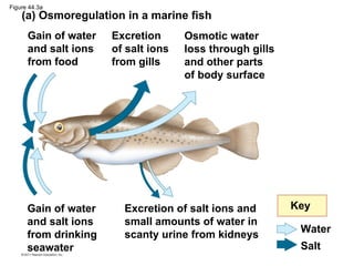
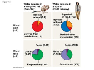
Osmoregulation
and Excretion


 Dr. Kristen Walker
Osmoregulation balances the uptake and loss
of water and solutes
 • Relative concentrations of water and solutes
   must be maintained within fairly narrow limits
 • Osmoregulation is based largely on controlled
   movement of solutes between internal fluids
   and the external environment
 • Excretion gets rid of nitrogenous metabolites
   and other waste products
Osmosis and Osmolarity
 • Cells require a balance between uptake and
   loss of water
 • Osmolarity, the solute concentration of a
   solution, determines movement of water
   across a selectively permeable membrane
 • If two solutions are isoosmotic, the movement
   of water is equal in both directions
 • If two solutions differ in osmolarity, the net
   flow of water is from the hypoosmotic to the
   hyperosmotic solution
Figure 44.2


                   Selectively permeable
                         membrane
                 Solutes              Water


 Hyperosmotic side:                       Hypoosmotic side:
 • Higher solute                          • Lower solute
   concentration                            concentration
 • Lower free H2O                         • Higher free H2O
   concentration                            concentration


                       Net water flow
Osmotic Challenges
 • Osmoconformers, consisting only of some
   marine animals, are isoosmotic with their
   surroundings and do not regulate their
   osmolarity
   – marine invertebrates
 • Osmoregulators expend energy to control
   water uptake and loss in a hyperosmotic or
   hypoosmotic environment
   – marine vertebrates and some invertebrates
Marine Animals

 • Marine bony fishes are hypoosmotic to
   seawater
   – lose water by osmosis and gain salt by
     diffusion and from food
   – balance water loss by drinking seawater and
     excreting salts
Figure 44.3a
    (a) Osmoregulation in a marine fish
      Gain of water   Excretion      Osmotic water
      and salt ions   of salt ions   loss through gills
      from food       from gills     and other parts
                                     of body surface


      Gain of water     Excretion of salt ions and        Key
      and salt ions     small amounts of water in
                                                           Water
      from drinking     scanty urine from kidneys
      seawater                                             Salt
Freshwater Animals
 • Freshwater animals constantly take in water by
   osmosis from their hypoosmotic environment
 • lose salts by diffusion and maintain water
   balance by excreting large amounts of dilute
   urine
 • Salts lost by diffusion are replaced in foods and
   by uptake across the gills
Figure 44.3b
               (b) Osmoregulation in a freshwater fish
               Gain of water       Uptake of    Osmotic water
               and some ions       salt ions    gain through
               in food             by gills     gills and other
                                                parts of body
                                                surface


                 Key                 Excretion of salt ions and
                                     large amounts of water in
                   Water             dilute urine from kidneys
                   Salt
Land Animals
 • Body coverings of most terrestrial animals help
   prevent dehydration
 • Desert animals get major water savings from
   simple anatomical features and behaviors such
   as a nocturnal lifestyle
 • Land animals maintain water balance by eating
   moist food and producing water metabolically
   through cellular respiration
Figure 44.6
                      Water balance in          Water balance in
                      a kangaroo rat            a human
                      (2 mL/day)                (2,500 mL/day)

                               Ingested                   Ingested
                               in food (0.2)              in food (750)
                                                Ingested
                                                in liquid
              Water                             (1,500)
              gain
              (mL)
                      Derived from                     Derived from
                      metabolism (1.8)                 metabolism (250)


                                 Feces (0.09)               Feces (100)

              Water   Urine                     Urine
              loss    (0.45)                    (1,500)
              (mL)


                        Evaporation (1.46)            Evaporation (900)
Energetics of Osmoregulation
 • Osmoregulators must expend energy to
   maintain osmotic gradients
 • The amount of energy differs based on
     – How different the animal’s osmolarity is from
       its surroundings
     – How easily water and solutes move across
       the animal’s surface
     – The work required to pump solutes across the
       membrane
Transport Epithelia in Osmoregulation
 • Animals regulate the solute content of body
   fluid that bathes their cells
 • Transport epithelia are epithelial cells that
   are specialized for moving solutes in specific
   directions
 • They are typically arranged in complex tubular
   networks
    – e.g., nasal glands of marine birds, which remove
      excess sodium chloride from the blood
Figure 44.7


                                                                       Secretory cell Lumen of
                                                                       of transport   secretory
                                                           Vein Artery
 Nasal salt                                                            epithelium     tubule
 gland
 Ducts
                                    Nasal gland
                                                                                             Salt
                                        Capillary                                           ions
                                        Secretory tubule
                                        Transport
              Nostril with salt
                                        epithelium
              secretions

 (a) Location of nasal glands     (b) Secretory
     in a marine bird                 tubules
                                                                      Blood flow   Salt secretion
   Key                                                                (c) Countercurrent
                                                                          exchange
         Salt movement
         Blood flow
                                                           Central duct
An animal’s nitrogenous wastes reflect its
phylogeny and habitat
  • Type and quantity of an animal’s waste
    products may greatly affect its water balance
  • Among the most significant wastes are
    nitrogenous breakdown products of proteins
    and nucleic acids
  • Some animals convert toxic ammonia (NH3)
    to less toxic compounds prior to excretion
Figure 44.8
                                   Proteins        Nucleic acids


                                    Amino          Nitrogenous
                                    acids             bases


                                           —NH2
                                        Amino groups


                 Most aquatic          Mammals, most             Many reptiles
              animals, including     amphibians, sharks,       (including birds),
              most bony fishes        some bony fishes        insects, land snails


                  Ammonia                   Urea                   Uric acid
Forms of Nitrogenous Wastes
 • Animals excrete nitrogenous wastes in different
   forms: ammonia, urea, or uric acid
 • These differ in toxicity and the energy costs of
   producing them
Ammonia
 • Animals that excrete nitrogenous wastes as
   ammonia need access to lots of water
 • They release ammonia across the whole body
   surface or through gills
Urea
 • The liver of mammals and most adult
   amphibians converts ammonia to the less toxic
   urea
 • The circulatory system carries urea to the
   kidneys, where it is excreted
 • Conversion of ammonia to urea is energetically
   expensive; excretion of urea requires less
   water than ammonia
Uric Acid
 • Insects, land snails, and many reptiles, including
   birds, mainly excrete uric acid
 • Uric acid is relatively nontoxic and does not
   dissolve readily in water
 • It can be secreted as a paste with little water
   loss
 • Uric acid is more energetically expensive to
   produce than urea
Excretory Processes
 • Most excretory systems produce urine by
   refining a filtrate derived from body fluids
 • Key functions of most excretory systems
     – Filtration: Filtering of body fluids
     – Reabsorption: Reclaiming valuable solutes
     – Secretion: Adding nonessential solutes and
       wastes from the body fluids to the filtrate
     – Excretion: Processed filtrate containing
       nitrogenous wastes, released from the body
Figure 44.10


                                        1 Filtration
               Capillary


                           Filtrate
                                      Excretory
                                      tubule


                                        2 Reabsorption


                                        3 Secretion
                           Urine


                                        4 Excretion
Malpighian Tubules
 • In insects and other terrestrial arthropods,
   Malpighian tubules remove nitrogenous
   wastes from hemolymph and function in
   osmoregulation
 • Insects produce a relatively dry waste matter,
   mainly uric acid, an important adaptation to
   terrestrial life
 • Some terrestrial insects can also take up water
   from the air
Figure 44.13
                         Digestive tract


                                                 Rectum
                                                           Hindgut
                                                 Intestine

                Midgut         Malpighian
                (stomach)       tubules
               Salt, water, and
                                         Feces       To anus
                 nitrogenous
                                         and urine
                    wastes

                             Malpighian
                               tubule
                                                 Rectum
                                                Reabsorption


                                  HEMOLYMPH
Kidneys
 • Kidneys, the excretory organs of vertebrates,
   function in both excretion and osmoregulation
Figure 44.14-a


 Excretory Organs                 Kidney Structure             Nephron Types
                                                                       Cortical Juxtamedullary
                                  Renal                                nephron     nephron
 Posterior                        cortex
 vena cava                        Renal
                                  medulla
                               Renal artery
 Renal                     Kidney
 artery
 and vein                   Renal vein
                                                          Renal
                                                          cortex
 Aorta
 Ureter                           Ureter
                                                                   Renal
 Urinary                                                           medulla
 bladder
                 Urethra                        Renal pelvis
Figure 44.14-b
 Nephron Organization
            Afferent arteriole
            from renal artery      Glomerulus
                                      Bowman’s
                                      capsule
                                                 Proximal
                                                 tubule


                                                 Peritubular
 Distal
                                                 capillaries
 tubule


 Efferent
 arteriole
 from
 glomerulus

                      Branch of
                      renal vein
                                            Descending
                                            limb
                                                         Loop
                       Vasa
                                                          of
 Collecting            recta
                                                         Henle
                                                                 200 µm
 duct
                                            Ascending
                                            limb                          Blood vessels from a human
                                                                          kidney. Arterioles and peritubular
                                                                          capillaries appear pink; glomeruli
                                                                          appear yellow.
The nephron is organized for stepwise
processing of blood filtrate
 • The filtrate produced in Bowman’s capsule
   contains salts, glucose, amino acids, vitamins,
   nitrogenous wastes, and other small molecules
From Blood Filtrate to Urine: A Closer Look
  Proximal Tubule
  • Reabsorption of ions, water, and nutrients takes
    place in the proximal tubule
  • Molecules are transported actively and passively
    from the filtrate into the interstitial fluid and then
    capillaries
  • Some toxic materials are actively secreted into the
    filtrate
  • As the filtrate passes through the proximal tubule,
    materials to be excreted become concentrated
Figure 44.15

                                 Proximal tubule      Distal tubule
                                    NaCl Nutrients           H2O
                                 HCO3−  H2O     K+    NaCl         HCO3−


                                       H+   NH3          K+        H+
                      Filtrate
                       CORTEX

                                            Loop of
                                            Henle
                                                      NaCl
                                              H2O
                            OUTER                                            NaCl
                            MEDULLA
                                                                           Collecting
                                                                           duct
           Key                                                             Urea

        Active transport                              NaCl                 H2O
        Passive transport
                             INNER
                             MEDULLA
Descending Limb of the Loop of Henle
• Reabsorption of water continues through
  channels formed by aquaporin proteins
• Movement is driven by the high osmolarity of
  the interstitial fluid, which is hyperosmotic to the
  filtrate
• The filtrate becomes increasingly concentrated
Ascending Limb of the Loop of Henle
• In the ascending limb of the loop of Henle, salt
  but not water is able to diffuse from the tubule
  into the interstitial fluid
• The filtrate becomes increasingly dilute
Distal Tubule
• The distal tubule regulates the K+ and NaCl
  concentrations of body fluids
• The controlled movement of ions contributes
  to pH regulation
Collecting Duct
• The collecting duct carries filtrate through the
  medulla to the renal pelvis
• One of the most important tasks is reabsorption
  of solutes and water
• Urine is hyperosmotic to body fluids
Solute Gradients and Water Conservation
 • The mammalian kidney’s ability to conserve
   water is a key terrestrial adaptation
 • Hyperosmotic urine can be produced only
   because considerable energy is expended to
   transport solutes against concentration
   gradients
 • The two primary solutes affecting osmolarity are
   NaCl and urea
Adaptations of the Vertebrate Kidney to
Diverse Environments
 • The form and function of nephrons in various
   vertebrate classes are related to requirements
   for osmoregulation in the animal’s habitat
Mammals

 • Mammals that inhabit dry environments have
   long loops of Henle, while those in fresh water
   have relatively short loops
Birds and Other Reptiles
 • Birds have shorter loops of Henle but
   conserve water by excreting uric acid instead
   of urea
 • Other reptiles have only cortical nephrons but
   also excrete nitrogenous waste as uric acid
Freshwater Fishes and Amphibians
 • Freshwater fishes conserve salt in their distal
   tubules and excrete large volumes of dilute
   urine
 • Kidney function in amphibians is similar to
   freshwater fishes
 • Amphibians conserve water on land by
   reabsorbing water from the urinary bladder
Marine Bony Fishes
 • Marine bony fishes are hypoosmotic compared
   with their environment
 • Their kidneys have small glomeruli and some
   lack glomeruli entirely
 • Filtration rates are low, and very little urine is
   excreted
Hormonal circuits link kidney function,
water balance, and blood pressure
  • Mammals control the volume and osmolarity of
    urine
  • Osmolarity of the urine is regulated by nervous
    and hormonal control
  • Antidiuretic hormone (ADH) makes the
    collecting duct epithelium more permeable to
    water
  • An increase in osmolarity triggers the release of
    ADH, which helps to conserve water
• Mutation in ADH production causes severe
  dehydration and results in diabetes insipidus
• Alcohol is a diuretic as it inhibits the release of
  ADH
Figure 44.UN01a


           Animal        Inflow/Outflow       Urine

    Freshwater         Does not drink water   Large volume
    fish. Lives in     Salt in      H2O in    of urine
    water less         (active trans-
    concentrated       port by gills)         Urine is less
    than body                                 concentrated
    fluids; fish                              than body
    tends to gain                             fluids
    water, lose salt


                            Salt out
Figure 44.UN01b


           Animal      Inflow/Outflow        Urine

    Marine bony         Drinks water         Small volume
    fish. Lives in     Salt in H2O out       of urine
    water more
    concentrated                             Urine is
    than body                                slightly less
    fluids; fish                             concentrated
    tends to lose                            than body
    water, gain salt                         fluids
                       Salt out (active
                       transport by gills)
Figure 44.UN01c


           Animal   Inflow/Outflow   Urine

   Terrestrial      Drinks water     Moderate
   vertebrate.          Salt in      volume
   Terrestrial          (by mouth)   of urine
   environment;
   tends to lose                     Urine is
   body water                        more
   to air                            concentrated
                                     than body
                        H2O and      fluids
                        salt out

Osmoregulation

  • 1.
  • 2.
    Osmoregulation balances theuptake and loss of water and solutes • Relative concentrations of water and solutes must be maintained within fairly narrow limits • Osmoregulation is based largely on controlled movement of solutes between internal fluids and the external environment • Excretion gets rid of nitrogenous metabolites and other waste products
  • 3.
    Osmosis and Osmolarity • Cells require a balance between uptake and loss of water • Osmolarity, the solute concentration of a solution, determines movement of water across a selectively permeable membrane • If two solutions are isoosmotic, the movement of water is equal in both directions • If two solutions differ in osmolarity, the net flow of water is from the hypoosmotic to the hyperosmotic solution
  • 4.
    Figure 44.2 Selectively permeable membrane Solutes Water Hyperosmotic side: Hypoosmotic side: • Higher solute • Lower solute concentration concentration • Lower free H2O • Higher free H2O concentration concentration Net water flow
  • 5.
    Osmotic Challenges •Osmoconformers, consisting only of some marine animals, are isoosmotic with their surroundings and do not regulate their osmolarity – marine invertebrates • Osmoregulators expend energy to control water uptake and loss in a hyperosmotic or hypoosmotic environment – marine vertebrates and some invertebrates
  • 6.
    Marine Animals •Marine bony fishes are hypoosmotic to seawater – lose water by osmosis and gain salt by diffusion and from food – balance water loss by drinking seawater and excreting salts
  • 7.
    Figure 44.3a (a) Osmoregulation in a marine fish Gain of water Excretion Osmotic water and salt ions of salt ions loss through gills from food from gills and other parts of body surface Gain of water Excretion of salt ions and Key and salt ions small amounts of water in Water from drinking scanty urine from kidneys seawater Salt
  • 8.
    Freshwater Animals •Freshwater animals constantly take in water by osmosis from their hypoosmotic environment • lose salts by diffusion and maintain water balance by excreting large amounts of dilute urine • Salts lost by diffusion are replaced in foods and by uptake across the gills
  • 9.
    Figure 44.3b (b) Osmoregulation in a freshwater fish Gain of water Uptake of Osmotic water and some ions salt ions gain through in food by gills gills and other parts of body surface Key Excretion of salt ions and large amounts of water in Water dilute urine from kidneys Salt
  • 10.
    Land Animals •Body coverings of most terrestrial animals help prevent dehydration • Desert animals get major water savings from simple anatomical features and behaviors such as a nocturnal lifestyle • Land animals maintain water balance by eating moist food and producing water metabolically through cellular respiration
  • 11.
    Figure 44.6 Water balance in Water balance in a kangaroo rat a human (2 mL/day) (2,500 mL/day) Ingested Ingested in food (0.2) in food (750) Ingested in liquid Water (1,500) gain (mL) Derived from Derived from metabolism (1.8) metabolism (250) Feces (0.09) Feces (100) Water Urine Urine loss (0.45) (1,500) (mL) Evaporation (1.46) Evaporation (900)
  • 12.
    Energetics of Osmoregulation • Osmoregulators must expend energy to maintain osmotic gradients • The amount of energy differs based on – How different the animal’s osmolarity is from its surroundings – How easily water and solutes move across the animal’s surface – The work required to pump solutes across the membrane
  • 13.
    Transport Epithelia inOsmoregulation • Animals regulate the solute content of body fluid that bathes their cells • Transport epithelia are epithelial cells that are specialized for moving solutes in specific directions • They are typically arranged in complex tubular networks – e.g., nasal glands of marine birds, which remove excess sodium chloride from the blood
  • 14.
    Figure 44.7 Secretory cell Lumen of of transport secretory Vein Artery Nasal salt epithelium tubule gland Ducts Nasal gland Salt Capillary ions Secretory tubule Transport Nostril with salt epithelium secretions (a) Location of nasal glands (b) Secretory in a marine bird tubules Blood flow Salt secretion Key (c) Countercurrent exchange Salt movement Blood flow Central duct
  • 15.
    An animal’s nitrogenouswastes reflect its phylogeny and habitat • Type and quantity of an animal’s waste products may greatly affect its water balance • Among the most significant wastes are nitrogenous breakdown products of proteins and nucleic acids • Some animals convert toxic ammonia (NH3) to less toxic compounds prior to excretion
  • 16.
    Figure 44.8 Proteins Nucleic acids Amino Nitrogenous acids bases —NH2 Amino groups Most aquatic Mammals, most Many reptiles animals, including amphibians, sharks, (including birds), most bony fishes some bony fishes insects, land snails Ammonia Urea Uric acid
  • 17.
    Forms of NitrogenousWastes • Animals excrete nitrogenous wastes in different forms: ammonia, urea, or uric acid • These differ in toxicity and the energy costs of producing them
  • 18.
    Ammonia • Animalsthat excrete nitrogenous wastes as ammonia need access to lots of water • They release ammonia across the whole body surface or through gills
  • 19.
    Urea • Theliver of mammals and most adult amphibians converts ammonia to the less toxic urea • The circulatory system carries urea to the kidneys, where it is excreted • Conversion of ammonia to urea is energetically expensive; excretion of urea requires less water than ammonia
  • 20.
    Uric Acid •Insects, land snails, and many reptiles, including birds, mainly excrete uric acid • Uric acid is relatively nontoxic and does not dissolve readily in water • It can be secreted as a paste with little water loss • Uric acid is more energetically expensive to produce than urea
  • 21.
    Excretory Processes •Most excretory systems produce urine by refining a filtrate derived from body fluids • Key functions of most excretory systems – Filtration: Filtering of body fluids – Reabsorption: Reclaiming valuable solutes – Secretion: Adding nonessential solutes and wastes from the body fluids to the filtrate – Excretion: Processed filtrate containing nitrogenous wastes, released from the body
  • 22.
    Figure 44.10 1 Filtration Capillary Filtrate Excretory tubule 2 Reabsorption 3 Secretion Urine 4 Excretion
  • 23.
    Malpighian Tubules •In insects and other terrestrial arthropods, Malpighian tubules remove nitrogenous wastes from hemolymph and function in osmoregulation • Insects produce a relatively dry waste matter, mainly uric acid, an important adaptation to terrestrial life • Some terrestrial insects can also take up water from the air
  • 24.
    Figure 44.13 Digestive tract Rectum Hindgut Intestine Midgut Malpighian (stomach) tubules Salt, water, and Feces To anus nitrogenous and urine wastes Malpighian tubule Rectum Reabsorption HEMOLYMPH
  • 25.
    Kidneys • Kidneys,the excretory organs of vertebrates, function in both excretion and osmoregulation
  • 26.
    Figure 44.14-a ExcretoryOrgans Kidney Structure Nephron Types Cortical Juxtamedullary Renal nephron nephron Posterior cortex vena cava Renal medulla Renal artery Renal Kidney artery and vein Renal vein Renal cortex Aorta Ureter Ureter Renal Urinary medulla bladder Urethra Renal pelvis
  • 27.
    Figure 44.14-b NephronOrganization Afferent arteriole from renal artery Glomerulus Bowman’s capsule Proximal tubule Peritubular Distal capillaries tubule Efferent arteriole from glomerulus Branch of renal vein Descending limb Loop Vasa of Collecting recta Henle 200 µm duct Ascending limb Blood vessels from a human kidney. Arterioles and peritubular capillaries appear pink; glomeruli appear yellow.
  • 28.
    The nephron isorganized for stepwise processing of blood filtrate • The filtrate produced in Bowman’s capsule contains salts, glucose, amino acids, vitamins, nitrogenous wastes, and other small molecules
  • 29.
    From Blood Filtrateto Urine: A Closer Look Proximal Tubule • Reabsorption of ions, water, and nutrients takes place in the proximal tubule • Molecules are transported actively and passively from the filtrate into the interstitial fluid and then capillaries • Some toxic materials are actively secreted into the filtrate • As the filtrate passes through the proximal tubule, materials to be excreted become concentrated
  • 30.
    Figure 44.15 Proximal tubule Distal tubule NaCl Nutrients H2O HCO3− H2O K+ NaCl HCO3− H+ NH3 K+ H+ Filtrate CORTEX Loop of Henle NaCl H2O OUTER NaCl MEDULLA Collecting duct Key Urea Active transport NaCl H2O Passive transport INNER MEDULLA
  • 31.
    Descending Limb ofthe Loop of Henle • Reabsorption of water continues through channels formed by aquaporin proteins • Movement is driven by the high osmolarity of the interstitial fluid, which is hyperosmotic to the filtrate • The filtrate becomes increasingly concentrated
  • 32.
    Ascending Limb ofthe Loop of Henle • In the ascending limb of the loop of Henle, salt but not water is able to diffuse from the tubule into the interstitial fluid • The filtrate becomes increasingly dilute
  • 33.
    Distal Tubule • Thedistal tubule regulates the K+ and NaCl concentrations of body fluids • The controlled movement of ions contributes to pH regulation
  • 34.
    Collecting Duct • Thecollecting duct carries filtrate through the medulla to the renal pelvis • One of the most important tasks is reabsorption of solutes and water • Urine is hyperosmotic to body fluids
  • 35.
    Solute Gradients andWater Conservation • The mammalian kidney’s ability to conserve water is a key terrestrial adaptation • Hyperosmotic urine can be produced only because considerable energy is expended to transport solutes against concentration gradients • The two primary solutes affecting osmolarity are NaCl and urea
  • 36.
    Adaptations of theVertebrate Kidney to Diverse Environments • The form and function of nephrons in various vertebrate classes are related to requirements for osmoregulation in the animal’s habitat
  • 37.
    Mammals • Mammalsthat inhabit dry environments have long loops of Henle, while those in fresh water have relatively short loops
  • 38.
    Birds and OtherReptiles • Birds have shorter loops of Henle but conserve water by excreting uric acid instead of urea • Other reptiles have only cortical nephrons but also excrete nitrogenous waste as uric acid
  • 39.
    Freshwater Fishes andAmphibians • Freshwater fishes conserve salt in their distal tubules and excrete large volumes of dilute urine • Kidney function in amphibians is similar to freshwater fishes • Amphibians conserve water on land by reabsorbing water from the urinary bladder
  • 40.
    Marine Bony Fishes • Marine bony fishes are hypoosmotic compared with their environment • Their kidneys have small glomeruli and some lack glomeruli entirely • Filtration rates are low, and very little urine is excreted
  • 41.
    Hormonal circuits linkkidney function, water balance, and blood pressure • Mammals control the volume and osmolarity of urine • Osmolarity of the urine is regulated by nervous and hormonal control • Antidiuretic hormone (ADH) makes the collecting duct epithelium more permeable to water • An increase in osmolarity triggers the release of ADH, which helps to conserve water
  • 42.
    • Mutation inADH production causes severe dehydration and results in diabetes insipidus • Alcohol is a diuretic as it inhibits the release of ADH
  • 43.
    Figure 44.UN01a Animal Inflow/Outflow Urine Freshwater Does not drink water Large volume fish. Lives in Salt in H2O in of urine water less (active trans- concentrated port by gills) Urine is less than body concentrated fluids; fish than body tends to gain fluids water, lose salt Salt out
  • 44.
    Figure 44.UN01b Animal Inflow/Outflow Urine Marine bony Drinks water Small volume fish. Lives in Salt in H2O out of urine water more concentrated Urine is than body slightly less fluids; fish concentrated tends to lose than body water, gain salt fluids Salt out (active transport by gills)
  • 45.
    Figure 44.UN01c Animal Inflow/Outflow Urine Terrestrial Drinks water Moderate vertebrate. Salt in volume Terrestrial (by mouth) of urine environment; tends to lose Urine is body water more to air concentrated than body H2O and fluids salt out

Editor's Notes

  • #3 Freshwater animals show adaptations that reduce water uptake and conserve solutes Desert and marine animals face desiccating environments that can quickly deplete body water
  • #5 Figure 44.2 Solute concentration and osmosis.
  • #8 Figure 44.3 Osmoregulation in marine and freshwater bony fishes: a comparison.
  • #10 Figure 44.3 Osmoregulation in marine and freshwater bony fishes: a comparison.
  • #12 Figure 44.6 Water balance in two terrestrial mammals.
  • #15 Figure 44.7 Countercurrent exchange in salt-excreting nasal glands.
  • #17 Figure 44.8 Forms of nitrogenous waste.
  • #23 Figure 44.10 Key steps of excretory system function: an overview.
  • #25 Figure 44.13 Malpighian tubules of insects.
  • #27 Figure 44.14 Exploring: the Mammalian Excretory System
  • #28 Figure 44.14 Exploring: the Mammalian Excretory System
  • #31 Figure 44.15 The nephron and collecting duct: regional functions of the transport epithelium.
  • #44 Figure 44.UN01 Summary figure, Concept 44.1
  • #45 Figure 44.UN01 Summary figure, Concept 44.1
  • #46 Figure 44.UN01 Summary figure, Concept 44.1