Downloaded 110 times

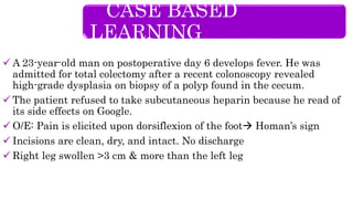
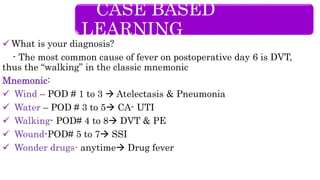
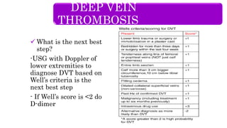


A 23-year-old man presents with fever on postoperative day 6 after undergoing total colectomy for colon polyps. On examination, he has right leg swelling and a positive Homan's sign. Doppler ultrasound is recommended to diagnose deep vein thrombosis (DVT), which is a common cause of fever at this postoperative time due to immobility risk. Risk factors for DVT include recent surgery and refusal of prophylactic heparin.


![CASE_PRESENTATION_ON_subdural_hematoma(SDH)[1 FINAL PPT]-1.pptx](https://cdn.slidesharecdn.com/ss_thumbnails/casepresentationonsubduralhematomasdh1finalppt-1-260129172522-d405d375-thumbnail.jpg?width=640&height=640&fit=bounds)


