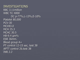
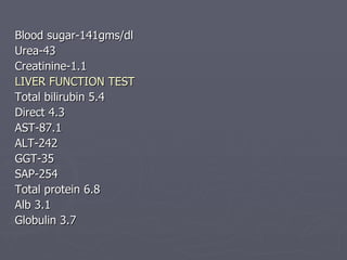
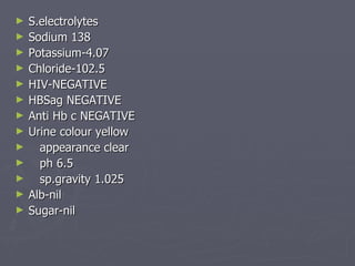
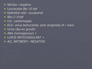
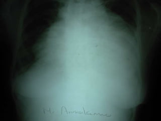
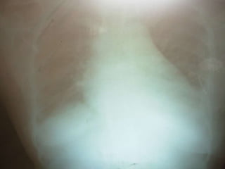
A CASE OF DEEP VEIN THROMBOSIS FOR DISCUSSION PROF. P. VIJAYARAGHAVAN’S UNIT Dr.P.ARUL P.G.
45 years old Mrs. Ammakanu was admitted with chief complaints of Left leg swelling 30 days right sided abdominal pain 20 days and breathlessness 4 days
History of presenting illness: Patient apparently normal before 30 days. developed left leg swelling and pain. she was admitted in a private hospital, following treatment the swelling and pain subsided. Later she had abdomen pain–dull aching, more over the right hypochondrium, no aggravating or relieving factors. Breathlessness present for past 4 days grade 3 initially and now grade 4. H/o orthopnea+ H/o vomiting once No h/o fever or headache. No h/o chest pain or palpitation.
No h/o cough No trauma No h/o altered bladder or bowel habits. No h/o altered sensorium. Past history: h/o arthritis for which she takes NSAIDS Not a diabetic or hypertensive or asthmatic or epileptic or tuberculous patient
Treatment history: Patient was started on warfarin 15 days back in a private hospital. Personal history :  Takes mixed diet. Not an alcoholic or tobacco chewer. Regular 3/30 days menstrual cycle. H/o 3 spontaneous abortions +, 2 live children. Family history : No similar illness in the family. Contact history :  No history of contact with patients with tuberculosis.
General examination Pt conscious, oriented, afebrile, dyspneic, tachypneic pallor+, malar rash + no icterus/cyanosis/clubbing no significant lymphadenopathy bilateral pitting pedal edema generalised swelling of the left lower limb JVP raised
Vital signs: Bp 110/60mm hg  Pulse 76/min RR 24/min Temperature 98.4 F
EXAMINATION OF CVS: S1 S2 heard but muffled RV-S3 heard  EXAMINATION OF RS; NVBS+  . no added sounds. EXAMINATION OF ABDOMEN: Soft ,bowel sounds heard, liver enlarged 7X3.5 cms size, soft, tender . EXAMINATION OF CNS: No FND.
EXAMINATION OF THE LEFT LL: Limb is swollen through out, tender, dusky appearance. Peripheral pulses felt.
INVESTIGATIONS RBC 3.11million WBC TC 4000 DC p-77%,L-13%,E-10% Platelet 80,000 PCV-30 MCV83.0 MCH 25.3 MCHC 30.5  HB-9.4 gm% ESR 36mm. Blood group A+ PT control 12-15 sec, test 38 APTT control 26.test 38 INR 2.2
Blood sugar-141gms/dl Urea-43 Creatinine-1.1 LIVER FUNCTION TEST Total bilirubin 5.4 Direct 4.3 AST-87.1 ALT-242 GGT-35 SAP-254 Total protein 6.8 Alb 3.1 Globulin 3.7
S.electrolytes Sodium 138 Potassium-4.07 Chloride-102.5 HIV-NEGATIVE HBSag NEGATIVE Anti Hb c NEGATIVE Urine colour yellow appearance clear ph 6.5 sp.gravity 1.025 Alb-nil Sugar-nil
Nitrites –negative Leucocytes 8to 10 hpf Epithelial cells –occasional Rbc-2-3/hpf Cxr- cardiomegaly ECG- sinus tachycardia, poor prognosis of r wave Urine c&s-no growth ANA (homogenous) + LUPUS ANTICOAGULANT +  ACL ANTIBODY –NEGATIVE.


Echo Pericardial effusion+ Fibrous strands+ RA collapse+ Normal lv systolic function Mild diastolic dysfunction Mild hypokinesia of IVS AND LV APEX No evidence of tamponade Normal chamber dimension, Doppler study Left popliteal vein thrombosis extending into superficial femoral vein.
Ultra sound abdomen Minimal free fluid in morrison pouch Hepatomegaly Minimal ascites Left pleural effusion Pericardial effusion
Hematologist and rheumatologist opinion ?APLAS Vascular surgeon opinion – DVT Left LL Elastocrepe bandage, leg elevation, tablet folic acid, in view of jaundice and abnormal PT and INR Heparin therapy is deferred. Cardiologist opinion to start acitrom
DIAGNOSIS A Case of DVT left lower limb probably due to  hyper coagulable state (anti phospholipid antibody syndrome secondary to SLE) with pericardial effusion, pleural effusion, ascites due to polyserositis.
 
DIAGNOSIS – ARA CRITERIA Malar rash Discoid rash photosensitivity Oral ulcers Arthritis Serositis Renal disorder Neurological disorder Hematological disorder Imm. disorder Antinuclear Abs 4  or >likely to be SLE
 
DEEP VEIN THROMBOSIS Commonly affects-leg veins or deep veins of pelvis. Causes and risk factors: Conditions that cause compression of veins, physical trauma, cancer, infections , certain inflammatory diseases, specific conditions like stroke, heart failure and nephrotic syndrome.
Surgery, hospitalisation, Immobilisation, smoking,obesity, drugs like estrogens, erythropoietin and diseases like thrombophilia. Women have risk during pregnancy and postnatal period, Air travel May thurner syndrome
MECHANISM -VIRCHOWS TRIAD Decreased flow rate of blood, Damage of blood vessel, Increased tendency of blood  to clot
SYMPTOMS AND SIGNS No symptoms sometime Pain,swelling and redness of leg Superficial veins may enlarge.
COMPLICATIONS Pulmonary embolism Post phlebitic syndrome INVESTIGATIONS Gold standard test-IV venography. Physical examination Homans test Pratts sign
Wells score Active cancer-1point Calf swelling>3cms-1pt Collateral veins-1pt Pitting edema-1pt Swelling of entire leg-1pt
Localised pain along distribution of deep venous system-1pt Paralysis,paresis or recent immobilisation of leg-1pt Recently bedridden or surgeryor pts requirina GA-1pt  INTERPRATATION. 2 OR > PTS-DVT LIKELY <2 DVT UNLIKELY BLOOD TEST D-DIMER TEST
OTHER TESTS CBC, pt APTT, fibrinogen, liver enzymes, renal function test and electrolytes Imaging leg veins Impedance plythesmography Doppler ultra sonogram
TREATMENT ANTI COAGULATION MEASURES Unfractionated heparin LMW Heparin Warfarin Thrombolysis  Compression stockings IVC filter
 

A Case of DVT for Discussion

  • 1.
    A CASE OFDEEP VEIN THROMBOSIS FOR DISCUSSION PROF. P. VIJAYARAGHAVAN’S UNIT Dr.P.ARUL P.G.
  • 2.
    45 years oldMrs. Ammakanu was admitted with chief complaints of Left leg swelling 30 days right sided abdominal pain 20 days and breathlessness 4 days
  • 3.
    History of presentingillness: Patient apparently normal before 30 days. developed left leg swelling and pain. she was admitted in a private hospital, following treatment the swelling and pain subsided. Later she had abdomen pain–dull aching, more over the right hypochondrium, no aggravating or relieving factors. Breathlessness present for past 4 days grade 3 initially and now grade 4. H/o orthopnea+ H/o vomiting once No h/o fever or headache. No h/o chest pain or palpitation.
  • 4.
    No h/o coughNo trauma No h/o altered bladder or bowel habits. No h/o altered sensorium. Past history: h/o arthritis for which she takes NSAIDS Not a diabetic or hypertensive or asthmatic or epileptic or tuberculous patient
  • 5.
    Treatment history: Patientwas started on warfarin 15 days back in a private hospital. Personal history : Takes mixed diet. Not an alcoholic or tobacco chewer. Regular 3/30 days menstrual cycle. H/o 3 spontaneous abortions +, 2 live children. Family history : No similar illness in the family. Contact history : No history of contact with patients with tuberculosis.
  • 6.
    General examination Ptconscious, oriented, afebrile, dyspneic, tachypneic pallor+, malar rash + no icterus/cyanosis/clubbing no significant lymphadenopathy bilateral pitting pedal edema generalised swelling of the left lower limb JVP raised
  • 7.
    Vital signs: Bp110/60mm hg Pulse 76/min RR 24/min Temperature 98.4 F
  • 8.
    EXAMINATION OF CVS:S1 S2 heard but muffled RV-S3 heard EXAMINATION OF RS; NVBS+ . no added sounds. EXAMINATION OF ABDOMEN: Soft ,bowel sounds heard, liver enlarged 7X3.5 cms size, soft, tender . EXAMINATION OF CNS: No FND.
  • 9.
    EXAMINATION OF THELEFT LL: Limb is swollen through out, tender, dusky appearance. Peripheral pulses felt.
  • 10.
    INVESTIGATIONS RBC 3.11millionWBC TC 4000 DC p-77%,L-13%,E-10% Platelet 80,000 PCV-30 MCV83.0 MCH 25.3 MCHC 30.5 HB-9.4 gm% ESR 36mm. Blood group A+ PT control 12-15 sec, test 38 APTT control 26.test 38 INR 2.2
  • 11.
    Blood sugar-141gms/dl Urea-43Creatinine-1.1 LIVER FUNCTION TEST Total bilirubin 5.4 Direct 4.3 AST-87.1 ALT-242 GGT-35 SAP-254 Total protein 6.8 Alb 3.1 Globulin 3.7
  • 12.
    S.electrolytes Sodium 138Potassium-4.07 Chloride-102.5 HIV-NEGATIVE HBSag NEGATIVE Anti Hb c NEGATIVE Urine colour yellow appearance clear ph 6.5 sp.gravity 1.025 Alb-nil Sugar-nil
  • 13.
    Nitrites –negative Leucocytes8to 10 hpf Epithelial cells –occasional Rbc-2-3/hpf Cxr- cardiomegaly ECG- sinus tachycardia, poor prognosis of r wave Urine c&s-no growth ANA (homogenous) + LUPUS ANTICOAGULANT + ACL ANTIBODY –NEGATIVE.
  • 14.
  • 15.
  • 16.
  • 17.
  • 18.
    Echo Pericardial effusion+Fibrous strands+ RA collapse+ Normal lv systolic function Mild diastolic dysfunction Mild hypokinesia of IVS AND LV APEX No evidence of tamponade Normal chamber dimension, Doppler study Left popliteal vein thrombosis extending into superficial femoral vein.
  • 19.
    Ultra sound abdomenMinimal free fluid in morrison pouch Hepatomegaly Minimal ascites Left pleural effusion Pericardial effusion
  • 20.
    Hematologist and rheumatologistopinion ?APLAS Vascular surgeon opinion – DVT Left LL Elastocrepe bandage, leg elevation, tablet folic acid, in view of jaundice and abnormal PT and INR Heparin therapy is deferred. Cardiologist opinion to start acitrom
  • 21.
    DIAGNOSIS A Caseof DVT left lower limb probably due to hyper coagulable state (anti phospholipid antibody syndrome secondary to SLE) with pericardial effusion, pleural effusion, ascites due to polyserositis.
  • 22.
  • 23.
    DIAGNOSIS – ARACRITERIA Malar rash Discoid rash photosensitivity Oral ulcers Arthritis Serositis Renal disorder Neurological disorder Hematological disorder Imm. disorder Antinuclear Abs 4 or >likely to be SLE
  • 24.
  • 25.
    DEEP VEIN THROMBOSISCommonly affects-leg veins or deep veins of pelvis. Causes and risk factors: Conditions that cause compression of veins, physical trauma, cancer, infections , certain inflammatory diseases, specific conditions like stroke, heart failure and nephrotic syndrome.
  • 26.
    Surgery, hospitalisation, Immobilisation,smoking,obesity, drugs like estrogens, erythropoietin and diseases like thrombophilia. Women have risk during pregnancy and postnatal period, Air travel May thurner syndrome
  • 27.
    MECHANISM -VIRCHOWS TRIADDecreased flow rate of blood, Damage of blood vessel, Increased tendency of blood to clot
  • 28.
    SYMPTOMS AND SIGNSNo symptoms sometime Pain,swelling and redness of leg Superficial veins may enlarge.
  • 29.
    COMPLICATIONS Pulmonary embolismPost phlebitic syndrome INVESTIGATIONS Gold standard test-IV venography. Physical examination Homans test Pratts sign
  • 30.
    Wells score Activecancer-1point Calf swelling>3cms-1pt Collateral veins-1pt Pitting edema-1pt Swelling of entire leg-1pt
  • 31.
    Localised pain alongdistribution of deep venous system-1pt Paralysis,paresis or recent immobilisation of leg-1pt Recently bedridden or surgeryor pts requirina GA-1pt INTERPRATATION. 2 OR > PTS-DVT LIKELY <2 DVT UNLIKELY BLOOD TEST D-DIMER TEST
  • 32.
    OTHER TESTS CBC,pt APTT, fibrinogen, liver enzymes, renal function test and electrolytes Imaging leg veins Impedance plythesmography Doppler ultra sonogram
  • 33.
    TREATMENT ANTI COAGULATIONMEASURES Unfractionated heparin LMW Heparin Warfarin Thrombolysis Compression stockings IVC filter
  • 34.