SlideShare a Scribd company logo
CANDIDATE INFORMATION AND TASKS
You are working in a medical outpatient clinic. Your next
patient is a single 25-year-old sales assistant who has
consulted you about general malaise, anorexia, nausea and
fever gradually worsening over the last 10 days. Yesterday he
noted dark urine and pale stools.
On examination you have found icteric conjunctivae, a
temperature of 38.8 °C and tender enlargement of the liver.
Urinalysis was positive for bilirubin and urobilinogen. There
are no other abnormal findings on examination.
CANDIDATE INFORMATION AND TASKS
YOUR TASKS ARE TO:
1. Take any further relevant history you require.
2. Tell the examiner what investigations you would
arrange.
3. Explain the nature of the illness and possible
cause to the patient, indicating which you
consider to be the most likely cause.
4. Advise the patient concerning immediate
management.
EVALUATION OF JAUNDICE
Dr. Sookun Rajeev K
(MD)
Dept of General Medicine
Anna Medical College
Definition:
• Jaundice is the Yellow colouration of the
skin, sclerae and mucous membrane
which is caused by the deposition of bile
pigment and usually apparent when the
serum bilirubin is more than 35 mmol/l
Classification of Jaundice
• Pre Hepatic Jaundice ( causes lemon-yellow skin
colouration) e.g Hemolytic Jaundice
• Hepatic Jaundice (causes deep yellow jaundice
progressing to a greenish tinge)
• Post Hepatic Jaundice
• The sclerae are not coloured in those with yellow skin
caused by hypercarotinaemia.
Common Causes of Jaundice
Viral hepatitis
Biliary obstruction from gallstones or carcinoma of the
head of the pancreas
Drugs (Alcohol,Acetaminophen,psy meds,diuretics,etc..
Metastases
Intrahepatic cholestasis (including ascending
cholangitis and primary biliary cirrhosis)
Infectious mononucleosis
Gilbert syndrome
Uncommon Causes of Jaundice (cont)
Haemolytic anaemia
Congenital hyperbilirubinaemia
Stricture or carcinoma of the major bile ducts or
ampulla
History
• Sudden Onset : Viral hepatitis,acute biliary obstruction,
trauma or toxin-mediated fulminant liver failure.
• Gradual onset : chronic liver disease (including alcoholic
cirrhosis) or malignancy.
• A lifelong history : suggests an inherited metabolic or
haemolytic cause.
History (cont)
• Hx of Right Upper Quadrant pain especially after
fatty meal, should suggest Cholestasis or
Cholelithiasis
• Hx of Fever or prior biliary surgery, points to
Cholangitis
• Hx of Anorexia, malaise, or myalgia, a viral etiology
is most probable
History (cont)
• Hx of Pruritus or weight loss associated with noninfectious
etiologies.
• Blood transfusion, IVD use, sexual contacts, travel to
endemic areas or ingestion of contaminated foods expose
patients to viral related Hepatocellular injury.
• PMHx,PSHx,FHx of inherited defect in bilirubin conjugation
or transport,wilson’s disease,hemochromatoses
Signs & Symptoms in Jaundice
 Dark urine
Signs & Symptoms in Jaundice (cont)
Yellow coloration of the skin – should be
examined in natural white light
Steatorrhea – suggests no breakdown of fats
Pruritus - suggests chronic disease
Abdominal pain - suggests biliary tract
obstruction from stone or pancreatic tumour
(obstructive jaundice)
Signs & Symptoms in Jaundice (cont)
Clay coloured stool
Painless jaundice in the elderly - think of
pancreatic cancer, although most patients with
pancreatic cancer have pain
Kernicterus - rarely seen in adults due to
maturation of blood brain barrier
Signs & Symptoms in Jaundice (cont)
Spider Angioma
Caput medusa
Gynecomastia
Testicular Atrophy
Palmar Erythema
Dupuytren’s contracture
• All suggest
Chronic Liver
Disease and
Portal
Hypertension
Specific features on examination of the
abdomen in a jaundiced patient include
• Recent operation scars: suggesting
cholecystectomy or surgery for intra-
abdominal carcinoma
• Palpable Enlarged Gall Bladder suggesting bile
duct obstruction caused by carcinoma of the
pancreas (rather than gallstones)
Specific features on examination of the
abdomen in a jaundiced patient include (cont)
• Hepatomegaly: irregular
when infiltrated with
carcinoma or in early
cirrhosis, tender in
infectious and acute
alcoholic hepatitis and
occasionally in
congestive heart failure
Specific features on examination of the
abdomen in a jaundiced patient include (cont)
• Splenomegaly
in portal
hypertension,
spherocytosis
and infectious
mononucleosis
Specific features on examination of the
abdomen in a jaundiced patient include (cont)
• Ascites.
Aims for Investigations
1. Investigation aims to discover the site of any
biliary outflow obstruction
2. Determine the degree of impairment of liver cell
function and its cause
3. Eliminate rare causes such as haemolysis
4. Establish potential for treatment.
1. Haematology
Full blood count, reticulocyte count and Coombs’
test
A normal reticulocyte count virtually excludes
haemolytic jaundice.
Leukocytosis may suggest infection or carcinoma.
Abnormal mononuclear cells suggest infectious
mononucleosis or viral hepatitis.
2. Liver Function Tests
 Measure the ability of the liver to perform normal functions (e.g.
serum albumin is a measure of protein synthesis; prothrombin
time is a measure of synthetic function; bilirubin is a measure of
bile salt conjugation and excretion).
 Liver enzymes (alkaline phosphatase, transaminases) are
indicators of ductal or liver cell damage.
 In obstructive jaundice the alkaline phosphatase is greatly
elevated compared with transaminases; in hepatocellular disease
transaminases are predominantly raised.
3. Bilirubin
Bilirubin derived from red cell breakdown is transported to
the liver where it is conjugated to glucuronic acid.
Conjugated bilirubin is secreted in the bile and degraded
in the gut by bacteria to form urobilinogen. Urobilinogen is
either excreted in the stool or reabsorbed from the gut and
excreted by the kidneys.
Serum bilirubin is predominantly unconjugated in
haemolytic jaundice and the other liver function tests are
usually normal. It is mainly conjugated in obstructive
jaundice.
3. Bilirubin (cont)
Causes of increased bilirubin
Hepatocellular failure
Biliary obstruction
Haemolysis
Gilbert syndrome
4. Alkaline Phosphatase
Elevated in
Obstructive jaundice
Hepatocellular jaundice
Growth in adolescence
Pregnancy
Normal in
Gilbert syndrome
Myeloma
Alkaline phosphatase is found in high levels in biliary
Canaliculi, osteoblasts, intestinal mucosa and placenta.
4. Alkaline Phosphatase (cont)
• A raised level in the absence of other signs of liver
disease or abnormal liver function tests suggests
the presence of malignant secondary deposits in
the bone or Paget’s disease. Consider measuring
isoenzymes if there is doubt.
4. Alkaline Phosphatase (cont)
Causes of increased hepatic alkaline phosphatase
 Extra-hepatic cholestasis
 Obstructive jaundice
 Intra-hepatic cholestasis (e.g. cirrhosis, drugs, cholangitis, primary biliary
cirrhosis)
 Obstructive phase of hepatitis
Causes of increased bone alkaline phosphatase (osteoblastic activity)
 Paget’s disease
 Bone metastases
 Osteomalacia
 Hyperparathyroidism
 Normal growth in puberty
 Fractures
5. Transaminases
Elevated serum transaminases (alanine
aminotransferase, aspartate aminotransferase and
gamma glutamyl transferase) indicate
hepatocellular damage. Slight elevation is
consistent with obstructive jaundice.
5. Transaminases (cont)
Causes of elevated alanine aminotransferase
 Active liver cell damage, including drugs, hepatitis and metastatic
infiltration
 Acute myocardial infarction (peaks at 24–48 h, may fall to normal
by 72h). The degree of elevation reflects the amount of muscle
damage
 Acute pancreatitis
 Haemolysis
5. Transaminases (cont)
Elevated aspartate aminotransferase levels parallel the alanine
aminotransferase
-glutamyl transferase
 Inducible microsomal enzyme
 Most sensitive index of alcohol ingestion
 Raised in most forms of liver disease including acute and chronic
hepatitis, cirrhosis and following drugs that induce microsomal
enzymes
6. Urinalysis
 Conjugated bilirubin renders the urine dark yellow
 Urobilinogen is colourless but on standing the urine turns brown
as urobilinogen is converted to urobilin by oxidation.
 Haemolytic jaundice is acholuric (no bilirubin in the urine) but the
urine contains excess urobilinogen because excess bilirubin
reaches the intestine and is re-excreted as urobilinogen.
 Obstructive jaundice produces dark brown urine with excess
bilirubin but a reduction of urinary urobilinogen (little or no
bilirubin reaches the gut because of the obstruction and
therefore cannot be reabsorbed and re-excreted).
6. Urinalysis (cont)
In the early stages of acute viral hepatitis, excess
urobilinogen may sometimes be present before clinical
jaundice becomes apparent.
This is a result of failure of the liver to take up the excess
urobilinogen absorbed from the gut.
With increasing severity, biliary obstruction develops and
as conjugated bilirubin appears in the urine it disappears
from the gut and urobilinogen disappears from the urine.
The reciprocal effect also occurs during recovery.
7. Serology
 Viral hepatitis serology – hepatitis A–E,
cytomegalo- virus (CMV) and Epstein–Barr
virus (EBV)
Antimitochondrial antibodies (primary biliary
cirrhosis)
Antinuclear factor and smooth-muscle
antibodies (chronic active hepatitis)
8. Abdominal Radiology in jaundice
Plain X-ray and ultrasound may show gallstones.
8. Abdominal Radiology in jaundice (cont)
Ultrasound, CT and MRI may show prImary or
secondary tumours, pancreatic carcinoma, stones
in the gall bladder and dilated biliary ducts in
obstruction.
Isotope liver scans may demonstrate secondary
deposits.
9. Needle Liver Biopsy
Biliary obstruction is a relative contraindication
because of the potential danger of biliary peritonitis.
Ultrasound and CT-guided biopsy may provide the
histological diagnosis in focal lesions.
Check the prothrombin time and platelet counts are
normal. Fresh frozen plasma will quickly reverse the
prothrombin time for the duration of the procedure.
9. Needle Liver Biopsy (cont)
10. Other investigations
Endoscopic retrograde
cholangiopancreatography (ERCP) is valuable
to define obstruction of the
pancreaticoduodenal tree, for sphincterotomy,
to release stones and to relieve obstruction by
insertion of a stent.
-fetoprotein is raised in hepatocellular
carcinoma.
ERCP
Evaluation of Jaundice by Dr. Sookun Rajeev Kumar

More Related Content

What's hot

Recurrent abdominal pain in children
Recurrent abdominal pain in childrenRecurrent abdominal pain in children
Recurrent abdominal pain in children
samialbdairat
 
Approach to child with generalized edema
Approach to child with generalized edemaApproach to child with generalized edema
Approach to child with generalized edema
Ahmed Bahamid
 
Approach to a child with hepatosplenomegaly
Approach to a child with hepatosplenomegalyApproach to a child with hepatosplenomegaly
Approach to a child with hepatosplenomegaly
Yousif Murtada
 
Approach To A Patient With Jaundice
Approach To A Patient With JaundiceApproach To A Patient With Jaundice
Approach To A Patient With Jaundice
Tanuj Bhatia
 

What's hot (20)

An approach to jaundice
An approach to jaundiceAn approach to jaundice
An approach to jaundice
 
Approach to hypoglycemia in infants and children
Approach to hypoglycemia in infants and childrenApproach to hypoglycemia in infants and children
Approach to hypoglycemia in infants and children
 
Recurrent abdominal pain in children
Recurrent abdominal pain in childrenRecurrent abdominal pain in children
Recurrent abdominal pain in children
 
Direct jaundice
Direct jaundiceDirect jaundice
Direct jaundice
 
Direct hyperbilirubinaemia in neonate by Dr. Tareq Rahman
Direct hyperbilirubinaemia in neonate by Dr. Tareq RahmanDirect hyperbilirubinaemia in neonate by Dr. Tareq Rahman
Direct hyperbilirubinaemia in neonate by Dr. Tareq Rahman
 
Neonatal Cholestasis
Neonatal CholestasisNeonatal Cholestasis
Neonatal Cholestasis
 
Recurrent abdominal pain
Recurrent abdominal painRecurrent abdominal pain
Recurrent abdominal pain
 
Approach to child with generalized edema
Approach to child with generalized edemaApproach to child with generalized edema
Approach to child with generalized edema
 
Approach to cholestatic jaundice
Approach to cholestatic jaundiceApproach to cholestatic jaundice
Approach to cholestatic jaundice
 
Budd chiari syndrome
Budd chiari syndromeBudd chiari syndrome
Budd chiari syndrome
 
Approach to a child with hepatosplenomegaly
Approach to a child with hepatosplenomegalyApproach to a child with hepatosplenomegaly
Approach to a child with hepatosplenomegaly
 
Chronic diarrhoea
Chronic diarrhoeaChronic diarrhoea
Chronic diarrhoea
 
Approach to jaundice
Approach to jaundiceApproach to jaundice
Approach to jaundice
 
Hepato&spleenomegaly
Hepato&spleenomegalyHepato&spleenomegaly
Hepato&spleenomegaly
 
Approach To A Patient With Jaundice
Approach To A Patient With JaundiceApproach To A Patient With Jaundice
Approach To A Patient With Jaundice
 
1 Hx & PE in pediatrics
1 Hx & PE in pediatrics 1 Hx & PE in pediatrics
1 Hx & PE in pediatrics
 
Reductive adaptation in sam
Reductive adaptation in samReductive adaptation in sam
Reductive adaptation in sam
 
Chronic diarrhea
Chronic diarrheaChronic diarrhea
Chronic diarrhea
 
Hepatospleenomegaly in children
Hepatospleenomegaly in childrenHepatospleenomegaly in children
Hepatospleenomegaly in children
 
Chronic diarrhoea in children
Chronic diarrhoea in childrenChronic diarrhoea in children
Chronic diarrhoea in children
 

Similar to Evaluation of Jaundice by Dr. Sookun Rajeev Kumar

03251163_jaundice_asc_port_hypertension.pptx
03251163_jaundice_asc_port_hypertension.pptx03251163_jaundice_asc_port_hypertension.pptx
03251163_jaundice_asc_port_hypertension.pptx
ReshmaShajiPns1
 
All about Jaundice
All about JaundiceAll about Jaundice
All about Jaundice
ozhin araz
 
Chronic hepatitis ppt
Chronic hepatitis pptChronic hepatitis ppt
Chronic hepatitis ppt
Manoj Ghoda
 
hepatitis lecture 2021.pptdgvhehnvnjvfvlfv
hepatitis lecture 2021.pptdgvhehnvnjvfvlfvhepatitis lecture 2021.pptdgvhehnvnjvfvlfv
hepatitis lecture 2021.pptdgvhehnvnjvfvlfv
interaman123
 

Similar to Evaluation of Jaundice by Dr. Sookun Rajeev Kumar (20)

evaluation of jaundice
evaluation of jaundiceevaluation of jaundice
evaluation of jaundice
 
03251163_jaundice_asc_port_hypertension.pptx
03251163_jaundice_asc_port_hypertension.pptx03251163_jaundice_asc_port_hypertension.pptx
03251163_jaundice_asc_port_hypertension.pptx
 
All about Jaundice
All about JaundiceAll about Jaundice
All about Jaundice
 
Jaundice.pptx
Jaundice.pptxJaundice.pptx
Jaundice.pptx
 
Bile: a review of the biliary system
Bile: a review of the biliary systemBile: a review of the biliary system
Bile: a review of the biliary system
 
Surgical jaundice
Surgical jaundiceSurgical jaundice
Surgical jaundice
 
Approach to jaundice bikal
Approach to jaundice bikalApproach to jaundice bikal
Approach to jaundice bikal
 
Jaundice
JaundiceJaundice
Jaundice
 
Clinical Case on Jaundice
Clinical Case on JaundiceClinical Case on Jaundice
Clinical Case on Jaundice
 
Chronic hepatitis ppt
Chronic hepatitis pptChronic hepatitis ppt
Chronic hepatitis ppt
 
‫ Jaundice
‫  Jaundice‫  Jaundice
‫ Jaundice
 
Jaundice
Jaundice Jaundice
Jaundice
 
jaundice-(three types)- pre hepatic, hepatic , post hepatic
jaundice-(three types)- pre hepatic, hepatic , post hepaticjaundice-(three types)- pre hepatic, hepatic , post hepatic
jaundice-(three types)- pre hepatic, hepatic , post hepatic
 
hepatitis lecture 2021.pptdgvhehnvnjvfvlfv
hepatitis lecture 2021.pptdgvhehnvnjvfvlfvhepatitis lecture 2021.pptdgvhehnvnjvfvlfv
hepatitis lecture 2021.pptdgvhehnvnjvfvlfv
 
Postoperative jaundice
Postoperative jaundicePostoperative jaundice
Postoperative jaundice
 
jaundice
jaundice jaundice
jaundice
 
jaundice-160414200805.pptx
jaundice-160414200805.pptxjaundice-160414200805.pptx
jaundice-160414200805.pptx
 
Diarrhea and IBD When to be woeeied ?
Diarrhea and IBD When to be woeeied ?Diarrhea and IBD When to be woeeied ?
Diarrhea and IBD When to be woeeied ?
 
Approach patient with juandice
Approach patient with juandiceApproach patient with juandice
Approach patient with juandice
 
34936_Evaluation of a patient with Jaundice.ppt
34936_Evaluation of a patient with Jaundice.ppt34936_Evaluation of a patient with Jaundice.ppt
34936_Evaluation of a patient with Jaundice.ppt
 

More from Dr. Sookun Rajeev Kumar

More from Dr. Sookun Rajeev Kumar (20)

Endocrinology part IIi + Charts by Dr. Sookun Rajeev Kumar
Endocrinology part IIi + Charts by Dr. Sookun Rajeev KumarEndocrinology part IIi + Charts by Dr. Sookun Rajeev Kumar
Endocrinology part IIi + Charts by Dr. Sookun Rajeev Kumar
 
Endocrinology part I by Dr. Sookun Rajeev Kumar
Endocrinology part I by Dr. Sookun Rajeev KumarEndocrinology part I by Dr. Sookun Rajeev Kumar
Endocrinology part I by Dr. Sookun Rajeev Kumar
 
Entomology part 3 by Dr. Sookun Rajeev K
Entomology part 3 by Dr. Sookun Rajeev KEntomology part 3 by Dr. Sookun Rajeev K
Entomology part 3 by Dr. Sookun Rajeev K
 
Entomology part 2 by Dr. Sookun Rajeev K
Entomology part 2 by Dr. Sookun Rajeev KEntomology part 2 by Dr. Sookun Rajeev K
Entomology part 2 by Dr. Sookun Rajeev K
 
Entomology part 1 Dr. Sookun Rajeev K
Entomology part 1 Dr. Sookun Rajeev KEntomology part 1 Dr. Sookun Rajeev K
Entomology part 1 Dr. Sookun Rajeev K
 
Yellow Fever by Dr. Sookun Rajeev Kumar
Yellow Fever by Dr. Sookun Rajeev KumarYellow Fever by Dr. Sookun Rajeev Kumar
Yellow Fever by Dr. Sookun Rajeev Kumar
 
Upper Respiratory Tract Infection by Dr. Sookun Rajeev Kumar
Upper Respiratory Tract Infection by Dr. Sookun Rajeev KumarUpper Respiratory Tract Infection by Dr. Sookun Rajeev Kumar
Upper Respiratory Tract Infection by Dr. Sookun Rajeev Kumar
 
Typhus Fever by Dr. Sookun Rajeev Kumar
Typhus Fever by Dr. Sookun Rajeev KumarTyphus Fever by Dr. Sookun Rajeev Kumar
Typhus Fever by Dr. Sookun Rajeev Kumar
 
Respiratory Failure by Dr. Sookun Rajeev Kumar
Respiratory Failure by Dr. Sookun Rajeev KumarRespiratory Failure by Dr. Sookun Rajeev Kumar
Respiratory Failure by Dr. Sookun Rajeev Kumar
 
Pulmonary Fibrosis by Dr. Sookun Rajeev Kumar
Pulmonary Fibrosis by Dr. Sookun Rajeev KumarPulmonary Fibrosis by Dr. Sookun Rajeev Kumar
Pulmonary Fibrosis by Dr. Sookun Rajeev Kumar
 
Polycythemia by Dr. Sookun Rajeev Kumar
Polycythemia by Dr. Sookun Rajeev KumarPolycythemia by Dr. Sookun Rajeev Kumar
Polycythemia by Dr. Sookun Rajeev Kumar
 
Pneumothorax by Dr. Sookun Rajeev Kumar
Pneumothorax by Dr. Sookun Rajeev KumarPneumothorax by Dr. Sookun Rajeev Kumar
Pneumothorax by Dr. Sookun Rajeev Kumar
 
Pneumonia by Dr. Sookun Rajeev Kumar
Pneumonia by Dr. Sookun Rajeev KumarPneumonia by Dr. Sookun Rajeev Kumar
Pneumonia by Dr. Sookun Rajeev Kumar
 
Peripheral Neuritis by Dr. Sookun Rajeev Kumar
Peripheral Neuritis by Dr. Sookun Rajeev KumarPeripheral Neuritis by Dr. Sookun Rajeev Kumar
Peripheral Neuritis by Dr. Sookun Rajeev Kumar
 
Megaloblastic Anemia by Dr. Sookun Rajeev Kumar
Megaloblastic Anemia by Dr. Sookun Rajeev KumarMegaloblastic Anemia by Dr. Sookun Rajeev Kumar
Megaloblastic Anemia by Dr. Sookun Rajeev Kumar
 
Lymphomas: Hodgkin & Non Hodgkin by Dr. Sookun Rajeev Kumar
Lymphomas: Hodgkin & Non Hodgkin by Dr. Sookun Rajeev KumarLymphomas: Hodgkin & Non Hodgkin by Dr. Sookun Rajeev Kumar
Lymphomas: Hodgkin & Non Hodgkin by Dr. Sookun Rajeev Kumar
 
Haemolytic Anemia by Dr. Sookun Rajeev Kumar
Haemolytic Anemia by Dr. Sookun Rajeev KumarHaemolytic Anemia by Dr. Sookun Rajeev Kumar
Haemolytic Anemia by Dr. Sookun Rajeev Kumar
 
Food Poisoning by Dr. Sookun Rajeev Kumar
Food Poisoning by Dr. Sookun Rajeev KumarFood Poisoning by Dr. Sookun Rajeev Kumar
Food Poisoning by Dr. Sookun Rajeev Kumar
 
Erythropoiesis,Anemia,Iron Deficiency Anemia by Dr. Sookun Rajeev Kumar
Erythropoiesis,Anemia,Iron Deficiency Anemia by Dr. Sookun Rajeev KumarErythropoiesis,Anemia,Iron Deficiency Anemia by Dr. Sookun Rajeev Kumar
Erythropoiesis,Anemia,Iron Deficiency Anemia by Dr. Sookun Rajeev Kumar
 
Enteric Fever by Dr. Sookun Rajeev Kumar
Enteric Fever by Dr. Sookun Rajeev KumarEnteric Fever by Dr. Sookun Rajeev Kumar
Enteric Fever by Dr. Sookun Rajeev Kumar
 

Recently uploaded

Alcohol_Dr. Jeenal Mistry MD Pharmacology.pdf
Alcohol_Dr. Jeenal Mistry MD Pharmacology.pdfAlcohol_Dr. Jeenal Mistry MD Pharmacology.pdf
Alcohol_Dr. Jeenal Mistry MD Pharmacology.pdf
Dr Jeenal Mistry
 

Recently uploaded (20)

Evaluation of antidepressant activity of clitoris ternatea in animals
Evaluation of antidepressant activity of clitoris ternatea in animalsEvaluation of antidepressant activity of clitoris ternatea in animals
Evaluation of antidepressant activity of clitoris ternatea in animals
 
How to Give Better Lectures: Some Tips for Doctors
How to Give Better Lectures: Some Tips for DoctorsHow to Give Better Lectures: Some Tips for Doctors
How to Give Better Lectures: Some Tips for Doctors
 
PT MANAGEMENT OF URINARY INCONTINENCE.pptx
PT MANAGEMENT OF URINARY INCONTINENCE.pptxPT MANAGEMENT OF URINARY INCONTINENCE.pptx
PT MANAGEMENT OF URINARY INCONTINENCE.pptx
 
US E-cigarette Summit: Taming the nicotine industrial complex
US E-cigarette Summit: Taming the nicotine industrial complexUS E-cigarette Summit: Taming the nicotine industrial complex
US E-cigarette Summit: Taming the nicotine industrial complex
 
TEST BANK For Advanced Practice Nursing in the Care of Older Adults, 2nd Edit...
TEST BANK For Advanced Practice Nursing in the Care of Older Adults, 2nd Edit...TEST BANK For Advanced Practice Nursing in the Care of Older Adults, 2nd Edit...
TEST BANK For Advanced Practice Nursing in the Care of Older Adults, 2nd Edit...
 
180-hour Power Capsules For Men In Ghana
180-hour Power Capsules For Men In Ghana180-hour Power Capsules For Men In Ghana
180-hour Power Capsules For Men In Ghana
 
Non-Invasive assessment of arterial stiffness in advanced heart failure patie...
Non-Invasive assessment of arterial stiffness in advanced heart failure patie...Non-Invasive assessment of arterial stiffness in advanced heart failure patie...
Non-Invasive assessment of arterial stiffness in advanced heart failure patie...
 
Relationship between vascular system disfunction, neurofluid flow and Alzheim...
Relationship between vascular system disfunction, neurofluid flow and Alzheim...Relationship between vascular system disfunction, neurofluid flow and Alzheim...
Relationship between vascular system disfunction, neurofluid flow and Alzheim...
 
Presentació "Advancing Emergency Medicine Education through Virtual Reality"
Presentació "Advancing Emergency Medicine Education through Virtual Reality"Presentació "Advancing Emergency Medicine Education through Virtual Reality"
Presentació "Advancing Emergency Medicine Education through Virtual Reality"
 
Book Trailer: PGMEE in a Nutshell (CEE MD/MS PG Entrance Examination)
Book Trailer: PGMEE in a Nutshell (CEE MD/MS PG Entrance Examination)Book Trailer: PGMEE in a Nutshell (CEE MD/MS PG Entrance Examination)
Book Trailer: PGMEE in a Nutshell (CEE MD/MS PG Entrance Examination)
 
Fundamental of Radiobiology -SABBU.pptx
Fundamental of Radiobiology  -SABBU.pptxFundamental of Radiobiology  -SABBU.pptx
Fundamental of Radiobiology -SABBU.pptx
 
The hemodynamic and autonomic determinants of elevated blood pressure in obes...
The hemodynamic and autonomic determinants of elevated blood pressure in obes...The hemodynamic and autonomic determinants of elevated blood pressure in obes...
The hemodynamic and autonomic determinants of elevated blood pressure in obes...
 
Contact dermaititis (irritant and allergic).pdf
Contact dermaititis (irritant and allergic).pdfContact dermaititis (irritant and allergic).pdf
Contact dermaititis (irritant and allergic).pdf
 
Alcohol_Dr. Jeenal Mistry MD Pharmacology.pdf
Alcohol_Dr. Jeenal Mistry MD Pharmacology.pdfAlcohol_Dr. Jeenal Mistry MD Pharmacology.pdf
Alcohol_Dr. Jeenal Mistry MD Pharmacology.pdf
 
DECIPHERING COMMON ECG FINDINGS IN ED.pptx
DECIPHERING COMMON ECG FINDINGS IN ED.pptxDECIPHERING COMMON ECG FINDINGS IN ED.pptx
DECIPHERING COMMON ECG FINDINGS IN ED.pptx
 
Multiple sclerosis diet.230524.ppt3.pptx
Multiple sclerosis diet.230524.ppt3.pptxMultiple sclerosis diet.230524.ppt3.pptx
Multiple sclerosis diet.230524.ppt3.pptx
 
Young at heart: Cardiovascular health stations to empower healthy lifestyle b...
Young at heart: Cardiovascular health stations to empower healthy lifestyle b...Young at heart: Cardiovascular health stations to empower healthy lifestyle b...
Young at heart: Cardiovascular health stations to empower healthy lifestyle b...
 
linearity concept of significance, standard deviation, chi square test, stude...
linearity concept of significance, standard deviation, chi square test, stude...linearity concept of significance, standard deviation, chi square test, stude...
linearity concept of significance, standard deviation, chi square test, stude...
 
Couples presenting to the infertility clinic- Do they really have infertility...
Couples presenting to the infertility clinic- Do they really have infertility...Couples presenting to the infertility clinic- Do they really have infertility...
Couples presenting to the infertility clinic- Do they really have infertility...
 
Arterial health throughout cancer treatment and exercise rehabilitation in wo...
Arterial health throughout cancer treatment and exercise rehabilitation in wo...Arterial health throughout cancer treatment and exercise rehabilitation in wo...
Arterial health throughout cancer treatment and exercise rehabilitation in wo...
 

Evaluation of Jaundice by Dr. Sookun Rajeev Kumar

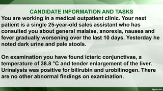
  • 1. CANDIDATE INFORMATION AND TASKS You are working in a medical outpatient clinic. Your next patient is a single 25-year-old sales assistant who has consulted you about general malaise, anorexia, nausea and fever gradually worsening over the last 10 days. Yesterday he noted dark urine and pale stools. On examination you have found icteric conjunctivae, a temperature of 38.8 °C and tender enlargement of the liver. Urinalysis was positive for bilirubin and urobilinogen. There are no other abnormal findings on examination.
  • 2. CANDIDATE INFORMATION AND TASKS YOUR TASKS ARE TO: 1. Take any further relevant history you require. 2. Tell the examiner what investigations you would arrange. 3. Explain the nature of the illness and possible cause to the patient, indicating which you consider to be the most likely cause. 4. Advise the patient concerning immediate management.
  • 3. EVALUATION OF JAUNDICE Dr. Sookun Rajeev K (MD) Dept of General Medicine Anna Medical College
  • 4. Definition: • Jaundice is the Yellow colouration of the skin, sclerae and mucous membrane which is caused by the deposition of bile pigment and usually apparent when the serum bilirubin is more than 35 mmol/l
  • 5. Classification of Jaundice • Pre Hepatic Jaundice ( causes lemon-yellow skin colouration) e.g Hemolytic Jaundice • Hepatic Jaundice (causes deep yellow jaundice progressing to a greenish tinge) • Post Hepatic Jaundice • The sclerae are not coloured in those with yellow skin caused by hypercarotinaemia.
  • 6. Common Causes of Jaundice Viral hepatitis Biliary obstruction from gallstones or carcinoma of the head of the pancreas Drugs (Alcohol,Acetaminophen,psy meds,diuretics,etc.. Metastases Intrahepatic cholestasis (including ascending cholangitis and primary biliary cirrhosis) Infectious mononucleosis Gilbert syndrome
  • 7. Uncommon Causes of Jaundice (cont) Haemolytic anaemia Congenital hyperbilirubinaemia Stricture or carcinoma of the major bile ducts or ampulla
  • 8. History • Sudden Onset : Viral hepatitis,acute biliary obstruction, trauma or toxin-mediated fulminant liver failure. • Gradual onset : chronic liver disease (including alcoholic cirrhosis) or malignancy. • A lifelong history : suggests an inherited metabolic or haemolytic cause.
  • 9. History (cont) • Hx of Right Upper Quadrant pain especially after fatty meal, should suggest Cholestasis or Cholelithiasis • Hx of Fever or prior biliary surgery, points to Cholangitis • Hx of Anorexia, malaise, or myalgia, a viral etiology is most probable
  • 10. History (cont) • Hx of Pruritus or weight loss associated with noninfectious etiologies. • Blood transfusion, IVD use, sexual contacts, travel to endemic areas or ingestion of contaminated foods expose patients to viral related Hepatocellular injury. • PMHx,PSHx,FHx of inherited defect in bilirubin conjugation or transport,wilson’s disease,hemochromatoses
  • 11. Signs & Symptoms in Jaundice  Dark urine
  • 12. Signs & Symptoms in Jaundice (cont) Yellow coloration of the skin – should be examined in natural white light Steatorrhea – suggests no breakdown of fats Pruritus - suggests chronic disease Abdominal pain - suggests biliary tract obstruction from stone or pancreatic tumour (obstructive jaundice)
  • 13. Signs & Symptoms in Jaundice (cont) Clay coloured stool Painless jaundice in the elderly - think of pancreatic cancer, although most patients with pancreatic cancer have pain Kernicterus - rarely seen in adults due to maturation of blood brain barrier
  • 14. Signs & Symptoms in Jaundice (cont)
  • 15. Signs & Symptoms in Jaundice (cont) Spider Angioma Caput medusa Gynecomastia Testicular Atrophy Palmar Erythema Dupuytren’s contracture • All suggest Chronic Liver Disease and Portal Hypertension
  • 16. Specific features on examination of the abdomen in a jaundiced patient include • Recent operation scars: suggesting cholecystectomy or surgery for intra- abdominal carcinoma • Palpable Enlarged Gall Bladder suggesting bile duct obstruction caused by carcinoma of the pancreas (rather than gallstones)
  • 17. Specific features on examination of the abdomen in a jaundiced patient include (cont) • Hepatomegaly: irregular when infiltrated with carcinoma or in early cirrhosis, tender in infectious and acute alcoholic hepatitis and occasionally in congestive heart failure
  • 18. Specific features on examination of the abdomen in a jaundiced patient include (cont) • Splenomegaly in portal hypertension, spherocytosis and infectious mononucleosis
  • 19. Specific features on examination of the abdomen in a jaundiced patient include (cont) • Ascites.
  • 20. Aims for Investigations 1. Investigation aims to discover the site of any biliary outflow obstruction 2. Determine the degree of impairment of liver cell function and its cause 3. Eliminate rare causes such as haemolysis 4. Establish potential for treatment.
  • 21. 1. Haematology Full blood count, reticulocyte count and Coombs’ test A normal reticulocyte count virtually excludes haemolytic jaundice. Leukocytosis may suggest infection or carcinoma. Abnormal mononuclear cells suggest infectious mononucleosis or viral hepatitis.
  • 22. 2. Liver Function Tests  Measure the ability of the liver to perform normal functions (e.g. serum albumin is a measure of protein synthesis; prothrombin time is a measure of synthetic function; bilirubin is a measure of bile salt conjugation and excretion).  Liver enzymes (alkaline phosphatase, transaminases) are indicators of ductal or liver cell damage.  In obstructive jaundice the alkaline phosphatase is greatly elevated compared with transaminases; in hepatocellular disease transaminases are predominantly raised.
  • 23. 3. Bilirubin Bilirubin derived from red cell breakdown is transported to the liver where it is conjugated to glucuronic acid. Conjugated bilirubin is secreted in the bile and degraded in the gut by bacteria to form urobilinogen. Urobilinogen is either excreted in the stool or reabsorbed from the gut and excreted by the kidneys. Serum bilirubin is predominantly unconjugated in haemolytic jaundice and the other liver function tests are usually normal. It is mainly conjugated in obstructive jaundice.
  • 24. 3. Bilirubin (cont) Causes of increased bilirubin Hepatocellular failure Biliary obstruction Haemolysis Gilbert syndrome
  • 25. 4. Alkaline Phosphatase Elevated in Obstructive jaundice Hepatocellular jaundice Growth in adolescence Pregnancy Normal in Gilbert syndrome Myeloma Alkaline phosphatase is found in high levels in biliary Canaliculi, osteoblasts, intestinal mucosa and placenta.
  • 26. 4. Alkaline Phosphatase (cont) • A raised level in the absence of other signs of liver disease or abnormal liver function tests suggests the presence of malignant secondary deposits in the bone or Paget’s disease. Consider measuring isoenzymes if there is doubt.
  • 27. 4. Alkaline Phosphatase (cont) Causes of increased hepatic alkaline phosphatase  Extra-hepatic cholestasis  Obstructive jaundice  Intra-hepatic cholestasis (e.g. cirrhosis, drugs, cholangitis, primary biliary cirrhosis)  Obstructive phase of hepatitis Causes of increased bone alkaline phosphatase (osteoblastic activity)  Paget’s disease  Bone metastases  Osteomalacia  Hyperparathyroidism  Normal growth in puberty  Fractures
  • 28. 5. Transaminases Elevated serum transaminases (alanine aminotransferase, aspartate aminotransferase and gamma glutamyl transferase) indicate hepatocellular damage. Slight elevation is consistent with obstructive jaundice.
  • 29. 5. Transaminases (cont) Causes of elevated alanine aminotransferase  Active liver cell damage, including drugs, hepatitis and metastatic infiltration  Acute myocardial infarction (peaks at 24–48 h, may fall to normal by 72h). The degree of elevation reflects the amount of muscle damage  Acute pancreatitis  Haemolysis
  • 30. 5. Transaminases (cont) Elevated aspartate aminotransferase levels parallel the alanine aminotransferase -glutamyl transferase  Inducible microsomal enzyme  Most sensitive index of alcohol ingestion  Raised in most forms of liver disease including acute and chronic hepatitis, cirrhosis and following drugs that induce microsomal enzymes
  • 31. 6. Urinalysis  Conjugated bilirubin renders the urine dark yellow  Urobilinogen is colourless but on standing the urine turns brown as urobilinogen is converted to urobilin by oxidation.  Haemolytic jaundice is acholuric (no bilirubin in the urine) but the urine contains excess urobilinogen because excess bilirubin reaches the intestine and is re-excreted as urobilinogen.  Obstructive jaundice produces dark brown urine with excess bilirubin but a reduction of urinary urobilinogen (little or no bilirubin reaches the gut because of the obstruction and therefore cannot be reabsorbed and re-excreted).
  • 32. 6. Urinalysis (cont) In the early stages of acute viral hepatitis, excess urobilinogen may sometimes be present before clinical jaundice becomes apparent. This is a result of failure of the liver to take up the excess urobilinogen absorbed from the gut. With increasing severity, biliary obstruction develops and as conjugated bilirubin appears in the urine it disappears from the gut and urobilinogen disappears from the urine. The reciprocal effect also occurs during recovery.
  • 33. 7. Serology  Viral hepatitis serology – hepatitis A–E, cytomegalo- virus (CMV) and Epstein–Barr virus (EBV) Antimitochondrial antibodies (primary biliary cirrhosis) Antinuclear factor and smooth-muscle antibodies (chronic active hepatitis)
  • 34. 8. Abdominal Radiology in jaundice Plain X-ray and ultrasound may show gallstones.
  • 35. 8. Abdominal Radiology in jaundice (cont) Ultrasound, CT and MRI may show prImary or secondary tumours, pancreatic carcinoma, stones in the gall bladder and dilated biliary ducts in obstruction. Isotope liver scans may demonstrate secondary deposits.
  • 36. 9. Needle Liver Biopsy Biliary obstruction is a relative contraindication because of the potential danger of biliary peritonitis. Ultrasound and CT-guided biopsy may provide the histological diagnosis in focal lesions. Check the prothrombin time and platelet counts are normal. Fresh frozen plasma will quickly reverse the prothrombin time for the duration of the procedure.
  • 37. 9. Needle Liver Biopsy (cont)
  • 38. 10. Other investigations Endoscopic retrograde cholangiopancreatography (ERCP) is valuable to define obstruction of the pancreaticoduodenal tree, for sphincterotomy, to release stones and to relieve obstruction by insertion of a stent. -fetoprotein is raised in hepatocellular carcinoma.
  • 39. ERCP