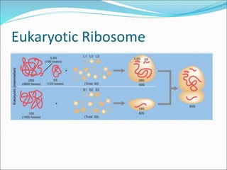
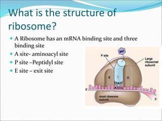
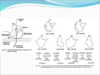
Dr.P.Nithiya
Assistant Professor
Department of Botany
Seethalakshmi Ramaswami College
Trichirapalli-2
Introduction
• Cells need to make proteins.
• Enzymes made of proteins are used to help speed up
biological processes.
• Other proteins support cell functions and are found
embedded in membranes.
• Proteins even make up most of your hair.
• When a cell needs to make proteins, it looks for
ribosomes.
• Ribosomes are the protein builders or the
protein synthesizers of the cell.
• They are like construction guys who connect one amino
acid at a time and build long chains.
Ribosome
• The ribosome is a complex molecular machine, found
within all living cells, that serves as the site
of biological protein synthesis (translation).
• Ribosomes link amino acids together in the order
specified by messenger RNA (mRNA) molecules.
• Ribosomes consist of two major components: the small
ribosomal subunits, which read the RNA, and the large
subunits, which join amino acids to form
a polypeptide chain.
• Each subunit comprises one or more ribosomal
RNA (rRNA) molecules and a variety of ribosomal
proteins (r-protein or rProtein).
• The ribosomes and associated molecules are also
known as the translational apparatus.
Discovery
 Ribosomes were first observed in the mid-1950s
by Romanian-American cell biologist George Emil
Palade, using an electron microscope, as dense
particles or granules. So it is also called Palade particle
 The term "ribosome" was proposed by scientist
Richard B. Roberts in the end of 1950s
George Emil Palade
Svedberg
 It is the centrifugal unit
depending on the density
of the object (and in the
cage of cell, organelles)
determining that how
quickly they sink to the
depth when centrifuged
Centrifugal force
The structures of ribosomes
include:
• Situated in two areas of the cytoplasm.
• They are seen scattered in the cytoplasm and a few are
connected to the endoplasmic reticulum.
• Whenever joined to the ER they are called the rough
endoplasmic reticulum.
• The free and the bound ribosomes are very much alike
in structure and are associated with protein synthesis.
• Around 37 to 62% of RNA is comprised of RNA and
the rest is proteins.
General structure of Ribosome
 The general structures of prokaryotic and eukaryotic
ribosomes are similar.
 Each ribosome is porous, hydrated and composed of
two subunits.
 One subunit is large in size and has a dome-like shape,
while the other subunit is smaller in size and occurring
above the larger subunit, forming a cap-like structure.
 When the concentration of Mg++ reduces in the
matrix, both ribosomal subunits get separated.
 In bacterial cells, the two subunits are found to occur
freely in the cytoplasm and they unite only during
protein synthesis.
Prokaryotic Ribosome
• Prokaryotes have 70S ribosomes, each consisting of a
small (30S) and a large (50S) subunit.
• Their small subunit has a 16S rRNA subunit
(consisting of 1540 nucleotides) bound to 21 proteins.
• The large subunit is composed of a 5S rRNA subunit
(120 nucleotides), a 23S rRNA subunit (2900
nucleotides) and 31 proteins
Prokaryotic ribosome
Models of 70S Ribosome:
• A. Quasi-symmetrical Model (Stoffler and Wittmann,
1970):
• According to this model, the smaller 30S subunit of prokaryote
ribosomes has a bipartite structure with an elongated, slightly
bent pro-late shape.
• A transverse cleft divides the subunit into two parts; a smaller
head and a larger body, giving it the appearance of a ‘telephone
receiver or embryo’.
• Under electron microscope, the frontal view of larger subunit
shows three protuberances (Fig. 3.17) arising from a rounded
base; the central being the most prominent and often gives the
larger subunit the appearance of an arm chair (the rounded base
forms the vaulted seat, the central protuberance forms the back
and the lateral protuberances the arm of the chair) or a maple
leaf.
ii. Asymmetrical Model (J. A. Lake,
1981):
• According to this model, the smaller subunit has a
head, a base and a platform (Fig. 3.18).
• The platform separates the head from the base by a
cleft.
• The cleft is an important functional region the site of
codonanticodon interaction and part of binding site
for the initiation factor of protein synthesis.
• On the other hand, the large subunit consists of a
ridge, a central protuberance and a stalk.
• The first two are separated with the help of a valley.
Eukaryotic ribosome
• Eukaryotes have 80S ribosomes, each consisting of a
small (40S) and large (60S) subunit.
• Their 40S subunit has an 18S rRNA (1900 nucleotides)
and 33 proteins.
• The large subunit is composed of a 5S rRNA (120
nucleotides), 28S rRNA (4700 nucleotides), a 5.8S
rRNA (160 nucleotides) subunits and 46 proteins
Eukaryotic Ribosome
What is the structure of
ribosome?
 A Ribosome has an mRNA binding site and three
binding site
 A site- aminoacyl site
 P site –Peptidyl site
 E site – exit site
Three Dimensional Model of 80S
Ribosome:
• The cytoplasmic ribosomes of eukaryotes are remarkably
similar in morphology to those of the prokaryotes.
• Like the 30S subunit of prokaryotes, the 40S subunit of
eukaryotes is divided into head and base segments by a
transverse groove.
• The 60S subunit is rounded in shape, although its one side
is flattened and it becomes confluent with the small
subunit during the formation of the monomer.
• A ribosome contains three binding site for RNA molecules.
• One for mRNA and two for tRNAs; one of the last two is
called the peptidyl-tRNA binding site, or P-site and the
other is called the aminoacyl-tRNA binding site, or A-site.
• The 80S ribosomes contain four types of rRNA, viz.,
– 28S rRNA with 4,700 nucleotides,
– 5.8S rRNA with 160 nucleotides and
– 5S rRNA with 120 nucleotides
– in the larger 60S subunit and the smaller subunit of 40S contains 18S rRNA with
1,900 nucleotides
• The 55S ribosomes of mammalian mitochondria lack 5S rRNA but contain 21S
and 12S rRNAs of which 21S rRNA occurs in the larger or 35S subunit, while 12S
rRNA occurs in the smaller or 25S subunit.
• About 60 percent of the rRNA is helical or double stranded and contains paired
bases.
• These regions are due to hairpin loops between complementary regions of the
linear molecules.
• In ribosome, the RNA is exposed at the surface of the ribosomal subunits and
the protein is assumed to be in the interior, in relation to non-helical part of
the RNA.
Reference:
 http://www.notesonzoology.com/cytology/ribosome-
meaning-types-and-structure/2174

Ribosome ultra structure

  • 1.
    Dr.P.Nithiya Assistant Professor Department ofBotany Seethalakshmi Ramaswami College Trichirapalli-2
  • 2.
    Introduction • Cells needto make proteins. • Enzymes made of proteins are used to help speed up biological processes. • Other proteins support cell functions and are found embedded in membranes. • Proteins even make up most of your hair. • When a cell needs to make proteins, it looks for ribosomes. • Ribosomes are the protein builders or the protein synthesizers of the cell. • They are like construction guys who connect one amino acid at a time and build long chains.
  • 3.
    Ribosome • The ribosomeis a complex molecular machine, found within all living cells, that serves as the site of biological protein synthesis (translation). • Ribosomes link amino acids together in the order specified by messenger RNA (mRNA) molecules. • Ribosomes consist of two major components: the small ribosomal subunits, which read the RNA, and the large subunits, which join amino acids to form a polypeptide chain.
  • 4.
    • Each subunitcomprises one or more ribosomal RNA (rRNA) molecules and a variety of ribosomal proteins (r-protein or rProtein). • The ribosomes and associated molecules are also known as the translational apparatus.
  • 5.
    Discovery  Ribosomes werefirst observed in the mid-1950s by Romanian-American cell biologist George Emil Palade, using an electron microscope, as dense particles or granules. So it is also called Palade particle  The term "ribosome" was proposed by scientist Richard B. Roberts in the end of 1950s
  • 6.
  • 7.
    Svedberg  It isthe centrifugal unit depending on the density of the object (and in the cage of cell, organelles) determining that how quickly they sink to the depth when centrifuged
  • 8.
  • 9.
    The structures ofribosomes include: • Situated in two areas of the cytoplasm. • They are seen scattered in the cytoplasm and a few are connected to the endoplasmic reticulum. • Whenever joined to the ER they are called the rough endoplasmic reticulum. • The free and the bound ribosomes are very much alike in structure and are associated with protein synthesis. • Around 37 to 62% of RNA is comprised of RNA and the rest is proteins.
  • 10.
    General structure ofRibosome  The general structures of prokaryotic and eukaryotic ribosomes are similar.  Each ribosome is porous, hydrated and composed of two subunits.  One subunit is large in size and has a dome-like shape, while the other subunit is smaller in size and occurring above the larger subunit, forming a cap-like structure.
  • 11.
     When theconcentration of Mg++ reduces in the matrix, both ribosomal subunits get separated.  In bacterial cells, the two subunits are found to occur freely in the cytoplasm and they unite only during protein synthesis.
  • 12.
    Prokaryotic Ribosome • Prokaryoteshave 70S ribosomes, each consisting of a small (30S) and a large (50S) subunit. • Their small subunit has a 16S rRNA subunit (consisting of 1540 nucleotides) bound to 21 proteins. • The large subunit is composed of a 5S rRNA subunit (120 nucleotides), a 23S rRNA subunit (2900 nucleotides) and 31 proteins
  • 13.
  • 14.
    Models of 70SRibosome: • A. Quasi-symmetrical Model (Stoffler and Wittmann, 1970): • According to this model, the smaller 30S subunit of prokaryote ribosomes has a bipartite structure with an elongated, slightly bent pro-late shape. • A transverse cleft divides the subunit into two parts; a smaller head and a larger body, giving it the appearance of a ‘telephone receiver or embryo’. • Under electron microscope, the frontal view of larger subunit shows three protuberances (Fig. 3.17) arising from a rounded base; the central being the most prominent and often gives the larger subunit the appearance of an arm chair (the rounded base forms the vaulted seat, the central protuberance forms the back and the lateral protuberances the arm of the chair) or a maple leaf.
  • 16.
    ii. Asymmetrical Model(J. A. Lake, 1981): • According to this model, the smaller subunit has a head, a base and a platform (Fig. 3.18). • The platform separates the head from the base by a cleft. • The cleft is an important functional region the site of codonanticodon interaction and part of binding site for the initiation factor of protein synthesis. • On the other hand, the large subunit consists of a ridge, a central protuberance and a stalk. • The first two are separated with the help of a valley.
  • 18.
    Eukaryotic ribosome • Eukaryoteshave 80S ribosomes, each consisting of a small (40S) and large (60S) subunit. • Their 40S subunit has an 18S rRNA (1900 nucleotides) and 33 proteins. • The large subunit is composed of a 5S rRNA (120 nucleotides), 28S rRNA (4700 nucleotides), a 5.8S rRNA (160 nucleotides) subunits and 46 proteins
  • 19.
  • 20.
    What is thestructure of ribosome?  A Ribosome has an mRNA binding site and three binding site  A site- aminoacyl site  P site –Peptidyl site  E site – exit site
  • 21.
    Three Dimensional Modelof 80S Ribosome: • The cytoplasmic ribosomes of eukaryotes are remarkably similar in morphology to those of the prokaryotes. • Like the 30S subunit of prokaryotes, the 40S subunit of eukaryotes is divided into head and base segments by a transverse groove. • The 60S subunit is rounded in shape, although its one side is flattened and it becomes confluent with the small subunit during the formation of the monomer. • A ribosome contains three binding site for RNA molecules. • One for mRNA and two for tRNAs; one of the last two is called the peptidyl-tRNA binding site, or P-site and the other is called the aminoacyl-tRNA binding site, or A-site.
  • 23.
    • The 80Sribosomes contain four types of rRNA, viz., – 28S rRNA with 4,700 nucleotides, – 5.8S rRNA with 160 nucleotides and – 5S rRNA with 120 nucleotides – in the larger 60S subunit and the smaller subunit of 40S contains 18S rRNA with 1,900 nucleotides • The 55S ribosomes of mammalian mitochondria lack 5S rRNA but contain 21S and 12S rRNAs of which 21S rRNA occurs in the larger or 35S subunit, while 12S rRNA occurs in the smaller or 25S subunit. • About 60 percent of the rRNA is helical or double stranded and contains paired bases. • These regions are due to hairpin loops between complementary regions of the linear molecules. • In ribosome, the RNA is exposed at the surface of the ribosomal subunits and the protein is assumed to be in the interior, in relation to non-helical part of the RNA.
  • 24.