Coronal advanced flap in combination with a connective tissue graft. Is the t...MD Abdul Haleem
Coronal advanced flap in combination with a connective tissue graft. Is the thickness of the flap a predictor for root coverage? - A prospective clinical study.
Department of Periodontology and Oral Implantology.
"A Journal Club Presentation"
Determination of root canal working length /certified fixed orthodontic cours...Indian dental academy
Welcome to Indian Dental Academy
The Indian Dental Academy is the Leader in continuing dental education , training dentists in all aspects of dentistry and offering a wide range of dental certified courses in different formats.
Indian dental academy has a unique training program & curriculum that provides students with exceptional clinical skills and enabling them to return to their office with high level confidence and start treating patients
State of the art comprehensive training-Faculty of world wide repute &Very affordable.
The denture-wearing history should provide information on the age of existing dentures, the frequency of denture replacement, the patient's experiences and expectations. It is important to identify whether any previous dentures have been successful as it may be suitable to copy features from a previously successful set. It will be important to manage expectations for those patients with a history of denture intolerance, yet technically satisfactory prostheses.
Clinical examination
Clinical examination should fully evaluate both the patient's anatomy and previous dentures to anticipate challenges and the potential to improve upon retention, stability, support, appearance and/or other factors. This should be undertaken in a systematic manner and would typically involve assessment of anatomy followed by an assessment of any existing dentures. This should follow a diagnostic process to determine if the patient presents with:
Technically adequate dentures on a favourable tissue base
Technically adequate dentures on an unfavourable tissue base
Technically inadequate dentures on a favourable tissue base
Technically inadequate dentures on an unfavourable tissue base.
Intentional replantation of maxillary second molar; case report and 15-year f...Abu-Hussein Muhamad
Abstract: Intentional reimplantation is a procedure in which tooth extraction is performed followed by reinsertion of the extracted tooth into its own socket after performing the desired procedure. In this article, intentional reimplantation is described and discussed as a treatment approach for aperiapical lesion that is in maxillary second molar. After 15 years, the patient was asymptomatic, the tooth was still functional and a recall intraoral periapical radiograph showed an intact periodontal ligament space and lamina dura with no evidence of gross root resorption or ankylosis.
Keywords: Intentional replantation, calcified canals, mineral trioxide aggregate
Objectives and rationale
Indications
Contraindications
False indications
Treatment planning and presurgical notes
Classification
Gutmann’s
Kim’s
Steps in endosurgery
Treatment planning & Presurgical notes
Mandatory investigations
Premedication
Local anaesthesia and hemostasis
Flap
Requirements of an ideal flap
Flap design
Semilunar flap
Vertical flaps
Horizontal flap
Ochsenbein-Luebke flap
Two-step or filling first technique
Disinfection immediately prior to filling
Preparation of surgical site
Soft tissue management
Opening the flap
Flap elevation
Flap retraction
Hard tissue considerations
Locating root apex
Osteotomy
Apical curettage
Apical rood end resection
Surgery from palatal access
Post-resection filling
Root end preparation
Root end filling materials
Reverse filling
Surgery for root fractures
Surgical management of internal resorption
Radisectomy and hemisection
Intentional replantation
Closure of surgical area
Repositioning of flap and compression
Needle selection
Suturing
Post surgical care
Being a Periodontist, what necessary is to know what actually periodontal flaps are. So this presentation might provide you an insight into the field of periodontics as well as periodontal flaps.
Mahendra Azad et al. GAINT ODONTOGENIC KERATOCYST OF MANDIBLE OPERATED UNDER LOCAL ANESTHESIA- A CASE REPORT. JOURNAL OF DENTAL HEALTH & RESEARCH (VOL. 1, ISSUE 2, JUL - DEC 2020): 24-2
Reconstructive periodontal surgery aims to treat deep pockets which have not be reduced after non surgical periodontal therapy. periodontal regenerative procedures mainly include the use of modified flap techniques , use of bone grafts and newer gene therapies. Biologic mediators play key role in the regeneration process. Guided tissue regeneration and Guided Bone regeneration are commonly used methods for periodontal regeneration. Minimally invasive surgical techniques are preferred surgical methods for treating deep infrabony pockets
Ethanol (CH3CH2OH), or beverage alcohol, is a two-carbon alcohol
that is rapidly distributed in the body and brain. Ethanol alters many
neurochemical systems and has rewarding and addictive properties. It
is the oldest recreational drug and likely contributes to more morbidity,
mortality, and public health costs than all illicit drugs combined. The
5th edition of the Diagnostic and Statistical Manual of Mental Disorders
(DSM-5) integrates alcohol abuse and alcohol dependence into a single
disorder called alcohol use disorder (AUD), with mild, moderate,
and severe subclassifications (American Psychiatric Association, 2013).
In the DSM-5, all types of substance abuse and dependence have been
combined into a single substance use disorder (SUD) on a continuum
from mild to severe. A diagnosis of AUD requires that at least two of
the 11 DSM-5 behaviors be present within a 12-month period (mild
AUD: 2–3 criteria; moderate AUD: 4–5 criteria; severe AUD: 6–11 criteria).
The four main behavioral effects of AUD are impaired control over
drinking, negative social consequences, risky use, and altered physiological
effects (tolerance, withdrawal). This chapter presents an overview
of the prevalence and harmful consequences of AUD in the U.S.,
the systemic nature of the disease, neurocircuitry and stages of AUD,
comorbidities, fetal alcohol spectrum disorders, genetic risk factors, and
pharmacotherapies for AUD.
Title: Sense of Smell
Presenter: Dr. Faiza, Assistant Professor of Physiology
Qualifications:
MBBS (Best Graduate, AIMC Lahore)
FCPS Physiology
ICMT, CHPE, DHPE (STMU)
MPH (GC University, Faisalabad)
MBA (Virtual University of Pakistan)
Learning Objectives:
Describe the primary categories of smells and the concept of odor blindness.
Explain the structure and location of the olfactory membrane and mucosa, including the types and roles of cells involved in olfaction.
Describe the pathway and mechanisms of olfactory signal transmission from the olfactory receptors to the brain.
Illustrate the biochemical cascade triggered by odorant binding to olfactory receptors, including the role of G-proteins and second messengers in generating an action potential.
Identify different types of olfactory disorders such as anosmia, hyposmia, hyperosmia, and dysosmia, including their potential causes.
Key Topics:
Olfactory Genes:
3% of the human genome accounts for olfactory genes.
400 genes for odorant receptors.
Olfactory Membrane:
Located in the superior part of the nasal cavity.
Medially: Folds downward along the superior septum.
Laterally: Folds over the superior turbinate and upper surface of the middle turbinate.
Total surface area: 5-10 square centimeters.
Olfactory Mucosa:
Olfactory Cells: Bipolar nerve cells derived from the CNS (100 million), with 4-25 olfactory cilia per cell.
Sustentacular Cells: Produce mucus and maintain ionic and molecular environment.
Basal Cells: Replace worn-out olfactory cells with an average lifespan of 1-2 months.
Bowman’s Gland: Secretes mucus.
Stimulation of Olfactory Cells:
Odorant dissolves in mucus and attaches to receptors on olfactory cilia.
Involves a cascade effect through G-proteins and second messengers, leading to depolarization and action potential generation in the olfactory nerve.
Quality of a Good Odorant:
Small (3-20 Carbon atoms), volatile, water-soluble, and lipid-soluble.
Facilitated by odorant-binding proteins in mucus.
Membrane Potential and Action Potential:
Resting membrane potential: -55mV.
Action potential frequency in the olfactory nerve increases with odorant strength.
Adaptation Towards the Sense of Smell:
Rapid adaptation within the first second, with further slow adaptation.
Psychological adaptation greater than receptor adaptation, involving feedback inhibition from the central nervous system.
Primary Sensations of Smell:
Camphoraceous, Musky, Floral, Pepperminty, Ethereal, Pungent, Putrid.
Odor Detection Threshold:
Examples: Hydrogen sulfide (0.0005 ppm), Methyl-mercaptan (0.002 ppm).
Some toxic substances are odorless at lethal concentrations.
Characteristics of Smell:
Odor blindness for single substances due to lack of appropriate receptor protein.
Behavioral and emotional influences of smell.
Transmission of Olfactory Signals:
From olfactory cells to glomeruli in the olfactory bulb, involving lateral inhibition.
Primitive, less old, and new olfactory systems with different path
Ozempic: Preoperative Management of Patients on GLP-1 Receptor Agonists Saeid Safari
Preoperative Management of Patients on GLP-1 Receptor Agonists like Ozempic and Semiglutide
ASA GUIDELINE
NYSORA Guideline
2 Case Reports of Gastric Ultrasound
Lung Cancer: Artificial Intelligence, Synergetics, Complex System Analysis, S...Oleg Kshivets
RESULTS: Overall life span (LS) was 2252.1±1742.5 days and cumulative 5-year survival (5YS) reached 73.2%, 10 years – 64.8%, 20 years – 42.5%. 513 LCP lived more than 5 years (LS=3124.6±1525.6 days), 148 LCP – more than 10 years (LS=5054.4±1504.1 days).199 LCP died because of LC (LS=562.7±374.5 days). 5YS of LCP after bi/lobectomies was significantly superior in comparison with LCP after pneumonectomies (78.1% vs.63.7%, P=0.00001 by log-rank test). AT significantly improved 5YS (66.3% vs. 34.8%) (P=0.00000 by log-rank test) only for LCP with N1-2. Cox modeling displayed that 5YS of LCP significantly depended on: phase transition (PT) early-invasive LC in terms of synergetics, PT N0—N12, cell ratio factors (ratio between cancer cells- CC and blood cells subpopulations), G1-3, histology, glucose, AT, blood cell circuit, prothrombin index, heparin tolerance, recalcification time (P=0.000-0.038). Neural networks, genetic algorithm selection and bootstrap simulation revealed relationships between 5YS and PT early-invasive LC (rank=1), PT N0—N12 (rank=2), thrombocytes/CC (3), erythrocytes/CC (4), eosinophils/CC (5), healthy cells/CC (6), lymphocytes/CC (7), segmented neutrophils/CC (8), stick neutrophils/CC (9), monocytes/CC (10); leucocytes/CC (11). Correct prediction of 5YS was 100% by neural networks computing (area under ROC curve=1.0; error=0.0).
CONCLUSIONS: 5YS of LCP after radical procedures significantly depended on: 1) PT early-invasive cancer; 2) PT N0--N12; 3) cell ratio factors; 4) blood cell circuit; 5) biochemical factors; 6) hemostasis system; 7) AT; 8) LC characteristics; 9) LC cell dynamics; 10) surgery type: lobectomy/pneumonectomy; 11) anthropometric data. Optimal diagnosis and treatment strategies for LC are: 1) screening and early detection of LC; 2) availability of experienced thoracic surgeons because of complexity of radical procedures; 3) aggressive en block surgery and adequate lymph node dissection for completeness; 4) precise prediction; 5) adjuvant chemoimmunoradiotherapy for LCP with unfavorable prognosis.
263778731218 Abortion Clinic /Pills In Harare ,sisternakatoto
263778731218 Abortion Clinic /Pills In Harare ,ABORTION WOMEN’S CLINIC +27730423979 IN women clinic we believe that every woman should be able to make choices in her pregnancy. Our job is to provide compassionate care, safety,affordable and confidential services. That’s why we have won the trust from all generations of women all over the world. we use non surgical method(Abortion pills) to terminate…Dr.LISA +27730423979women Clinic is committed to providing the highest quality of obstetrical and gynecological care to women of all ages. Our dedicated staff aim to treat each patient and her health concerns with compassion and respect.Our dedicated group ABORTION WOMEN’S CLINIC +27730423979 IN women clinic we believe that every woman should be able to make choices in her pregnancy. Our job is to provide compassionate care, safety,affordable and confidential services. That’s why we have won the trust from all generations of women all over the world. we use non surgical method(Abortion pills) to terminate…Dr.LISA +27730423979women Clinic is committed to providing the highest quality of obstetrical and gynecological care to women of all ages. Our dedicated staff aim to treat each patient and her health concerns with compassion and respect.Our dedicated group of receptionists, nurses, and physicians have worked together as a teamof receptionists, nurses, and physicians have worked together as a team wwww.lisywomensclinic.co.za/
HOT NEW PRODUCT! BIG SALES FAST SHIPPING NOW FROM CHINA!! EU KU DB BK substit...GL Anaacs
Contact us if you are interested:
Email / Skype : kefaya1771@gmail.com
Threema: PXHY5PDH
New BATCH Ku !!! MUCH IN DEMAND FAST SALE EVERY BATCH HAPPY GOOD EFFECT BIG BATCH !
Contact me on Threema or skype to start big business!!
Hot-sale products:
NEW HOT EUTYLONE WHITE CRYSTAL!!
5cl-adba precursor (semi finished )
5cl-adba raw materials
ADBB precursor (semi finished )
ADBB raw materials
APVP powder
5fadb/4f-adb
Jwh018 / Jwh210
Eutylone crystal
Protonitazene (hydrochloride) CAS: 119276-01-6
Flubrotizolam CAS: 57801-95-3
Metonitazene CAS: 14680-51-4
Payment terms: Western Union,MoneyGram,Bitcoin or USDT.
Deliver Time: Usually 7-15days
Shipping method: FedEx, TNT, DHL,UPS etc.Our deliveries are 100% safe, fast, reliable and discreet.
Samples will be sent for your evaluation!If you are interested in, please contact me, let's talk details.
We specializes in exporting high quality Research chemical, medical intermediate, Pharmaceutical chemicals and so on. Products are exported to USA, Canada, France, Korea, Japan,Russia, Southeast Asia and other countries.
Tom Selleck Health: A Comprehensive Look at the Iconic Actor’s Wellness Journeygreendigital
Tom Selleck, an enduring figure in Hollywood. has captivated audiences for decades with his rugged charm, iconic moustache. and memorable roles in television and film. From his breakout role as Thomas Magnum in Magnum P.I. to his current portrayal of Frank Reagan in Blue Bloods. Selleck's career has spanned over 50 years. But beyond his professional achievements. fans have often been curious about Tom Selleck Health. especially as he has aged in the public eye.
Follow us on: Pinterest
Introduction
Many have been interested in Tom Selleck health. not only because of his enduring presence on screen but also because of the challenges. and lifestyle choices he has faced and made over the years. This article delves into the various aspects of Tom Selleck health. exploring his fitness regimen, diet, mental health. and the challenges he has encountered as he ages. We'll look at how he maintains his well-being. the health issues he has faced, and his approach to ageing .
Early Life and Career
Childhood and Athletic Beginnings
Tom Selleck was born on January 29, 1945, in Detroit, Michigan, and grew up in Sherman Oaks, California. From an early age, he was involved in sports, particularly basketball. which played a significant role in his physical development. His athletic pursuits continued into college. where he attended the University of Southern California (USC) on a basketball scholarship. This early involvement in sports laid a strong foundation for his physical health and disciplined lifestyle.
Transition to Acting
Selleck's transition from an athlete to an actor came with its physical demands. His first significant role in "Magnum P.I." required him to perform various stunts and maintain a fit appearance. This role, which he played from 1980 to 1988. necessitated a rigorous fitness routine to meet the show's demands. setting the stage for his long-term commitment to health and wellness.
Fitness Regimen
Workout Routine
Tom Selleck health and fitness regimen has evolved. adapting to his changing roles and age. During his "Magnum, P.I." days. Selleck's workouts were intense and focused on building and maintaining muscle mass. His routine included weightlifting, cardiovascular exercises. and specific training for the stunts he performed on the show.
Selleck adjusted his fitness routine as he aged to suit his body's needs. Today, his workouts focus on maintaining flexibility, strength, and cardiovascular health. He incorporates low-impact exercises such as swimming, walking, and light weightlifting. This balanced approach helps him stay fit without putting undue strain on his joints and muscles.
Importance of Flexibility and Mobility
In recent years, Selleck has emphasized the importance of flexibility and mobility in his fitness regimen. Understanding the natural decline in muscle mass and joint flexibility with age. he includes stretching and yoga in his routine. These practices help prevent injuries, improve posture, and maintain mobilit
New Drug Discovery and Development .....NEHA GUPTA
The "New Drug Discovery and Development" process involves the identification, design, testing, and manufacturing of novel pharmaceutical compounds with the aim of introducing new and improved treatments for various medical conditions. This comprehensive endeavor encompasses various stages, including target identification, preclinical studies, clinical trials, regulatory approval, and post-market surveillance. It involves multidisciplinary collaboration among scientists, researchers, clinicians, regulatory experts, and pharmaceutical companies to bring innovative therapies to market and address unmet medical needs.
These simplified slides by Dr. Sidra Arshad present an overview of the non-respiratory functions of the respiratory tract.
Learning objectives:
1. Enlist the non-respiratory functions of the respiratory tract
2. Briefly explain how these functions are carried out
3. Discuss the significance of dead space
4. Differentiate between minute ventilation and alveolar ventilation
5. Describe the cough and sneeze reflexes
Study Resources:
1. Chapter 39, Guyton and Hall Textbook of Medical Physiology, 14th edition
2. Chapter 34, Ganong’s Review of Medical Physiology, 26th edition
3. Chapter 17, Human Physiology by Lauralee Sherwood, 9th edition
4. Non-respiratory functions of the lungs https://academic.oup.com/bjaed/article/13/3/98/278874
Flu Vaccine Alert in Bangalore Karnatakaaddon Scans
As flu season approaches, health officials in Bangalore, Karnataka, are urging residents to get their flu vaccinations. The seasonal flu, while common, can lead to severe health complications, particularly for vulnerable populations such as young children, the elderly, and those with underlying health conditions.
Dr. Vidisha Kumari, a leading epidemiologist in Bangalore, emphasizes the importance of getting vaccinated. "The flu vaccine is our best defense against the influenza virus. It not only protects individuals but also helps prevent the spread of the virus in our communities," he says.
This year, the flu season is expected to coincide with a potential increase in other respiratory illnesses. The Karnataka Health Department has launched an awareness campaign highlighting the significance of flu vaccinations. They have set up multiple vaccination centers across Bangalore, making it convenient for residents to receive their shots.
To encourage widespread vaccination, the government is also collaborating with local schools, workplaces, and community centers to facilitate vaccination drives. Special attention is being given to ensuring that the vaccine is accessible to all, including marginalized communities who may have limited access to healthcare.
Residents are reminded that the flu vaccine is safe and effective. Common side effects are mild and may include soreness at the injection site, mild fever, or muscle aches. These side effects are generally short-lived and far less severe than the flu itself.
Healthcare providers are also stressing the importance of continuing COVID-19 precautions. Wearing masks, practicing good hand hygiene, and maintaining social distancing are still crucial, especially in crowded places.
Protect yourself and your loved ones by getting vaccinated. Together, we can help keep Bangalore healthy and safe this flu season. For more information on vaccination centers and schedules, residents can visit the Karnataka Health Department’s official website or follow their social media pages.
Stay informed, stay safe, and get your flu shot today!
Acute scrotum is a general term referring to an emergency condition affecting the contents or the wall of the scrotum.
There are a number of conditions that present acutely, predominantly with pain and/or swelling
A careful and detailed history and examination, and in some cases, investigations allow differentiation between these diagnoses. A prompt diagnosis is essential as the patient may require urgent surgical intervention
Testicular torsion refers to twisting of the spermatic cord, causing ischaemia of the testicle.
Testicular torsion results from inadequate fixation of the testis to the tunica vaginalis producing ischemia from reduced arterial inflow and venous outflow obstruction.
The prevalence of testicular torsion in adult patients hospitalized with acute scrotal pain is approximately 25 to 50 percent
Couples presenting to the infertility clinic- Do they really have infertility...Sujoy Dasgupta
Dr Sujoy Dasgupta presented the study on "Couples presenting to the infertility clinic- Do they really have infertility? – The unexplored stories of non-consummation" in the 13th Congress of the Asia Pacific Initiative on Reproduction (ASPIRE 2024) at Manila on 24 May, 2024.
micro teaching on communication m.sc nursing.pdfAnurag Sharma
Microteaching is a unique model of practice teaching. It is a viable instrument for the. desired change in the teaching behavior or the behavior potential which, in specified types of real. classroom situations, tends to facilitate the achievement of specified types of objectives.
New Directions in Targeted Therapeutic Approaches for Older Adults With Mantl...i3 Health
i3 Health is pleased to make the speaker slides from this activity available for use as a non-accredited self-study or teaching resource.
This slide deck presented by Dr. Kami Maddocks, Professor-Clinical in the Division of Hematology and
Associate Division Director for Ambulatory Operations
The Ohio State University Comprehensive Cancer Center, will provide insight into new directions in targeted therapeutic approaches for older adults with mantle cell lymphoma.
STATEMENT OF NEED
Mantle cell lymphoma (MCL) is a rare, aggressive B-cell non-Hodgkin lymphoma (NHL) accounting for 5% to 7% of all lymphomas. Its prognosis ranges from indolent disease that does not require treatment for years to very aggressive disease, which is associated with poor survival (Silkenstedt et al, 2021). Typically, MCL is diagnosed at advanced stage and in older patients who cannot tolerate intensive therapy (NCCN, 2022). Although recent advances have slightly increased remission rates, recurrence and relapse remain very common, leading to a median overall survival between 3 and 6 years (LLS, 2021). Though there are several effective options, progress is still needed towards establishing an accepted frontline approach for MCL (Castellino et al, 2022). Treatment selection and management of MCL are complicated by the heterogeneity of prognosis, advanced age and comorbidities of patients, and lack of an established standard approach for treatment, making it vital that clinicians be familiar with the latest research and advances in this area. In this activity chaired by Michael Wang, MD, Professor in the Department of Lymphoma & Myeloma at MD Anderson Cancer Center, expert faculty will discuss prognostic factors informing treatment, the promising results of recent trials in new therapeutic approaches, and the implications of treatment resistance in therapeutic selection for MCL.
Target Audience
Hematology/oncology fellows, attending faculty, and other health care professionals involved in the treatment of patients with mantle cell lymphoma (MCL).
Learning Objectives
1.) Identify clinical and biological prognostic factors that can guide treatment decision making for older adults with MCL
2.) Evaluate emerging data on targeted therapeutic approaches for treatment-naive and relapsed/refractory MCL and their applicability to older adults
3.) Assess mechanisms of resistance to targeted therapies for MCL and their implications for treatment selection
New Directions in Targeted Therapeutic Approaches for Older Adults With Mantl...
Periodontal flaps
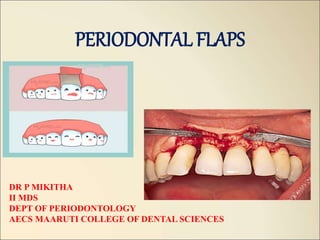
1. PERIODONTAL FLAPS
DR P MIKITHA
II MDS
DEPT OF PERIODONTOLOGY
AECS MAARUTI COLLEGE OF DENTAL SCIENCES
2. Contents
• Introduction
• Definition
• Historical background
• Objectives of flap surgery
• Indications and contraindications
• Advantages and disadvantages
• Principle of flap design
• Classification of flap
• Properties of ideal flap
• Types of incisions
• Different flap techniques
• Healing after flap surgery
• Factors affecting the outcome of
flap surgery
• Conclusion
• references
3. INTRODUCTION
• The ultimate aim of periodontal therapy is to establish a healthy dentition
with sound attachment appartus resulting in proper frrm, function and
esthetics.
• To achieve this goal many non-surgical and surgical techniques have been
proposed to treat a variety of periodontal conditions most commonly the
periodontal pocket.
• Periodontal therapy comprises of initial non-surgical debridement followed
by a re-evaluation at which stage the need for further treatment, usually
surgical in nature is established.
4. DEFINITIONS
• Periodontal flap is defined as a section of gingiva and/or mucosa surgically
separated from the underlying tissues to provide visibility of and access to
the bone and root surface. -(Carranza 10th edition).
• Flap is defined as the separation of a section of tissue from the surrounding
tissue except at its base, -(Glossary of periodontal terms)
• A flap is defined as a mass of tissue, usually including skin, only partially
removed from one part of the body so that it retains its own blood supply
during transfer to another site. –(Dorland’s medical dictionary).
Takei H, Carranza FA, Shin K. the flap technique for pocket therapy In: carranza’s clinical periodontolgy, Elsevier, 12, 2012; 593- 603.
5. Carl Partch- 19th century -1907 Partch incison
Robert Neumann- 1912 1st introduced mucoperiosteal flap –”Neumann
flap”
Leonard Widman – 1918 Modified the Neumann flap – “Widmann flap”
Cieszynski- 1918 Reverse bevel incision
Kirkland – 1931 Modified flap procedure
Carranza – 1939 Surgical treatment of periodontitis
Nabers – 1954 Introduced “repositioning of attached gingiva”
Ariaudo and Tyrrell – 1957 Modified Nabers procedure
Friedman- 1962 Apically positioned flap
Oschenbein and Bohannan – 1964 Palatal flap
Morris – 1965 Unrepositioned mucoperiosteal flap
Ramjford and Nissle – 1974 Modified Widmann flap
Takei et al- 1985 Pappila preservation flap
Trombelli et al – 2007 Single flap approach (SFA)
Bianchi and Bassetti - 2009 Whales technique
Takei H, Carranza FA, Shin K. the flap technique for pocket therapy In: carranza’s clinical periodontolgy, Elsevier, 12, 2012; 593- 603.
6. Objectives of flap surgery
MAIN OBJECTIVE of periodontal
surgery is to contribute to the long-
term preservation of the
periodontium by facilitating plaque
removal and plaque control.
-Jan Lindhe
To enable visual
instrumentation of root
surfaces
To re-establish the healthy,
clinical status of
periodontium with long term
maintenance
To restore the periodontal
apparatus when attachment
loss has occurred
Cohen SE. fundamental of surgical therapy. In: Atlas of cosmetic and reconstructive periodontal surgery, BC Decker Inc, 3, 2007; 56-72.
7. 1. Access to roots and alveolar bone
2. Modification of osseous defects:
• estabilish physiologic architecture of hard tissues through regeneration or
resection
• Augment alveolar ridge defects
3. Repair or regeneration of the periodontium
4. Pocket reduction:
• Enhance maintenance by patient and therapist
• Improves long term stability
5. Provide acceptable soft tissue contours
• Enhances plaque control measures
• Improve esthetics
Cohen SE. fundamental of surgical therapy. In: Atlas of cosmetic and reconstructive periodontal surgery, BC Decker Inc, 3, 2007; 56-72.
8. indications
• Irregular bony contours
• Deep craters
• Pockets on teeth in which a complete removal of root irritants is not clinically
possible
• Grade II or III furcation involvemnet
• Root resection/ hemisection
• Intrabony pockets on distal areas of last molars
Takei H, Carranza FA, Shin K. the flap technique for pocket therapy In: carranza’s clinical periodontolgy, Elsevier, 12, 2012; 593- 603.
9. • Persistent inflammation in aresas with moderate to deep pockets
• Unaccesible areas like root concavities, furctaion areas, etc
• Deep periodontal pockets: Waerhaug stated that pocket depth greater tha
5mm demonstrated onlu an 11% efficacy in removal of plaque and
calculus.
• Osseous defect: morphology of osseous defects can limit the effectiveness
of non-surgical therapy
Takei H, Carranza FA, Shin K. the flap technique for pocket therapy In: carranza’s clinical periodontolgy, Elsevier, 12, 2012; 593- 603.
10. contraindications
A. Patient non co-operation
B. Poor plaque control
C. High caries rate
D. Systemic conditions:
Cardiovascular diseases:
Arterial HTN: patients consent should be taken and LA with adrenaline or
without adrenaline must be used
Angina pectoris: premedication with sedatives and LA, low in adrenaline is
recommended.
Takei H, Carranza FA, Shin K. the flap technique for pocket therapy In: carranza’s clinical periodontolgy, Elsevier, 12, 2012; 593- 603.
11. Myocardial infarction
Anticoagulant treatment: the range in which SRP and surgical procedures
can be safely performed is one and half to 2 times the avarage normal
prothrombin time (12-14 sec)
Aspirin and other NSAID drugs should not be used for post-op pain control
Rheumatic endicarditis, congenital heart lesions and heart/vascukakr
implants involve risk of transient bacteremia that follows manipulation of
infection periodontal pockets,
ADA- recommended antibiotic prophylaxis and antiseptic mouthrinsing
0.2% CHX prior to surgery
Cohen SE. fundamental of surgical therapy. In: Atlas of cosmetic and reconstructive periodontal surgery, BC Decker Inc, 3, 2007; 56-72.
12. Organ transplantation:
Prophylactic antibiotics are recommended in transplant patient taking
immunosuppressive drugs
Blood disorders:
Patients suffering from acute leukemia, agranulocytosis and
lymphogranulomatosis must not be subjected to periodontal surgery
Diabetes
Neurological disorders:
Multiple sclerosis and parkinsons disease make periodontal surgery impossible
Epilepsy
Drugs used to treat epilepsy may cause gingival enlargements. Theese patients
may withspecial restrictions be subjected to periodontal surgery.
Cohen SE. fundamental of surgical therapy. In: Atlas of cosmetic and reconstructive periodontal surgery, BC Decker Inc, 3, 2007; 56-72.
13. advantages
• Pocket epithelium is removed by the
inverse bevel incision
• The inter dental bone or infrabony
defects can be covered by the flaps
• No open wound persists
postoperatively
• Rapid healing.
• Less post operative discomfort and
fewer complications.
• Less post operative gingival
recession, therefore esthetic
• Less dentin exposure.
• Short surgical time
• Direct healing
disadvantages
• When flaps are repositioned
apically, the cervical areas of the
teeth are often exposed, long and
sensitive, due also to shrinkage of
the tissues.
• Possibility of deep periodontal
pockets remaining after surgery.
• Possibility of formation of post-
operative gingival craters in
proximal surface areas (especially
in molars).
• New attachment is unpredictable.
• Less regeneration achieved
compared to other regenerative
procedures.
Takei H, Carranza FA, Shin K. the flap technique for pocket therapy In: carranza’s clinical periodontolgy, Elsevier, 12, 2012; 593- 603.
14. Principles of flap design
• According to HUPP 1933 following principles should be followed:
Prevention of flap necrosis:
The apex of flap should never be wider than the base
Flap should either run parallel to each other or preferably converge from the
base of the flap to its apex
Flap length to base ratio should be no greater than 2:1
The major blood supply to a flap was found to exist at its base and travels in an
apical to coronal direction. So, it was also determined that the greater the ratio
of flap length to flap base, the greater the vascular compromise at the flap
margins.
Cohen SE. fundamental of surgical therapy. In: Atlas of cosmetic and reconstructive periodontal surgery, BC Decker Inc, 3, 2007; 56-72.
15. Whenever possible axial blood supply should be included in the base of the
flap
The base of the flap should not be excessively twisted or stretched (as either of
these will compromise the supplying vessels)
• Prevention of flap tearing:
The access of the flap should be enough to avoid tearing
If an envelope flap doesn’t provide sufficient access, another incision should be
made
Cohen SE. fundamental of surgical therapy. In: Atlas of cosmetic and reconstructive periodontal surgery, BC Decker Inc, 3, 2007; 56-72.
16. Vertical releasing incisions should be placed one full tooth anterior to the
area of any anticipated bone removal
The incision should be started at the line angle of the tooth and carried
obliquely apically into the unattached gingiva.
Takei H, Carranza FA, Shin K. the flap technique for pocket therapy In: carranza’s clinical periodontolgy, Elsevier, 12, 2012; 593- 603.
17. Properties of ideal flap
Ideal flap/section of soft tissue:
• Is outlined by a surgical incision
• Carries its own blood supply
• Allows surgical access to the underlying tissues
• Can be placed in the original position
• Can be maintained with sutures in a particular desired position
• Expected to heal
• Sharp incisions heal rapidly
• Flap extension- 2 teeth anterior and 1 tooth posterior to the area of surgery
• Incisions- over intact bone/ 6-8mm away from the diseased bone (Peterson)
Takei H, Carranza FA, Shin K. the flap technique for pocket therapy In: carranza’s clinical periodontolgy, Elsevier, 12, 2012; 593- 603.
18. Types of incision
Principles governing incision placement
According to LASKIN 1980:
• The incision should not be made over the operative site but in the adjacent, undisturbed areas
so that the flap will be supported by normal tissue and the potential for rapid
revascularization is preserved
• The incision should be placed do that major nerves are not transected unless necessary
• An adequate blood supply should be maintained by incising parallel to the major vessels
• Incisions should not be made in areas of thinned mucosa like that found over an exostosis
because the blood supply is reduced, suturing is difficult and rate of dehiscence is very high.
Cohen SE. fundamental of surgical therapy. In: Atlas of cosmetic and reconstructive periodontal surgery, BC Decker Inc, 3, 2007; 56-72.
19. • When developing flaps around teeth, incisions should be made in gingival
crevice.
• Important to maintain the integrity of the interdental papillae.
• If access is inadequate, the surgeon may extend the length of the incision or
make a releasing incision. The releasing incision is usually made at about an
angle of 45 degrees from the direction of the parent incision
• If the flap is to include both mucosa and the periosteum the incision should be
made directly to the bone with one cut and it should be elevated in one piece
without tearing the periosteum
• After the necessary surgery, the clotted blood should be removed from beneath
the flap to lessen the possibility of infection and permits tissue fluid to penetrate
more readily.
Cohen SE. fundamental of surgical therapy. In: Atlas of cosmetic and reconstructive periodontal surgery, BC Decker Inc, 3, 2007; 56-72.
20. Seven main incision types are commonly used in
periodontal surgery
• External bevel or gingivectomy incision
• Horizontal incision:
a. internal bevel incision
b. Crevicular incision/ sulcular incision
c. Interdental incision
• Vertical/ oblique releasing incision
• Cutback incisions
• Thinning incisions
• Distal wedge incisions
• Periosteal releasing incisions
Cohen SE. fundamental of surgical therapy. In: Atlas of cosmetic and reconstructive periodontal surgery, BC Decker Inc, 3, 2007; 56-72.
21. THE EXTERNAL BEVEL OR GINGIVECTOMY
INCISIONS
• It is contained in the gingiva and coronally
directed with the surgical objectives of pocket
elimination, access to roots and improved
gingival contours.
• Indications:
1. To treat gingival enlargement and to perform
esthetic crow lengthening
2. Used in conjunction with flap surgery when
there is need to thin the tissues externally
before flap reflection. Eg: severe gingival
enlargement with lobulated gingiva and highly
irregular gingival margins
Takei H, Carranza FA, Shin K. the flap technique for pocket therapy In: carranza’s clinical periodontolgy, Elsevier, 12, 2012; 593- 603.
22. Typesof horizontalincisons
a. The internal bevel incision- starts at a
distance from the gngival margin and is
aimed at the bone crest.
b. The crevicular incision- starts at the bottom
of the pocket and is directed to the bone
margins
c. The interdental incision- performed after the
flap is elevated
Takei H, Carranza FA, Shin K. the flap technique for pocket therapy In: carranza’s clinical periodontolgy, Elsevier, 12, 2012; 593- 603.
23. Internalbevelincision • First incision- it is initial incision in the reflection of
the flap
• Reverse bevel incision- its bevel is in reverse direct
from the gingivectomy imcison
• #11 or #15 surgical scalpel is used mostly
Objectives of internal bevel incision:
1. Removes pocket lining and areas of tissue invaded by
microorganisms.
2. Chief advantage- eliminates the part of the gingival
margin which has been penetrated by the pathogens
3. Conserves the relatively less involved outter surface
of gingiva
4. Produces sharp, thin flap margin for adaptation to the
bone tooth junction
Takei H, Carranza FA, Shin K. the flap technique for pocket therapy In: carranza’s clinical periodontolgy, Elsevier, 12, 2012; 593- 603.
24. Indications:
• Primary incision of the flap surgery if there is sufficient band of attavhed
gingiva
• Desire to correct bone morphology
• Thick gingiva
• Deep periodontal pockets and bone defect
• Desire to lengthen clinical crown
Incision design:
The placement of primary incision is determined by the following factors:
1. Band of attached gingiva
2. Method of periodontal surgery
3. Periodontal pocket depth
Takei H, Carranza FA, Shin K. the flap technique for pocket therapy In: carranza’s clinical periodontolgy, Elsevier, 12, 2012; 593- 603.
25. 4. Whether osteoplasty and ostectomy are
necessary
5. Esthetics
6. Whether restorative treatment is necessary
after periodontal surgery
7. Clinical crown length needed for abutment
Takei H, Carranza FA, Shin K. the flap technique for pocket therapy In: carranza’s clinical periodontolgy, Elsevier, 12, 2012; 593- 603.
26. Variations in the type of
internal bevel incision for
different types of flaps:
Modified widmann flap doesn’t intend to
remove the pocket wall, but eliminates pocket
lining. Starts close, no more than 1-2mm
apically to the gingival margin and follows the
normal scalloping of the gingival margin.
Apically displaced flap, pocket wall is to be
preserved; so, incision is to be made as close to
the tooth as possible 0.5- 1mm
Undisplaced flap, incision is initiated at or near
a point just coronal to the projection of the
bottom of the pocket on the outer surface of
gingiva
Takei H, Carranza FA, Shin K. the flap technique for pocket therapy In: carranza’s clinical periodontolgy, Elsevier, 12, 2012; 593- 603.
27. Studies in favor of the benefits of removal of pocket
epithelium by internal bevel incision
Morris
1949
•Removal of pocket epithelium is necessary for new CT attachment
Stone
1966
•Any residual epithelium on the wound edge could serve as a seed area and result in rapid proliferation of the JE along the root
surface
Yukna
1976
•Successfully removed all epithelium with internal bevel incision as described by ENAP
Caffesse
at al
1968
•Observed that all pocket epithelium was removed with the reverse bevel incision as described in the modified widman flap
procedure
carranza
•Stated that placement of the scalloped internal bevel incision 1mm subcrestally will remove most of the granulation tissue
contained in the lateral wall of pocket
28. Sulcularor crevicularincision
• It is selected if preservation of all existing keratinized tissue is desirable
• Scalpel blade is inserted into the gingival crevice, aligned parallel to the
long axis of the tooth, and angled towards the alveolar crest. Interproximally
the incision is extended into the embrasure space to include as much papilla
as possible
Takei H, Carranza FA, Shin K. the flap technique for pocket therapy In: carranza’s clinical periodontolgy, Elsevier, 12, 2012; 593- 603.
29. Indications
• Narrow band of attached gingica
• Thin gingiva and alveolar process
• Shallow periodontal pocket
• Esthetic reason
• As a secondary incison of usual flap surgery
• Bone graft or GTR
• Facilitate the removal of inflammatory granulation tissue surrounding the
cervical area and the secondary flap of soft tissue walls of the periodontal
pocket
Takei H, Carranza FA, Shin K. the flap technique for pocket therapy In: carranza’s clinical periodontolgy, Elsevier, 12, 2012; 593- 603.
30. Interdentalincison • After first 2 incisions have been placed,
periosteal elevator is inserted into the initial
internal bevel incision, and the flap is
separated from the bone. With this access the
interdental incision is placed to separate the
collar of gingiva (around facial, lingual and
interdental areas that is left around the tooth)
• Orban knife is used
Takei H, Carranza FA, Shin K. the flap technique for pocket therapy In: carranza’s clinical periodontolgy, Elsevier, 12, 2012; 593- 603.
31. Verticalreleasingincisions
• They are normally perpendicular to the
gingival margins and placed at the line angles
of the tooth
Advantages:
• Increase access
• Decrease tension on retracted flap
• Allow apical and coronal positioning of flaps
• Vertical incisions in the lingual and palatal
areas are avoided
• Facial vertical incision shouls always be placed
at the line angles of the teeth and never over
the height of contour of the root
Takei H, Carranza FA, Shin K. the flap technique for pocket therapy In: carranza’s clinical periodontolgy, Elsevier, 12, 2012; 593- 603.
32. • As a rule, when trying to decide on what side of the interproximal space to
place the releasing incision, it is best to include papilla with the flap to
enhance the blood supply to the flap and to allow for ease of suturing
• Suture vertical incisions before horizontal portion of flap
Takei H, Carranza FA, Shin K. the flap technique for pocket therapy In: carranza’s clinical periodontolgy, Elsevier, 12, 2012; 593- 603.
33. Cutbackincisions
• Vertical incisions may be used to move the flap
lateraaly as in pedicle flap
• Vertical incision is made at an acute angle to
the horizontal incision, in the direction toward
which flap is moved, placing the base of the
pedicle at the recipient site. This is termed as
cutback incision.
• Care must be taken not to extended cutback
incisions more than 2-3mm to minimize
disruption of the remaining blood supply to the
flap.
Indicated to prevent tension in tissues during
healing, and to prevent the displacement of
laterally displaced flap
Takei H, Carranza FA, Shin K. the flap technique for pocket therapy In: carranza’s clinical periodontolgy, Elsevier, 12, 2012; 593- 603.
34. Thinningincisions
• Reduces the bulk of CT from the underside
of the flap and are used to reduce the
thickness of flaps before reflection
• Such incisions are used as a part of distal
wedge procedures and to thin the bulky
papillae.
Cohen SE. fundamental of surgical therapy. In: Atlas of cosmetic and reconstructive periodontal surgery, BC Decker Inc, 3, 2007; 56-72.
35. Distalwedgeincisions
• Triangular: placed creating the apex of the
triangel close to the hammular notch and the
base of the triangle next to the distal surface
of terminal tooth.
• The thinning or undermining incisions are
accomplished before full reflection of tissue
and are extended 2-3mm apical to the crestal
aspect of tuberosiy
Takei H, Carranza FA, Shin K. the flap technique for pocket therapy In: carranza’s clinical periodontolgy, Elsevier, 12, 2012; 593- 603.
36. • The linear distal wedge- 2 parallel incisions
over the crest of the tuberosity that extend
from the proximal surface of the terminal
molar to hammular notch area.
• The distance between 2 incisions is
determined by the thickness of the tissues,
with wider separation of the incisions in
thicker tissues.
Takei H, Carranza FA, Shin K. the flap technique for pocket therapy In: carranza’s clinical periodontolgy, Elsevier, 12, 2012; 593- 603.
37. Periostealreleasingincisions
• Used when coronal or lateral advamcement of a
flap onto the root or crown of the tooth is
indicated.
• The periosteum on the underside of the flap is
scored with a scalpel blade to increase flap
mobility allowing passive coronal advancement
of the flap.
• This incision which severs the underlying
periosteum at the base of full-thickness flap,
allows tension free coronal positioning of the flap
to cover exposed root surfaces and to provide
primary closure over barrier membranes used in
GTR and GBR procedures.
Cohen SE. fundamental of surgical therapy. In: Atlas of cosmetic and reconstructive periodontal surgery, BC Decker Inc, 3, 2007; 56-72.
38. Incisions Description Indication
External bevel Coronally directed Gingivectomy, crown
lengthening, gingivoplasty
Internal bevel Apically directed placed at crest of
the gingival margin or stepped back
from the margin 0.5-2mm
ENAP, Modified widman
flap, falp and curettage, crow
lengthening
Sulcular / crevicular Apically directed placed in gingival
crevice and directed towards the
alveolar crest
When preservation of
gingiva is critical, esthetic
areas, GTR procedures
Releasing Perpendicular to the gingical margin
at the line angles of teeth
Increase access, to allow
apical or coronal positioning
of flap
Thinning Internal or undermining incison
extemding from gingival margin
towards the base of flap to decrease
the bulk of CT underside of the flap
Palatal flaps, distal wedge
procedures, internal bevel
gingivectomy, bulky
paipillae
periosteal Incisions at the base of flap severing
the underlying periosteum
To release flap tension
allowing coronal
advancement of flap
Cohen SE. fundamental of surgical therapy. In: Atlas of cosmetic and reconstructive periodontal surgery, BC Decker Inc, 3, 2007; 56-72.
39. Classification of flaps
• Based on bone exposure after flap reflection (carranza 1979)
• Based on flap placement after surgery (carranza 1990)
• Based on management of papillae
• Based o presence/ absence of releasing incisions
• Based on the main purpose of procedure (Ramfjord 1979)
• Based on the anatomic type of mucosa
Cohen SE. fundamental of surgical therapy. In: Atlas of cosmetic and reconstructive periodontal surgery, BC Decker Inc, 3, 2007; 56-72.
40. • Mucoperiosteal or full thickness flap
• Partial thickness or mucosal flap
• Combination flap
Based on bone exposure
after flap reflection
• Non displaced flap
• Displaced flap/ positioned flap:
• apical displaced flap
• Coronal displaced flap
• Lateral displaced flap
Based on placement of
flap after surgery
• Conventional flap
• Pappila preservation flap
Based on management
of papillae
Based on presence /
absence of releasing
incisions
•Flap with releasing incisions
•Flaps without releasing incision
Takei H, Carranza FA, Shin K. the flap technique for pocket therapy In: carranza’s clinical periodontolgy, Elsevier, 12, 2012; 593- 603.
41. • Pocket elimination flap
• Reattachment flap surgery
• Mucogingival repair
Acc. To main purpose of
the procedure (Ramfjord
1979)
• Gingival flap
• Mucogingival flap: extends beyond
the mucogingival junction to include
alveolar mucosa
Based on the anatomic
type of mucosa
Cohen SE. fundamental of surgical therapy. In: Atlas of cosmetic and reconstructive periodontal surgery, BC Decker Inc, 3, 2007; 56-72.
42.
43. FullthicknessFlap
• In this flap all the soft tissue along with the
periosteum is reflected to expose the
underlying bone.
Advantages:
Improved visibility
Associated with less bleeding and post-op pain
Most common type of flap used when access to
the bone is indicated for resective or
regenerative procedure
Can be used to reduce or eliminate periodontal
pockets, but there must be a sufficient band of
attached gingiva and sufficient alveolar crest
width to achieve this
Cohen SE. fundamental of surgical therapy. In: Atlas of cosmetic and reconstructive periodontal surgery, BC Decker Inc, 3, 2007; 56-72.
44. Contraindications:
• Thin periodontal tissues with probable osseous dehiscence and osseous
fenestration
• Area where alveolar bone Is thin
Cohen SE. fundamental of surgical therapy. In: Atlas of cosmetic and reconstructive periodontal surgery, BC Decker Inc, 3, 2007; 56-72.
45. Partial/splitthicknessflap
• In this only the epithelium and a layer of the
underlying CT are included. The bone remains
covered by a layer of CT, including the
periosteum.
Indications:
• When the flap is to be positioned apically or
when the operator doesn’t want to expose the
bone.
• Indicated on buccal surfaces. Palatal and
lingual surfaces with their wide zones of
attached gingiva and thick alveolar bone do not
require split thickness flap
Cohen SE. fundamental of surgical therapy. In: Atlas of cosmetic and reconstructive periodontal surgery, BC Decker Inc, 3, 2007; 56-72.
46. Contraindications:
• Thin areas of gingiva
• Posterior areas of the mandible
Advantages:
• Favorable in augmentation of attached gingiva with thin bone (done by
positioning flap apically or laterally)
Disadvantages:
• The biggest problem- thickness of remaining periosteum-connective tissue
bed on bone.
• If it is less than 0.5-1mm the remaining periosteum-connective tissue may
become necrotic.
Cohen SE. fundamental of surgical therapy. In: Atlas of cosmetic and reconstructive periodontal surgery, BC Decker Inc, 3, 2007; 56-72.
47.
48. ComparisonbETwEEnfull thicknessand partial thickness
Cohen SE. fundamental of surgical therapy. In: Atlas of cosmetic and reconstructive periodontal surgery, BC Decker Inc, 3, 2007; 56-72.
49. Combination flap
• A useful variation of these 2 flaps is the combination or split-full-split flap.
• 1st a crevicular incision is made lateral to the periodontal pocket and down
to the crest of ther alveolar bone (split)
• 2nd periodontal elevator is used to bluntly dissect the flap down to the
approximate level of the mucogingival junction (full)
• 3rd scalpel is again used to split the alveolar mucosa apically beyond the
mucogingival junction (split)
Cohen SE. fundamental of surgical therapy. In: Atlas of cosmetic and reconstructive periodontal surgery, BC Decker Inc, 3, 2007; 56-72.
50. The original widman flap
• One of the first detailed descriptions of the use of a flap procedure for
pocket elimination was published in 1918 by Leonard Widman.
• Widman described a mucoperiosteal flap design aimed at removing the
pocket epithelium and the inflamed connective tissue, thereby facilitating
optimal cleaning of the root surfaces.
Cohen SE. fundamental of surgical therapy. In: Atlas of cosmetic and reconstructive periodontal surgery, BC Decker Inc, 3, 2007; 56-72.
51. Advantages
• Less discomfort for the oatient
since healing was by primary
intention
• Re-establish a proper contour of
the alveolar bone in sites with
angular bony defects
Disadvantages
• Exposure of root surfaces
• Vertical incisions
Cohen SE. fundamental of surgical therapy. In: Atlas of cosmetic and reconstructive periodontal surgery, BC Decker Inc, 3, 2007; 56-72.
52. PROCEDURE
• 2 releasing incisions demarcate the area
scheduled for surgical therapy. A scalloped
reverse bevel incision is made in the gingival
margin to connect the 2 releasing incisions.
• The collar of inflamed gingival tissue is
removed following the elevation of a
mucoperiosteal flap
• By bone recontouring a physiologic contour of
the alveolar bone may be reestablished
• The coronall ends of the buccal and lingual
flaps are placed at the alveolar bone crest and
secured in this position by interdentally placed
sutures.
Cohen SE. fundamental of surgical therapy. In: Atlas of cosmetic and reconstructive periodontal surgery, BC Decker Inc, 3, 2007; 56-72.
53. The Neumann Flap
• Neumann (1920, 26) suggested the use of a flaps procedure which in some
respects was different from the originally described by Widman.
Technique
• An intracrevicular incision was made through the base of the gingival
pockets, and the entire gingiva (and a part of the alveolar mucosa) was
elevated in a mucoperiosteal flap.
• Sectional releasing incisions well made to demarcate the area of surgery.
• Following flap elevation, the inside of the flap was curetted to remove the
pocket epithelium and the granulation tissue.
Cohen SE. fundamental of surgical therapy. In: Atlas of cosmetic and reconstructive periodontal surgery, BC Decker Inc, 3, 2007; 56-72.
54. • The root surfaces were subsequently carefully “cleaned”. Any
irregularities of the alveolar bone were corrected to give the bone crest a
horizontal outline.
• The flaps were trimmed to allow both an optimal adaptation to the teeth
and a proper coverage.
Cohen SE. fundamental of surgical therapy. In: Atlas of cosmetic and reconstructive periodontal surgery, BC Decker Inc, 3, 2007; 56-72.
55. Vertical incisonsi
Exposed root surfaces
subjected to mechanical
debridement
Suturing
Intra crevicular incision
Retracted gingiva to
expose the diseased
root surface
56. The modified flap or Kirkland Flap
• In a publication from 1931 Kirkland described a surgical procedure to be used in
the treatment of “Periodontal Pus Pockets”. The procedure was called the
modified flap operation, and is basically an access flap for proper root
debridement.
Advantages:
• Less extensive procedure
• Less postop pain and swelling
• More esthetic results
• No apical displacement of the gingival margins
• More chances of bone regeneration
Cohen SE. fundamental of surgical therapy. In: Atlas of cosmetic and reconstructive periodontal surgery, BC Decker Inc, 3, 2007; 56-72.
57. Technique:
• Intracrevicular incision
• The gingiva is retracted to expose the
diseased root surfaces
• Exposed root surfaces are subjected to
mechanical debridement
• Flaps are replced to their original position
and sutured.
Cohen SE. fundamental of surgical therapy. In: Atlas of cosmetic and reconstructive periodontal surgery, BC Decker Inc, 3, 2007; 56-72.
58. Modified widman flap
• Ramfjord and Nissle 1974 and it’s a open flap curettage technique
• Original widman flap= apical displacement+ osseous recontouring
• Modified widman flap= doesn’t meet above objectives
Indications :
• Whenever reattachment with minimal gingival recession is desired
• Especially effective with pocket depths of 5-7mm
• Moderate furcation involvement
• Patient with a high caries rate and root senstivity problem
Takei H, Carranza FA, Shin K. the flap technique for pocket therapy In: carranza’s clinical periodontolgy, Elsevier, 12, 2012; 593- 603.
59. Contraindications :
• Very thin and narrow attached gingiva
• Osseous surgical procedures with very deep osseous defects and irregular
bone loss.
Advantages :
• Tissue friendly
• Reparative with healing byb primary intention
• Minimal crestal bone resorption
• Lack of post-op discomfort
Disadvantages:
• Unfavourable proximal architecture immediately following surgery
• Pockets are not completely eliminated
• Cant be used for regenerative purposes
Takei H, Carranza FA, Shin K. the flap technique for pocket therapy In: carranza’s clinical periodontolgy, Elsevier, 12, 2012; 593- 603.
60. PRINCIPLES
1. Initial incision- continuous, scalloping, paramarginal (intragingival)
incisions; no vertical releasing incision
2. 2nd incision- sulcular incision
3. 3rd incision- horizontal incision, also interdentally; removal of the
delineated tissue and all granulation tissue
4. Root cleaning and planing with direct vision
5. Flap adaptation complete coverage interdentally
Takei H, Carranza FA, Shin K. the flap technique for pocket therapy In: carranza’s clinical periodontolgy, Elsevier, 12, 2012; 593- 603.
61. 1. Initial incision is an internal bevel incision to the alveolar crest starting
0.5-1mm away from the gingival margin
2. Gingiva is reflected
3. Crevicular incision is made from the bottom of the pocket to the bone
4. 3rd incision is mae in interdental spaces coronal to the bone with a curette or
an interproximal kife, and gingival collar is removed
5. Tissue tags and granulation tissue are removed
6. Adapt the facial and lingual interproximal tissue adjacent to each other in
such a way that no interproximal bone remains exposed at the time of
suturing. Interrupted direct sutures are placed
Takei H, Carranza FA, Shin K. the flap technique for pocket therapy In: carranza’s clinical periodontolgy, Elsevier, 12, 2012; 593- 603.
62. Takei H, Carranza FA, Shin K. the flap technique for pocket therapy In: carranza’s clinical periodontolgy, Elsevier, 12, 2012; 593- 603.
63. Differencesbetween modified and original widman flap
Takei H, Carranza FA, Shin K. the flap technique for pocket therapy In: carranza’s clinical periodontolgy, Elsevier, 12, 2012; 593- 603.
64. Laser assisted MWF
• Current alterations to the Modified Widman Flap include the use of diode laser
to aid in the removal of the epithelial lining of pockets and improve clinical
outcomes of flap surgery.
• Treatment protocols include the steps of incision and reflection of modified
widman flap
• In addition, the application of an 810- nm diode laser to all surfaces of the flap,
the exposed bone, and the tooth surface takes place
• While this study included only a small sample size over a rela tively short
follow-up period, they show promising results in decreasing postopera tive pain
while improving clinical measurements of probing depth and attachment level
65. Post op PD CAL at
baseline
Post debridement in
modified widman
flap A. using laser
b. alone
67. Apically repositioned flap
• In 1950s and 1960s new surgical techniques for the removal of soft tissue were
described
• Importance of maintaining an adequate zone of attached gingiva after surgery was
emphasized
• Apically positioned flap surgery, in which flaps are reflected with an internal bevel
incision and sutured apical to pre-op position
• Norberg (1926)- advocated this technique for mucogingival problems in periodontal
disease
• Nabers(1954)- described this technique for the preservation of the gingiva following
surgery
Cohen SE. fundamental of surgical therapy. In: Atlas of cosmetic and reconstructive periodontal surgery, BC Decker Inc, 3, 2007; 56-72.
68. ADVANTAGES
• Minimum pocket depth post
operatively.
• If optimal soft tissue coverage of the
alveolar bone is obtained, the post
surgical bone loss is minimal.
• Preserves attached gingiva and
increases its width.
• Establishes gingival morphology
facilitating good hygiene
• Ensures healthy root surface necessary
for the biologic width on alveolar
margin and lengthened clinical crown.
DISADVANTAGES
• May cause esthetic problems due to
root exposure
• May cause attachment loss due to
surgery.
• May cause hypersensitivity
• May increase risk of root caries.
• Unsuitable for treatment of deep
periodontal pockets.
• Possibility of exposure of furcations
and roots, which complicates post
operative supragingival plaque
control.
69. INDICATIONS
• To eliminate periodontal pockets.
• To increase the width of attached
gingiva.
• To lengthen the clinical crown for
prosthetic treatment.
• To improve gingival and gingival
alveolar bone morphology.
CONTRAINDICATIONS
• Periodontal pockets in severe
periodontal disease.
• Periodontal pockets in areas where
esthetics is critical
• Deep intrabony defects
• Patient at high risk for caries.
• Severe hypersensitivity.
• Tooth with marked mobility and
severe attachment loss
• Tooth with extremely unfavorable
clinical crown / root ratio.
Cohen SE. fundamental of surgical therapy. In: Atlas of cosmetic and reconstructive periodontal surgery, BC Decker Inc, 3, 2007; 56-72.
70. TECHNIQUE
• Following a vertical releasing incision the
reverse bevel incision is made through the
gingiva and the periosteum to separate the
inflamed tissue adjacent to the tooth from
the flap
• A mucoperiosteal flap is raised and the tissue
collar remaining around the teeth including
the pocket epithelium and inflamed CT is
removed with curette
• Osseous surgery is performed with the use of
a rotating bur
Cohen SE. fundamental of surgical therapy. In: Atlas of cosmetic and reconstructive periodontal surgery, BC Decker Inc, 3, 2007; 56-72.
71. • The flaps are repositioned in an apical direction to the level of the
recontoured alveolar bone crest and retained in this position by sutures
• A periodontal dressing is placed over the surgical area to ensure that the
flaps remain in the correct position during healing.
Cohen SE. fundamental of surgical therapy. In: Atlas of cosmetic and reconstructive periodontal surgery, BC Decker Inc, 3, 2007; 56-72.
72. Pappila preservation flap
• Proposed by Takei et al 1985, cortellini et al 1995, 1999; described
modifications of flap design to be used in combination with the
regenerative procedures
• For esthetic reasons, papillae preservation technique is often utilized in the
surgical treatment of anterior tooth regions
2 types:
Modified papilla preservation (cortellini et al 1995)
Simplified papilla preservation (cortellini et al 1999)
Cohen SE. fundamental of surgical therapy. In: Atlas of cosmetic and reconstructive periodontal surgery, BC Decker Inc, 3, 2007; 56-72.
73. TECHNIQUE
a. An intrasulcular incision is made along the
lingual/palatal aspects of the teeth with a
semi-lunar incision made across each
interdental area
b. Curette or interproximal knife is used to
carefully free the interdental papilla from
the underlying hard tissue
C-d. the detached interdental tissue is pushed
through the embrasure with a blunt
instrument to be included in the facial flap
e. The flap is replaced and sutures are placed on
the palatal aspect of the interdental areas.
Cohen SE. fundamental of surgical therapy. In: Atlas of cosmetic and reconstructive periodontal surgery, BC Decker Inc, 3, 2007; 56-72.
74. Modified papilla preservation (Cortellini et al 1995)
• Access to the interdental defects consists of a horizontal incision buccal
keratinized gingiva at the base of the papilla
• Connected with mesio-distal buccal intrasulcular incisions for elevation of
full-thickness buccal flap
• Residual interdental tissues are dissected from neighboring teeth and the
underlying bone and elevated towards the palatal aspect
• Elevation of full thickness palatal flap, including the interdental papilla,
interdental defect exposure
Cohen SE. fundamental of surgical therapy. In: Atlas of cosmetic and reconstructive periodontal surgery, BC Decker Inc, 3, 2007; 56-72.
75. Cohen SE. fundamental of surgical therapy. In: Atlas of cosmetic and reconstructive periodontal surgery, BC Decker Inc, 3, 2007; 56-72.
76. Simplified papillapreservation (Cortellini et al 1999)
• To overcome the technical problems encountered with MPPT:
• Difficult application in narrow interdental spaces and In posterior areas
• Suturing technique not appropriate for use with non supportive barriers
• Modified papilla preservation is used in wide interdental spaces(>2mm)
especially in anterior dentition.
Cohen SE. fundamental of surgical therapy. In: Atlas of cosmetic and reconstructive periodontal surgery, BC Decker Inc, 3, 2007; 56-72.
77. Distal molar surgery
• Described by Robinson and Braden and modified by several other
investigators.
Objectives of wedge procedure:
• Eliminate periodontal pockets
• Maintain and preserve the attached gingiva
• Make area accessible to instruments
• Lengthen clinical crown
• Create easily clearable gingival- alveolar form
Cohen SE. fundamental of surgical therapy. In: Atlas of cosmetic and reconstructive periodontal surgery, BC Decker Inc, 3, 2007; 56-72.
78. Maxillary molars:
Usually simpler than mandibular molars because:
• Tuberosity presents a greater amount of fibrous attached gingiva
• Anatomy of tuberosity extending distally is more adaptable to pocket
elimination than is that of mandibular molars
Cohen SE. fundamental of surgical therapy. In: Atlas of cosmetic and reconstructive periodontal surgery, BC Decker Inc, 3, 2007; 56-72.
79. Mandibular molars:
Differences from the treatment in the maxillary tuberosity region due to:
• Retromolar pad area doent usually present as much fibrous attached gingiva
• Keratinized gingiva if present may not be directly to the molar
• The greatest amount may be distolingual or distofacial and may be over the
bony crest
• The ascending ramus of the mandible may also create a short horizontal
area distal to the terminal molar.
Cohen SE. fundamental of surgical therapy. In: Atlas of cosmetic and reconstructive periodontal surgery, BC Decker Inc, 3, 2007; 56-72.
80. Modified distal wedge procedure
• Buccal and palatal flaps are elevated
a. Rectandular wedge is released from the tooth and underlying bone by
sharp dissection and removed.
b. Following bone recontouring and root debridement, the flaps are trimmed
and shortened to avoid overlappoing wound margins and sutured.
c. A close soft tisuue adaption should be accomplished to the distal surface
of the molar. The remaining fibrous tissue pad distal to the buccolingual
incision line is leveled by the use of gingivectomy incision
Cohen SE. fundamental of surgical therapy. In: Atlas of cosmetic and reconstructive periodontal surgery, BC Decker Inc, 3, 2007; 56-72.
81.
82. The palatal flap
• The surgical approach to the palatal area differs from that for the other areas
because of the character of the palatal tissue and the anatomy of the area.
• The palatal tissue is all attached, keratinized tissue and has none of the elastic
properties associated with other other gingival tissues. Therefore the palatal
tissues cant be apically displaced and a partial- thickness flap cant be
accomplished
2 methods for eliminating palatal flap:
• One incision is an internal bevel
• The other procedure uses a gingivectomy incision, which is followed by internal
bevel incision
Cohen SE. fundamental of surgical therapy. In: Atlas of cosmetic and reconstructive periodontal surgery, BC Decker Inc, 3, 2007; 56-72.
83. Procedure:
• Primary incison is made intracrevicularly through the bottom of the
periodontal pocket
• The palatal flap is replaced and osseous recontouring is performed in the
surgucal area
• A secondary, scalloped reverse bevel incision is made to adjust the length
of the flap to the height of the remianing alveolar bone
• The shortened and thinned flap is replaced over the alveolar bone and in
close contact with root surfaces
Cohen SE. fundamental of surgical therapy. In: Atlas of cosmetic and reconstructive periodontal surgery, BC Decker Inc, 3, 2007; 56-72.
84. The single flap approach (SFA)
• SFA is a simplified, minimally invasive surgical approach to access intra-osseous
periodontal defect. – Trombelli et al 2007
Advantages:
• Facilitate flap repositioning and suturing; flap can easily be stabilized to the
undetached papilla, thus optimizing wound closure for primary intention healing.
• By limiting surgical trauma on the vascular supply of the interproximal
supracrestal soft tissues due to a limited flap elevation, a faster wound-healing
process, particular;y at the level of the incision line
• Wound stabilization and preservation of an intact interdental papilla may laso
minimize the post-surgery shrinkage of gingival tissues and therefore limit the
esthetic improvement of the patient.
Trombelli L, Farina R et al. single flap approach with buccal access in periodontal reconstructive procedures. J Periodntol 2009; 80 (2): 353-60.
85. Trombelli L, Farina R et al. single flap approach with buccal access in periodontal reconstructive procedures. J Periodntol 2009; 80 (2): 353-60.
86. Buccal SFA and rh-pdgf-bband b-tcp
• When combined with rhPDGF-BB and b-TCP, the SFA ma result in similar
clinical outcomes, better quality of early wound healing and lower pain and
consumption of analegsics during the first post-op days compared to the
DFA . -Schincaglia GP, 2015
Trombelli L, Simonelli A et al. Single-flap approach for surgical debridement of deep intraosseous defects: A randomized controlled trial. J Periodontol. 2012; 83: 27-35.
87. CTG+SFA
• The adjunctive use of a CTG in the regenerative treatment of intraosseous
defects associated with buccal bone dehiscence accessed by buccal SFA
may support the stability of the gingival profile. –Leonardo Trombelli 2016
Trombelli L, Farina R et al. single flap approach with and without GTR and a hydroxyapatite biomaterial in the management of intraosseous periodontal defects. J Periodontal. 2010; 81: 1256-
1263.
88. Whales technique
• Bianchi and Bassetti (2009) described a new surgical technique- the
whale’s tail technique, which was designed for the treatment of wide
intrabony defects in the esthetic zone.
• This technique involves the elevation of large flap from the buccal to the
palatal side to facilitate access and visualization of the intrabony defects
and was created especially to perform regeneration while maintaining
interdental tissue over grafting materials
Vijay DM, Deepika P C, Sharma H M. Whale's tail technique. J Int Clin Dent Res Organ 2019; 11: 110-3.
89. Vijay DM, Deepika P C, Sharma H M. Whale's tail technique. J Int Clin Dent Res Organ 2019; 11: 110-3.
90. advantages
• Good access to defect
area
• Handling of
interdental papilla is
easier and more
convienient
indications
• Surgical treatment of
anterior teeth with
diastema present
• Therapies aimed at
regeneration of
periodontal defects
such as bone grafts,
membrane or both
Contraindication
• High frenal
attachments
• recession
• Diastema <2mm
Vijay DM, Deepika P C, Sharma H M. Whale's tail technique. J Int Clin Dent Res Organ 2019; 11: 110-3.
92. Factors affecting the outcome of flap surgery
• Pre therapeutic causes
• Therapeutic causes
• Post therapeutic cases
1. pre-threapeutic causes:
Incorrect patient selection
Improper diagnosis
Inappropriate dental restorations
Morphology of root surfaces
Habits
Occlusal trauma
Homlay W, Greenwell H. Periodontal surgery. J Periodontol 2000, 2001, 25: 89-105.
93. 2. Therapeutic causes:
Improper selection of surgical technique:
Width of attached gingiva
Height of remaining bone
Pocket depth
Mobility
Co-operation of patient
Patient systemic background
Improper asepsis of surgical field and patient, improper sterilization of the
imstruments
Homlay W, Greenwell H. Periodontal surgery. J Periodontol 2000, 2001, 25: 89-105.
94. • Improper flap design:
A properly designed flap will anatomically fall into correct position on its
bony base following surgery
If a mucoperiosteal flap is not designed correctly it may
Rise too high coronally- redundant tissue with subsequent repocketing
Fall for short of the osseous margin- resorption or sequestra formation
Inadequately cover the bone graft- minimizes the opportunity for ideal
healing
Incomplete debridement
Improper suturing
Homlay W, Greenwell H. Periodontal surgery. J Periodontol 2000, 2001, 25: 89-105.
95. • Improper incision: the rationale of periodontal flap surgery is to gain access
to underlying root and bone surfaces
• If incisions are not made upto the bone/root surfaces and a mucosal flap is
elevated which hinders in gaining proper access to the underlying root
surface, can cause increased amount of bone resorption
• Reflection of flap: elevation of flap shoud be such that only around 1 mm
of marginal bone is exposed
• Over reflection- bone resoprtion
• Under reflection- limited access to the underlying rot/bone surface
Homlay W, Greenwell H. Periodontal surgery. J Periodontol 2000, 2001, 25: 89-105.
96. • Debridement of the root surfaces and bone: complete debridement with
removal of plaque and calculus from the root surface
• Suturing of the flaps should be done to closely adapt the flap to tooth
margins
• Failure to properly place suture- gaping of the wound and hence recurrence
of the disease
Homlay W, Greenwell H. Periodontal surgery. J Periodontol 2000, 2001, 25: 89-105.
97. 3. post-therapeutic causes:
• Unsupervised healing
• Inadequate restorations post- surgically:
• Failure to replce the missing teeth
• Correction of overhanging restorations
• Correct carious lesions
Homlay W, Greenwell H. Periodontal surgery. J Periodontol 2000, 2001, 25: 89-105.
98. conclusion
• The periodontal flap is one of the most commonly employed procedures,
particularly for moderate and deep pockets in posterior areas.
• The design of the flap is dictated by the surgical judgement of the operator
and may depend on the objectives of the operation. The degree of access to
the underlying bone and root surfaces necessary and the final position of
the flap must be considered in designing the flap. Preservation of good
blood supply to the flap is an important consideration.
• The entire surgical procedure should be planned is every detail before the
intervention is begun.
99. References
• Takei H, Carranza FA, Shin K. the flap technique for pocket therapy In:
carranza’s clinical periodontology, Elsevier, 12, 2012; 593- 603.
• Cohen SE. fundamental of surgical therapy. In: Atlas of cosmetic and
reconstructive periodontal surgery, BC Decker Inc, 3, 2007; 56-72.
• Lindhe J, Karring T, Lang NP. Periodontal surgery: Access therapy In:
Clinical periodontology and implant dentistry, Wiley Blackwell, 6, 2015; 767-
803.
• Homlay W, Greenwell H. Periodontal surgery. J Periodontol 2000, 2001, 25:
89-105.
• Sato N. Periodontal surgery In: A clinical atlas of periodontology, 3, 2003;
353- 392.
100. • Vijay DM, Deepika P C, Sharma H M. Whale's tail technique. J Int Clin
Dent Res Organ 2019; 11: 110-3.
• Trombelli L, Farina R et al. single flap approach with buccal access in
periodontal reconstructive procedures. J Periodntol 2009; 80 (2): 353-60.
• Trombelli L, Simonelli A et al. Single-flap approach for surgical
debridement of deep intraosseous defects: A randomized controlled trial. J
Periodontol. 2012; 83: 27-35.
• Trombelli L, Farina R et al. single flap approach with and without GTR and
a hydroxyapatite biomaterial in the management of intraosseous
periodontal defects. J Periodontal. 2010; 81: 1256-1263.