2. CONTENTS
• Introduction
• Definitions of JE
• Historical concepts and terminologies
• Development of JE
• Anatomical structure of JE
• Microscopic feature of JE
• Ultrastructure of JE
• Permeability of JE
• Functional specificity of JE
• JE in anti-microbial defense
• Turnover of JE
• Expression of various molecules and their functions
• Role of JE in disease
3. • Role of JE in passive eruption
• Role of JE in Gingivitis
• Role of JE in NUG
• Role of JE in TFO
• Role of JE in intitiation of pocket formation
• Regeneration of JE
• JE adjacent to oral implants
• Conclusion
• References
4. INTRODUCTION
• There are three types of mucous membranes that line the oral cavity and
forms the structural boundary between the body and the external
enviornment.
1. Masticatory mucosa
2. Lining mucosa
3. Specialized mucosa.
• Epithelia exhibit considerable differences in their histology, thickness and
differentiation suitable for the functional demands of their location.
Newman GM, Takei HH, Klokkevold RP. Anatomy of periodontium. In: carranza’s clinical periodontology, Elsevier, 13,
15-21.
5. • Mucosal epithelia are composed of continuously dividing and
shedding populations of keratinocytes.
• The JE is attached to the tooth surface by a distinct mechanism
known as epithelial attachment apparatus.
• It is commonly accepted that JE exhibits several unique structural
and functional features that contribute to preventing pathogenic
bacterial flora from colonizing the subgingival tooth surface.
Newman GM, Takei HH, Klokkevold RP. Anatomy of periodontium. In: carranza’s clinical periodontology, Elsevier, 13,
15-21.
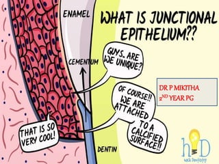
6. Definitions of JE
• JE is the non keratinised stratified squamous epithelium which
attaches and form a collar around the cervical portion of the tooth
that follows CEJ. – Joseph P. Fiorellini.
• JE represents the epithelial component of the dento-gingival
complex that lies in contact with the tooth surface, at the interface
between the gingival sulcus and the PDL fibers. – schroeder HE
1977.
• A single or multiple layers of non-keratinizing cells adhering to the
tooth surface at the base of gingival crevice. Formerly called
epithelial attachment. - GPT
7. Historical aspects
• G.V. BLACK 1915- opined that a “subgingival space” extends upto
CEJ, under loosely fitting gingiva.
• Gottlieb (1921) was the first to describe JE.
• Schroeder and Listgarten (1977) clarrified the anatomy and
histology of the dentogingival junction in their monograph: “fine
structure of developing epithelial attachment of human teeth”.
Newman GM, Takei HH, Klokkevold RP. Anatomy of periodontium. In: carranza’s clinical periodontology, Elsevier, 13,
15-21.
8. GOTTLIEB 1921
Epithelial
attachment is
organically united
to the tooth
surface.
Newman GM, Takei HH, Klokkevold RP. Anatomy of periodontium. In: carranza’s clinical periodontology, Elsevier, 13,
15-21.
9. WAERHUG 1952
Based on the animal
experiments in dogs he
postulated that the cells of
the epithelial attachment
adhere weakly to the tooth
surface and it forms the
lining of the physiologic
pocket.
Newman GM, Takei HH, Klokkevold RP. Anatomy of periodontium. In: carranza’s clinical periodontology, Elsevier, 13,
15-21.
10. WAERHUG CONCEPT 1960
• He presented the concept of epithelial cuff. This concept was based
on insertion of thin blades between the surface of the tooth and the
gingiva.
• Blades could be easily passed apically to the CT attachment at CEJ
without resistance.
• It was concluded that gingival tissues and the tooth are closely
adapted but are not organically united.
Newman GM, Takei HH, Klokkevold RP. Anatomy of periodontium. In: carranza’s clinical periodontology, Elsevier, 13,
15-21.
11. ORBAN’S CONCEPT 1953
He stated that the
separation of the
epithelial attachment
cells from the tooth
surface involved
preparatory
degenerative changes in
the epithelium.
Newman GM, Takei HH, Klokkevold RP. Anatomy of periodontium. In: carranza’s clinical periodontology, Elsevier, 13,
15-21.
12. MAX LISTGARTEN 1966-67
Based on transmission
electron microscopic studies
he proved the existence of a
hemidesmosomal basement
lamina attachment between
the tooth and the cells of the
so called epithelial attachment.
Newman GM, Takei HH, Klokkevold RP. Anatomy of periodontium. In: carranza’s clinical periodontology, Elsevier, 13,
15-21.
13. SCHROEDER AND LISTGARTEN
CONCEPT 1971
• The previoius controversy was resolved after evaluation of
transmission electron microscopy.
• Primary epithelial attachment refers to the epithelial attachment
lamina released by the REE. It lies in direct contact with enamel and
epithelial cells attached to it by hemi-desmosomes.
• When the REE cells transform into JE cells the primary epithelial
attachment becomes secondary epithelial attachment. It is made of
epithelial attachment between basal lamina and hemidesmosomes.
Newman GM, Takei HH, Klokkevold RP. Anatomy of periodontium. In: carranza’s clinical periodontology, Elsevier, 13,
15-21.
14. CURRENT CONCEPTS
KOBAYASHI ET AL. (1976)
• Studied the junctional epithelium of monkeys, reporting a highly
variable relationship between the junctional epithelium and the tooth
surface coronal to the CEJ.
• When present, a homogenous, electron dense dental cuticle occurred
between the afibrillar cementum and the junctional epithelium.
• When the dental cuticle was absent, a thin dense linear border was
noticed in the same position.
Pollanen TM. Structure and function of the tooth–epithelial interface in health and disease. Periodontology 2000, Vol. 31,
2003, 12–31.
15. • While the origin and function of the dental cuticle are unknown, the
linear border was determined to consist of serum proteins from
tissue fluids.
The author described a unit of adhesion consisting of following zones
(from tooth to JE):
• Sub lamina Lucida (95A0)
• Lamina Densa (400 Ao)
• Lamina Lucida (140 Ao) and
• the Hemi desmosomes
Pollanen TM. Structure and function of the tooth–epithelial interface in health and disease. Periodontology 2000, Vol. 31,
2003, 12–31.
16. SABAG ET AL 1981:
• described the attachment of epithelium to the cementum root surface
to be mediated by 4 to 8 hemidesmosomes/micron at the coronal
zone, and 2 hemidesmosomes /micron in the apical zone.
• Because of this arrangement, the authors suggested that the coronal
zone of the cemental surface may exhibit more adhesion of
epithelial attachment when compared to the apical zone
Pollanen TM. Structure and function of the tooth–epithelial interface in health and disease. Periodontology 2000, Vol. 31,
2003, 12–31.
17. FRIEDMAN 1993:
• Suggested that epithelial attachment to the tooth may occur without
the cuticle being present.
• He stated that the cuticle represents an accumulation of material
from plaque metabolites.
Pollanen TM. Structure and function of the tooth–epithelial interface in health and disease. Periodontology 2000, Vol. 31,
2003, 12–31.
18. Terminologies
• Gottlieb 1921- epithelial attachment
• Waerhaug 1952- epithelial cuff
• Orban 1956- attached epithelial cuff
• Grant, stern 1968- attachment epithelium
• Anderson and stern 1967- JE.
Saha AP, Saha S. junctional epithelium: a dynamic seal around the tooth. Journal of applied dental and medical sciences,
vol 4(3), 2018, 19-27.
19. Development of JE
• The junctional epithelium forms as the tooth crown erupts into the
oral cavity.
• Prior to the emergence of the tooth into the oral cavity, the enamel
surface is covered by the reduced enamel epithelium that consists of
reduced ameloblasts and the remaining cells of all other layers of the
enamel organ.
• The stratum intermedium cells of the reduced enamel epithelium
and the oral epithelial cells proliferate following breakdown of the
interposed connective tissue (Ten Cate, 1998).
Saha AP, Saha S. junctional epithelium: a dynamic seal around the tooth. Journal of applied dental and medical sciences,
vol 4(3), 2018, 19-27.
20.
21. • The 2 epithelia eventually fuse to form an epithelial cell mass.
• When the tips of the cusps or the incisal edge of the crown breaches
the oral mucosa (Ten Cate, 1998), or shortly before the
establishment of the first contact between the reduced enamel
epithelium and the oral gingival epithelium (Schroeder, 1996), a
slow cell transformation process develops.
Pollanen TM. Structure and function of the tooth–epithelial interface in health and disease. Periodontology 2000, Vol. 31,
2003, 12–31.
22. • Beginning orally and ending at the cemento-enamel junction 1 to 2
(Schroeder and Listgarten, 1977) or 3 to 4 (Ten Cate, 1998) yrs later,
the reduced enamel epithelium gradually converts into junctional
epithelium, a multilayer non-keratinizing squamous epithelium
(Glavind and Zander, 1970; Listgarten, 1972b; Schroeder and
Listgarten, 1977; Schroeder, 1996).
• During the transformation process, the reduced ameloblasts change
their morphology from short columnar to flattened cells that are
oriented parallel to the enamel surface.
Pollanen TM. Structure and function of the tooth–epithelial interface in health and disease. Periodontology 2000, Vol. 31,
2003, 12–31.
23. • Also, the cells external to the reduced ameloblasts undergo a
structural change.
• However, unlike the reduced and transformed ameloblasts, these
external cells regain mitotic activity.
• These transformed ameloblasts migrate in a coronal direction, are
exfoliated at the bottom of the sulcus, and eventually are replaced by
the cells external to the reduced/transformed ameloblasts
(Schroeder, 1996)
Pollanen TM. Structure and function of the tooth–epithelial interface in health and disease. Periodontology 2000, Vol. 31,
2003, 12–31.
24.
25. • It has been proposed that the junctional epithelium, which was
originally derived from the reduced enamel epithelium,may be
replaced in time by a junctional epithelium formed by basal cells
originating from the oral gingival epithelium (Ten Cate, 1996).
• This holds true, at least, for de novo formation of the junctional
epithelium following gingivectomy (Salonen, 1986; Salonen et al.,
1989).
• However, basal epithelial cells other than those of oral gingival
origin may also regenerate a junctional epithelium (Listgarten, 1967,
1972b; Braga and Squier, 1980; Freeman, 1981).
Pollanen TM. Structure and function of the tooth–epithelial interface in health and disease. Periodontology 2000, Vol. 31,
2003, 12–31.
26. Structure of JE
Anatomical features of JE
• JE forms a collar peripheral to
cervical region of tooth of about
0.75-1.35mm.
• Interproximally JE of adjacent
teeth fuse to form the lining of the
col area.
• JE is thick at bottom of sulcus and
tapers of in apical direction.
Nakamura M. histological and immunological characteristics of junctional epithelium. Japanese dental sciences, 2017, 1-7.
27. Microscopic features
• JE is a non-keratinized stratified squamous epithelium.
• It has 15-30 cell layers coronally and 1-3 layers at apical
termination.
• Stratum basale and stratum suprabasale.
• The basal and adjacent 1-2 suprabasal cells are cuboidal to slightly
spindle shaped and all the remaining cells are flat and oriented
parallel to the tooth surface.
Saha AP, Saha S. junctional epithelium: a dynamic seal around the tooth. Journal of applied dental and medical sciences,
vol 4(3), 2018, 19-27.
28. • The innnermost suprabasal cells (facing the tooth surface) also
called a DAT cells(Salonen et al 1994) form and maintain the
epithelial attachment apparatus.
• It has 2 basal lamina- external basal lamina and internal basal
lamina.
• EBL: JE attached to gingival CT.
• IBL: JE attached to the tooth surface.
Saha AP, Saha S. junctional epithelium: a dynamic seal around the tooth. Journal of applied dental and medical sciences,
vol 4(3), 2018, 19-27.
29.
30.
31. Functions
• Any structural/ molecular changes in the internal basal lamina can
influence the DAT cells homeostasis and also, its antimicrobial
defense or vice versa.
• The interaction between the IBL and cell surface macro-molecule is
fundamental for cell motility, adhesion, synthetic capacity, tissue
stability, regeneration and response to external signal.
• DAT cells possess the capacity to form and renew the components of
epithelial attachment
32. • The cells contain dense cytoplasm, adundant RER, golgi complex,
few tonofilaments, lysosomal bodies and numerous polyribosomes.
• Cytokeratins are the intermediate filament proteins of cytoskeletal
family and forms the main structural proteins of these junctional
epithelial cells. They express CK5, CK10, CK13. CK14, CK16,
CK19. CK19- high, found in almost all layers of the JE, have been
regarded as the characteristic histological marker of JE.
Ultrastructure of JE
Saha AP, Saha S. junctional epithelium: a dynamic seal around the tooth. Journal of applied dental and medical sciences,
vol 4(3), 2018, 19-27.
33. • The cells exhibit relatively loose intercellular junctions comprising
of few desomsomes, adherens junctions and occasional gap
junctions. The fluid filled spaces are responsible for remarkable
permeability.
• Intercellular spaces are occupied by mononuclear leukocytes of
varied nature. Different type of cells like PMN leucocytes,
lymphocytes, macrophages, APC cells, langerhans cells are seen to
exist in and around the JE cells.
• It is also being innnervated by sensory nerve fibers. Therefore, it
aids in neurosensary pain conduction, by releasing neuropeptides.
Saha AP, Saha S. junctional epithelium: a dynamic seal around the tooth. Journal of applied dental and medical sciences,
vol 4(3), 2018, 19-27.
34. Epithelial attachment apparatus
• The attachment of JE to the tooth is mediated through an
ultramicroscopic mechanism defined as the epithelial attachment
appartus.
• It consists of hemidesmosomes at the plasma membrane of the cells
Directly attached to tooth(DAT Cells) and a basal lamina like ECM,
termed the internal basal lamina on the tooth surface.
• By morphological criteria the IBL between JE DAT cells and the
enamel is quite similar to the basement membrane between the
epithelium and the CT.
Nakamura M. histological and immunological characteristics of junctional epithelium. Japanese dental sciences, 2017, 1-7.
35. Hemidesmosomes
• Hemidesmosomes have a decisive role
in the firm attachment of the cells to
the IBL on the tooth surface.
• It may also act as specific sites of
signal transduction and thus participate
in regulation of gene expression, cell
proliferation and cell differentiation.
• The intercellular part of
hemidesmosomes consists of atleast 2
distinct proteins BP230, Plectin.
Nakamura M. histological and immunological characteristics of junctional epithelium. Japanese dental sciences, 2017, 1-7.
36. • These proteins mediate the attachment of the epithelial cell
cytoplasmic keratin filaments to two transmembrane components of
hemidesomsomes.
• The interaction between the different components of ECM and cell
surface molecules linked to the intercellular cytoskeleton is
fundamental to cell adhesion, cell motility, synthetic capacity, tissue
stability, regeneration and responses to external signals.
Nakamura M. histological and immunological characteristics of junctional epithelium. Japanese dental sciences, 2017, 1-7.
37. Permeability of JE
• JE shows a higher permeability than the oral gingival epithelium and
provides a penetrating pathway into gingival CT for a variety of
bacterial products such as toxins and enzymes.
• The barrier function of JE plays a key role in the aetiology of
periodontal diseases. Membrane coating granules, ranging from 100-
400nm in diameter, have been found in the keratinized and non-
keratinized epithelia.
• Studies employing freeze-fracture and tracer methods have shown that
these membrane coating granules provide the rat oral epithelium with its
physiological permeability barrier.
Nakamura M. histological and immunological characteristics of junctional epithelium. Japanese dental sciences, 2017, 1-7.
38. Functional specificity of JE
• The JE has several specific functions that differ from those of other oral
epithelium. JE expresses defensive factors such as b-defensins, SLPI and
S100A8 against inflammation.
• JE constitutively produces chemokines and cytokines, such as
keratinocyte-derived chemokine (KC) MIP-2; which are upregulated by
the bacterial stimulation.
• The JE also expresses follicular dendritic cell secreted protein (FDC-SP)
and odontogenic ameloblast-associated protein; which appose at the
gene cluster for secretor calcium binding phospoproteins.
Shimona M et al. biological charcteristics of Junctional epithelium. Journal of electron microscopy, vol 52 (6), 2003, 627-
639.
39. • Secretory calcium-binding phosphoproteins interact with several
bioactive molecules to regulate cell adhesion, migration,
proliferation, and tumorigenesis.
• Histological and immunological investigation indicates massive
inflitration of lymphocyte function-associated antigen-1. The
interaction of LFA-1 and ICAM-1 play a key role in the migration of
these cells in JE.
Shimona M et al. biological charcteristics of Junctional epithelium. Journal of electron microscopy, vol 52 (6), 2003, 627-
639.
40. JE in Anti-microbial defense
• JE consists of active population of cells and antimicrobial functions,
which together form the first line of defense against microbial
invasion into tissue.
• Even though junctional epithelial cell layers provide a barrier
against bacteria many bacterial substances, such as
lipopolysaccharides, pass easily through the epithelium but have
only limited access through the EBL into the CT. ( Shwartz et al
1972).
• Rapid turnover as such is an imp factor in the microbial defense of
JE.
Roderick Marshall: Gingival defensins: linking the adaptive and immune responses to dental plaque. Periodontology 2000,
Vol. 35, 2004, 14-20
41. • The area covered by the
dividing cells in the JE is
atleast 50 times larger than the
area through which the
epithelial cells desquamate
into the gingival sulcus, there
is a strong funneling effect that
contributes to the flow of
epithelial cells (Schroeder et al
1967).
Roderick Marshall: Gingival defensins: linking the adaptive and immune responses to dental plaque. Periodontology 2000,
42. Role of enzymes in antimicrobial defense
• Recently, it has been found that junctional epithelial cells lateral to
DAT cells produce MMP-7 (uitto vj etal 2002).
• MMP-7 contributes to the mucosal defense by the release of
bioactive molecules from the cell srfaces which play a role in the
inflammatory reaction.
Roderick Marshall: Gingival defensins: linking the adaptive and immune responses to dental plaque. Periodontology 2000,
Vol. 35, 2004, 14-20
43. Several antimicrobial
mechanism exist in JE.
• In the coronal part of JE cell exfoliation
because of rapid cell division.
• Funnelling of junctional epithelial cells
towards the sulcus hinder bacterial
colonization. Laterally, external
basement membrane forms an effective
barrier against invading microbes.
• Active antimicrobial substances are
produced in JE cells.
Saha AP, Saha S. junctional epithelium: a dynamic seal around the tooth. Journal of applied dental and medical sciences,
vol 4(3), 2018, 19-27.
44. • Epithelial cells activated by anti-microbial substances secrete
chemokines. Eg: IL-8 and cytokine eg: IL-1,6, TNF-a that attract
and activate professional defense cells such as lymphocytes, PMN.
Their secreted product in turn cause further activation of JE cells.
45. Roderick Marshall: Gingival defensins: linking the adaptive and immune responses to dental plaque. Periodontology 2000,
Vol. 35, 2004, 14-20
46. Turnover of JE cells with DAT cells
• Turnover of JE is exceptionally rapid.
• At the coronal part of the JE, the DAT cells typically express a high
density of transferrin receptors.
• DAT cells have more imp role in tissue dynamic and reparative
capacity of JE than has previously been thought.
• Any structural or molecular changes in the IBL can potentially
influence the vital functions of the DAT cells and contribute to the
effectiveness or failure of the JE defense or vice versa.
Saha AP, Saha S. junctional epithelium: a dynamic seal around the tooth. Journal of applied dental and medical sciences,
vol 4(3), 2018, 19-27.
47. Saha AP, Saha S. junctional epithelium: a dynamic seal around the tooth. Journal of applied dental and medical sciences,
vol 4(3), 2018, 19-27.
52. detachment of DAT cells from toothsurface
• Role of GCF
• Role of polymorphonuclear leukocytes
• Role of proteinases and inflammatory mediators
• Role of bacterial products
• Role of risk factors for periodontal disease.
Roderick Marshall: Gingival defensins: linking the adaptive and immune responses to dental plaque. Periodontology 2000,
Vol. 35, 2004, 14-20
53. Role of GCF
• GCF is an exudate of varying composition found in the
sulcus/periodontal pocket between the tooth and marginal gingiva.
• It contains components of serum, inflammatory cells, CT,
epithelium, and microbial flora inhabiting the gingival margin or the
sulcus/pocket(Embery G et al 1994).
• The GCF passing through the JE determines the enviornmental
conditions and provides sufficient nutrients for the DAT cells to
grow.
Roderick Marshall: Gingival defensins: linking the adaptive and immune responses to dental plaque. Periodontology 2000,
Vol. 35, 2004, 14-20
54. • At the gingival margin the GCF may become contaminated so that
agents from the oral cavity or plaque bacteria challenge the most
coronal DAT cells.
• The conditions for DAT cell survival and adequate function at the
coronal part of the JE are different and more susceptible of
compromises than those for the basal cells living in the vicinty of
the CT and blood circulation.
Roderick Marshall: Gingival defensins: linking the adaptive and immune responses to dental plaque. Periodontology 2000,
Vol. 35, 2004, 14-20
55. • Main route of GCF diffusion is through
EBL and inter cellular spaces.
• GCF passing through JE gives nutrients
to DAT cells.
• Increased GCF flow during
inflammation will have a flushing action
against bacteria and its products.
• Biologically, active molecules in GCF
have protective as well as destructive
role.
Roderick Marshall: Gingival defensins: linking the adaptive and immune responses to dental plaque. Periodontology 2000,
Vol. 35, 2004, 14-20
56. ROLE OF POLYMORPHONUCLEAR
LEUKOCYTES
• Polymorphonuclear leukocytes form the most imp line of defense
against bacterial plaque at the gingival margin(page rc et al).
• PMN leukocytes are most effective in aerobic conditions close to the
gingival margin (dennison et al 1997) suggesting a different role for
them in anaerobic periodontal lesions.
• Lactoferrin is an imp antimicrobial protein present in the secretory
granules of PMN leukocytes.
Roderick Marshall: Gingival defensins: linking the adaptive and immune responses to dental plaque. Periodontology 2000,
Vol. 35, 2004, 14-20
57. • High con. Of lactoferrin hampers epithelial cell growth by
interfering with their adhesion and spreading. The molecule may
thus have a role in delaying the repair of JE cell populations during
severe inflammation.
Roderick Marshall: Gingival defensins: linking the adaptive and immune responses to dental plaque. Periodontology 2000,
Vol. 35, 2004, 14-20
58. ROLE OF HOST PROTEINASES AND
INFLAMMATORY MEDIATORS
• Degradation of extracellular matrix during periodontal inflammation
is a multistep process that involves several proteolytic enzymes.
• Different cell types of periodontal tissue produce MMP,
plasminogen activator, cathepsins and elastase (Brikedal H et al).
• Neutrophil elastase and cathepsin G are capable of degrading
basement membrane type IV collagen and laminin, and also type
VIII collagen, found in the IBL (Heck and Blackburn et al).
Roderick Marshall: Gingival defensins: linking the adaptive and immune responses to dental plaque. Periodontology 2000,
Vol. 35, 2004, 14-20
59. • However electron microscopic studies on DAT cells attached tp
teeth extracted because of advanced periodontitis don’t support idea
that enzymatic degradation of epithelial attachment appratus
precedes the degeneration of DAT cells. (Overman et al.)
Roderick Marshall: Gingival defensins: linking the adaptive and immune responses to dental plaque. Periodontology 2000,
Vol. 35, 2004, 14-20
60. Role of JE in passive eruption
• Passive eruption is the exposure of the teeth by apical migration of
the gingiva.
• This concept distinguish between the anatomic crown and the
anatomic root and between the clinical crown (the part of tooth that
has been denuded of its gingiva and projects into the oral cavity) and
clinical root (the portion of the tooth covered by periodontal tissue.)
• When the teeth reach their functional antagonists, the gingival
sulcus and JE are still on the enamel and clinical crown is approx
2/3rd of anatomic crown.
Nakamura M. histological and immunological characteristics of junctional epithelium. Japanese dental sciences, 2017, 1-7.
61. Passive eruption is divided into 4 stages:
Saha AP, Saha S. junctional epithelium: a dynamic seal around the tooth. Journal of applied dental and medical sciences,
vol 4(3), 2018, 19-27.
62. ROLE OF JE IN GINGIVITIS
• During the initial lesion- leucocytes leave the capillaries by
migrating through the walls (Lindhe J perio)
• They can be seen in increased quantitites in the CT and JE and
gingival sulcus
• The JE becomes densely infiltrated with neutrophils and it may
begin to show development of rete pegs in the early lesion of
gingivitis.
Dale B. Periodontal epithelium: a newly recognized role in health and disease. Periodontology 2000, Vol. 30, 2002, 70–78.
63.
64. • During established lesion: the JE reveals widened intercellular
spaces filled with granular cellular debris, including lysosomes,
lymphocytes and monocytes.
• JE forms rete pegs that proturde into the CT and basal lamina is
destroyed in some areas.
Shimona M et al. biological charcteristics of Junctional epithelium. Journal of electron microscopy, vol 52 (6), 2003, 627-
639.
65. Role of JE in NUG
• Surface epithelium is destroyed.
• Replaced by a network of fibrin, necrotic epithelial cells, PMNs and
neutrophils and various types of microorganisms.
• The epithelium becomes edematous and there is infiltration of
PMNs in the intercellular spaces.
Dale B. Periodontal epithelium: a newly recognized role in health and disease. Periodontology 2000, Vol. 30, 2002, 70–78.
66. JE IN TFO
• TFO causes widening of marginal PDL space, narrowing of the
interproximal alveolar bone.
• In case of TFO, JE will be intact and there will be no degeneration
of the epithelial tissues unless there is any plaque accumulation.
Dale B. Periodontal epithelium: a newly recognized role in health and disease. Periodontology 2000, Vol. 30, 2002, 70–78.
67. ROLE OF THE JE IN THE INITIATION OF
POCKET FORMATION
• A clinically healthy gingiva exhibits microscopic signs of slight
inflammation, including the presence of an inflammatory infiltrate
of very limited extent (Brecx et al., 1987).
• In that respect, the importance of the leukocytes, particularly PMNs,
migrating through the junctional epithelium has been recognized as
a significant factor contributing to the first line of peripheral host
defense (Schroeder and Listgarten, 1997).
Saha AP, Saha S. junctional epithelium: a dynamic seal around the tooth. Journal of applied dental and medical sciences,
vol 4(3), 2018, 19-27.
68. • Thus, such inflammatory cells in the subepithelial portion of the
lamina propria and in the junctional epithelium itself should be
regarded as a part of normal homeostasis and an essential element of
the defense system against continuous bacterial challenge
(Schroeder and Listgarten, 1997).
• Also, an acute gingivitis should not be interpreted as a first step in
the development of periodontitis.
Saha AP, Saha S. junctional epithelium: a dynamic seal around the tooth. Journal of applied dental and medical sciences,
vol 4(3), 2018, 19-27.
69. • Usually, the peripheral host defense system is efficient enough to
avoid exacerbation of the developing lesion and progression toward
connective tissue breakdown seen in periodontitis.
• Since the conversion of the junctional epithelium to pocket
epithelium is regarded as a hallmark in the development of
periodontitis, the potential factors contributing to the initiation of
pocket formation deserve particular attention.
Nakamura M. histological and immunological characteristics of junctional epithelium. Japanese dental sciences, 2017, 1-7.
70. JUNCTIONAL EPITHELIUMADJACENT TO
ORAL IMPLANTS
• The junctional epithelium around implants always originates from
epithelial cells of the oral mucosa, as opposed to the junctional
epithelium around teeth which originates from the reduced enamel
epithelium.
• Hence, it may be questioned whether or not the structural and
functional characteristics of these 2 junctional epithelia are identical.
Saha AP, Saha S. junctional epithelium: a dynamic seal around the tooth. Journal of applied dental and medical sciences,
vol 4(3), 2018, 19-27.
71. • Structurally, the periimplant
epithelium closely resembles the
junctional epithelium around
teeth (Berglundh et al., 1991;
Listgarten et al., 1991; Buser et
al., 1992; Listgarten, 1996;
Koka, 1998; Cochran, 2000),
• although dissimilarities have also
been reported (Inoue et al., 1997;
Ikeda et al., 2000, 2002; Fujiseki
et al., 2003; Shimono et al.,
2003).
Shimona M et al. biological charcteristics of Junctional epithelium. Journal of electron microscopy, vol 52 (6), 2003, 627-
639.
72.
73. • The biological attachment mechanism is thought to be mediated
through particular adhesins or integrins, which are fundamental in
cell to cell adhesion as well as cell to matrix adhesion.
• The junctional epithelium may be found on the implant itself or on
the abutment.
• This will be because of differences in the designs of implants, the
biological requirements of the attachment of the soft tissue cuff and
the level of the junction between abutment and implants.
Richard Palmer. Teeth and implants. British dental journal, volume 187, no. 4, August 28 1999.
74. • Peri-implant epithelium might be a poorly adhered and permeable
epithelium. (Masaki Shimono, Tatsuya Ishikawa, 2003).
• There is also evidence that several of the mentioned marker
molecules involved in the defense mechanisms against the bacterial
challenge are also expressed in the peri-implant epithelium.
• In that respect, the presence of t- PA (Schmid et al., 1992), ICAM-
1, and a cytokeratin profile resembling that of gingival junctional
epithelium (Mackenzie and Tonetti, 1995) has been documented.
Richard Palmer. Teeth and implants. British dental journal, volume 187, no. 4, August 28 1999.
75. • This, in turn, implies that, despite different origins of the 2 epithelia,
a functional adaptation occurs when oral epithelia form an epithelial
attachment around implants.
• Such an adaptive potential is also observed in the regenerating
junctional epithelium around teeth following gingivectomy.
Richard Palmer. Teeth and implants. British dental journal, volume 187, no. 4, August 28 1999.
76. REGENERATION OF THE JUNCTIONAL
EPITHELIUM
• Injury of the junctional epithelium may occur through accidental or
intentional trauma, toothbrushing, flossing, or clinical probing.
• Since the junctional epithelium is located at a strategically important
but also delicate site, it may be expected that it should be very well-
adapted to cope with mechanical insults.
• Clinical probing results in a mechanical disruption of the junctional
epithelial cells from the tooth.
Saha AP, Saha S. junctional epithelium: a dynamic seal around the tooth. Journal of applied dental and medical sciences,
vol 4(3), 2018, 19-27.
77. • Whether and how fast a new epithelial attachment reforms have
been the objectives of several studies.
• In an experimental study in marmosets, following probing, a new
and complete attachment indistinguishable from that in controls was
established 5 days after complete separation of the junctional
epithelium from the tooth surface (Taylor and Campbell, 1972).
• The reestablishment of the epithelial seal around implants after
clinical probing was shown to occur within about the same time
period (Etter et al., 2002).
Nakamura M. histological and immunological characteristics of junctional epithelium. Japanese dental sciences, 2017, 1-7.
78. • In both studies, persistence of tissue trauma and infection as a result of
probing were not observed.
• Based on these 2 studies, probing around teeth and implants does not
seem to cause irreversible damage to the soft tissue components.
• Oral hygiene practices may be accompanied by undesired trauma to
the junctional epithelium as well.
• Waerhaug (1981) studied healing of the junctional epithelium
following the use of dental floss at premolars in 12-year-old humans.
Detachment of cells persisted for 24 hrs after flossing ceased.
Shimona M et al. biological charcteristics of Junctional epithelium. Journal of electron microscopy, vol 52 (6), 2003, 627-
639.
79. • New attachment of junctional epithelial cells started 3 days after
flossing ceased.
• After 2 wks, the cell populations on the experimental and control
surfaces were again indistinguishable from each other.
• In the above studies, the junctional epithelium was never completely
removed from the tooth.
• However, the application of gingivectomy techniques would
completely remove the junctional epithelium.
Shimona M et al. biological charcteristics of Junctional epithelium. Journal of electron microscopy, vol 52 (6), 2003, 627-
639.
80. • Subsequently, the formation of a new junctional epithelium must occur
from basal cells of the oral gingival epithelium (Listgarten, 1967;
Innes, 1970; Frank et al., 1972; Listgarten and Ellegaard, 1973; Braga
and Squier, 1980).
• In humans, a new junctional epithelium after gingivectomy may form
within 20 days (Listgarten, 1972a,b; Schroeder and Listgarten, 1977).
• These studies show that the junctional epithelium is a highly dynamic
and adaptive tissue with a fast capacity for self renewal or de novo
formation from basal cells of the oral gingival epithelium.
Shimona M et al. biological charcteristics of Junctional epithelium. Journal of electron microscopy, vol 52 (6), 2003, 627-
639.
81. Long junctional epithelium
• Periodontal surgery, experimental surgery and the destruction of
periodontal tissues by insertion of a rubber piece have been reported
to produce long junctional epithelium on exposed root surfaces.
• Listagarten et al suggested that long junctional epithelium is a
transient feature of the healing process on the road to CT
attachment, but is not a final healing stage. JE is a rapidly
proliferating tissue and its regeneration appears as epithelail down-
growth producing long junctional epithelium.
Listagarten M A, Rosenberg S. progressive replacement of epithelial attachment by connective tissue junction after
experimental periodontal surgery in rats. J Periodontal, 1982, 53: 630-653.
82.
83. • The long junctional epithelium consists of 2 or 3 layers aligned parallel
to the tooth surface.
• This epithelium is connected to cementum by hemidesmosomes and
basal lamina.
• The epithelium is permeable due to its wide intercellular spaces.
• Fewer capillaries underneath the long junctional epithelium are evident
in normal JE.
• Function of long JE doesn’t require innervation and it does not make
neutrophic groth factors.
Listagarten M A, Rosenberg S. progressive replacement of epithelial attachment by connective tissue junction after
experimental periodontal surgery in rats. J Periodontal, 1982, 53: 630-653.
84. conclusion
• The Junctional epithelium is a unique tissue that fulfills a
challenging function at the border between the oral cavity, colonized
by bacteria, and the tooth attachment apparatus. It is structurally and
functionally very well-adapted to control the constant presence of
bacteria and their products.
• However, its antimicrobial defense mechanisms do not preclude the
development of inflammatory lesions in the gingiva.
• These defense mechanisms may be overwhelmed by bacterial
virulence factors, and the gingival lesion could progress to
periodontitis.
85. REFERENCES
• Newman GM, Takei HH, Klokkevold RP. Anatomy of
periodontium. In: carranza’s clinical periodontology, Elsevier, 13,
15-21.
• Bosshardt D and Lang NP :The Junctional Epithelium: from
Health to Disease. J Dent Res 84(1):9-20, 2005.
• Dale B. Periodontal epithelium: a newly recognized role in health
and disease. Periodontology 2000, Vol. 30, 2002, 70–78.
• Pollanen TM. Structure and function of the tooth–epithelial
interface in health and disease. Periodontology 2000, Vol. 31, 2003,
12–31.
86. • Roderick Marshall: Gingival defensins: linking the adaptive and
immune responses to dental plaque. Periodontology 2000, Vol. 35,
2004, 14-20
• Richard Palmer. Teeth and implants. British dental journal, volume
187, no. 4, August 28 1999.
• Lindhe J, Lang P N. anatomy of periodontal tissues. In: clinical
periodontolgy and implant dentistry. Wiley blackwell, 6th edition,
2015, 5-29.
87. • Saha AP, Saha S. junctional epithelium: a dynamic seal around the
tooth. Journal of applied dental and medical sciences, vol 4(3),
2018, 19-27.
• Nakamura M. histological and immunological characteristics of
junctional epithelium. Japanese dental sciences, 2017, 1-7.
• Shimona M et al. biological charcteristics of Junctional epithelium.
Journal of electron microscopy, vol 52 (6), 2003, 627-639.