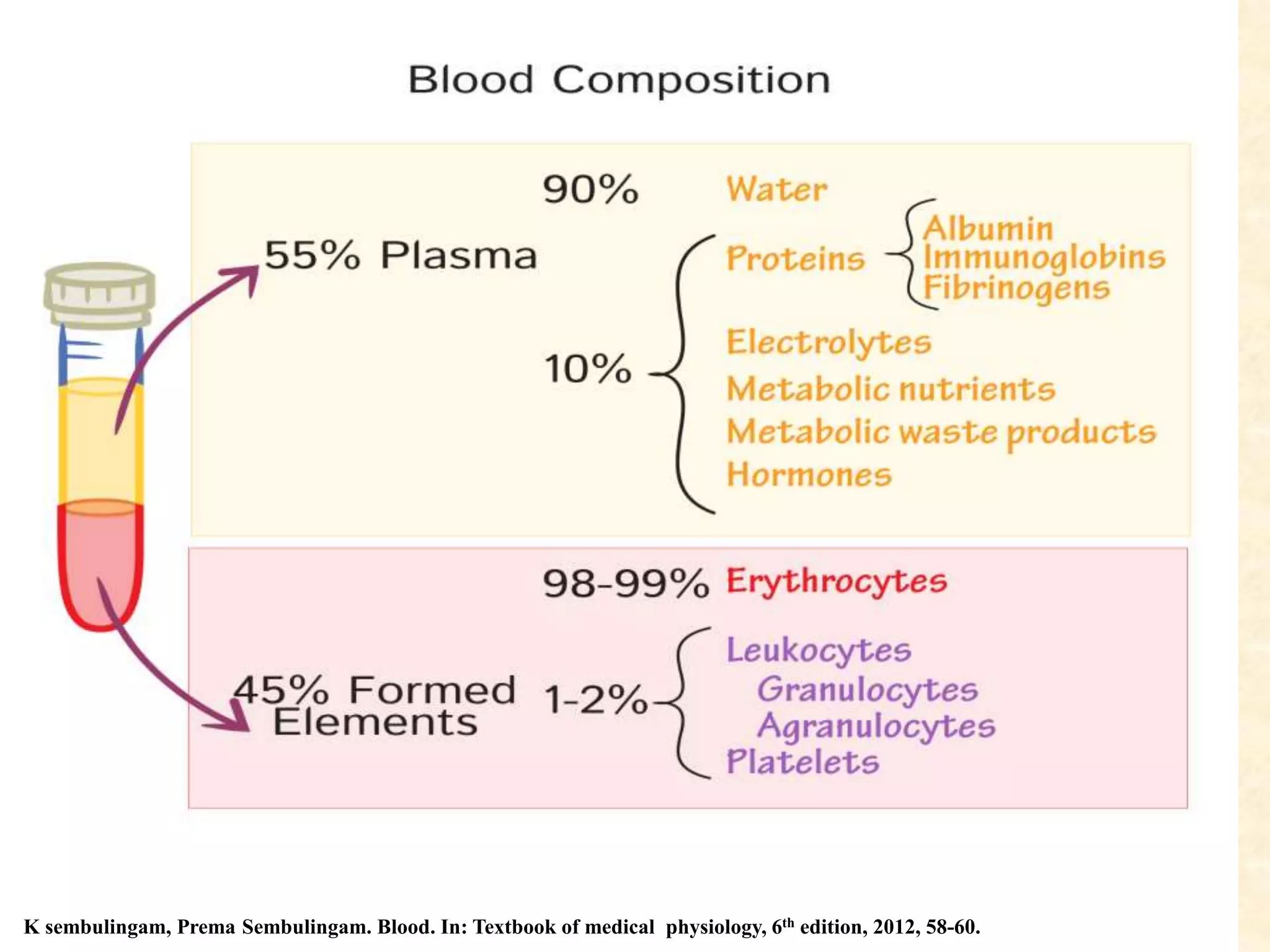
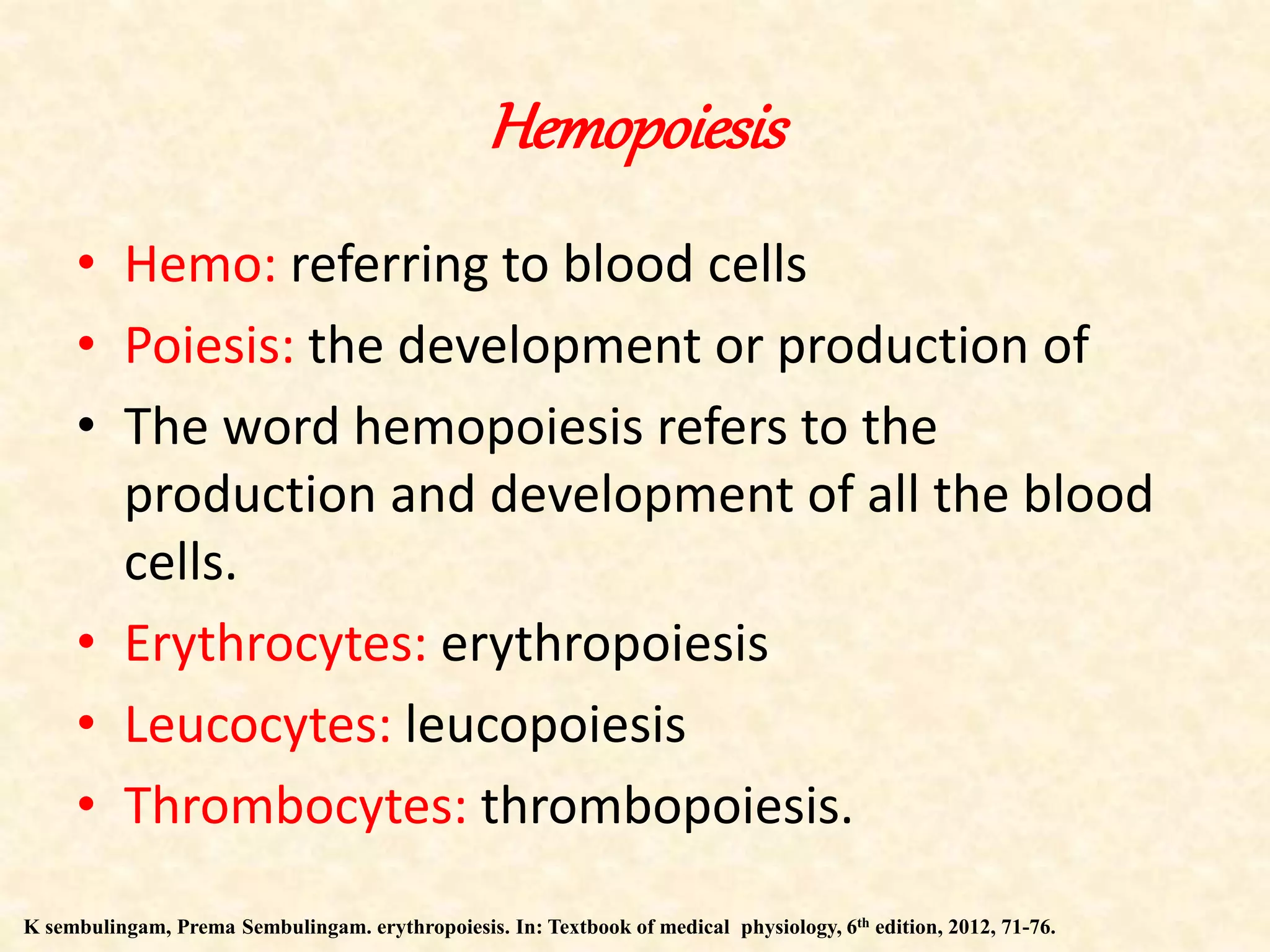
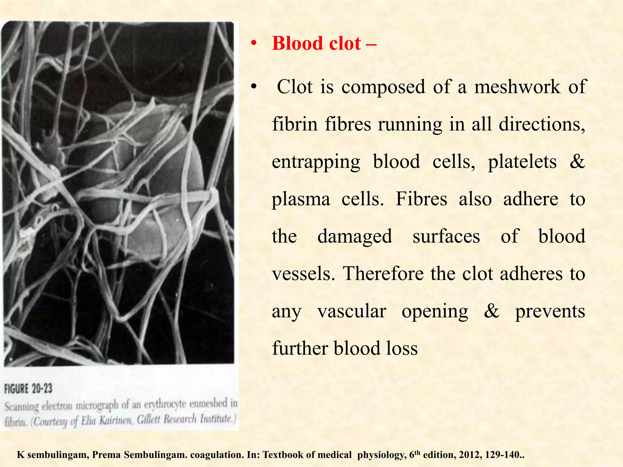
2. Contents
Introduction
Blood
Erythropoiesis
RBC
Clotting
Hemostasis
Coagulation
Lab investigations of bleeding and coagulation
Disorders of bleeding and coagulation
3. Pharmacology
Hemostasis in periodontics
Periodontal implications of bleeding disorders
Traditional recommendations of periodontal
treatment
Conclusion
References
4. DEFINITION
• Blood may be described, as a specialized connective tissue
in which there is a liquid intercellular substance known as
Plasma and Formed elements- RBC, WBC, Platelet
suspended in the plasma.
K sembulingam, Prema Sembulingam. Blood. In: Textbook of medical physiology, 6th edition, 2012, 58-60.
5. K sembulingam, Prema Sembulingam. Blood. In: Textbook of medical physiology, 6th edition, 2012, 58-60.
6. FUNCTIONSOFBLOOD
• Transport of respiratory gases
• Transport of nutrition
• Maintainence of water balance
• Maintainence of acid-base equilibrium
• Regulation of body temperature
• Defensive action
• Coagulative action
• Regulation of BP.
K sembulingam, Prema Sembulingam. Blood. In: Textbook of medical physiology, 6th edition, 2012, 58-60.
7. Hemopoiesis
• Hemo: referring to blood cells
• Poiesis: the development or production of
• The word hemopoiesis refers to the
production and development of all the blood
cells.
• Erythrocytes: erythropoiesis
• Leucocytes: leucopoiesis
• Thrombocytes: thrombopoiesis.
K sembulingam, Prema Sembulingam. erythropoiesis. In: Textbook of medical physiology, 6th edition, 2012, 71-76.
8. K sembulingam, Prema Sembulingam. erythropoiesis. In: Textbook of medical physiology, 6th edition, 2012, 71-76.
9. K sembulingam, Prema Sembulingam. erythropoiesis. In: Textbook of medical physiology, 6th edition, 2012, 71-76.
10. K sembulingam, Prema Sembulingam. erythropoiesis. In: Textbook of medical physiology, 6th edition, 2012, 71-76.
11. Factors necessary for erythropoiesis
GENERAL FACTORS:
• Erythropoietin
• Thyroxine
• Hemopoietic growth factors
• Vitamins.
Maturation factors:
• Vitamin B12
• Intrinsic factor of castle
• Folic acid
Factors necessary for HB formation
K sembulingam, Prema Sembulingam. erythropoiesis. In: Textbook of medical physiology, 6th edition, 2012, 71-76.
12. CLOTTING
When blood is shed it loses its fluidity in a few mins and sets
into a semisolid jelly. This phenomenon is called coagulation
or clotting.
If further undisturbed, clot retracts into a smaller volume &
presses out a clear straw coloured fluid, called serum.
Coagulation is a property of plasma alone. WBC’s & RBC’s
get caught up in the meshes of the clot, giving it a red colour.
Many important substances have been found in the blood &
tissues that affect coagulation
K sembulingam, Prema Sembulingam. Coagulation of blood In: Textbook of medical physiology, 6th edition, 2012, 129-140.
13. Pro-coagulants & Anticoagulants.
Blood coagulation depends on a balance between these two
groups.
In the blood stream, anticoagulants predominate, so that blood
does not coagulate during circulation. But when a blood vessel
ruptures procoagulants in the area of tissue damage become
activated & override the anticoagulants & a clot develops.
K sembulingam, Prema Sembulingam. Coagulation of blood In: Textbook of medical physiology, 6th edition, 2012, 129-140.
16. Alpha granules.
• These granules are oval or
sometimes round in shape. Often
seen enclosed in a membrane.
• The function: lysosomal function
which is important in platelet
release reaction and aggregation,
and in clot resolution or in the
platelets phagocytic capacity.
• They secrete fibronectin, platelet
factor 4, PGF, vWF.
Very dense granules.
These granules have 0.05 to 0.13µ
and is surrounded by a unit of
membrane. These granules have
been described to contain
serotonin.
Glycogen granules.
These granules are also distributed
in certain parts of the platelets.
K sembulingam, Prema Sembulingam. platelets In: Textbook of medical physiology, 6th edition, 2012, 122-124.
17. HEMOSTASIS
• Prevention of blood loss
Achieved by several mechanisms (when vessel ruptures)
1) Vascular spasm
2) Formation of platelet plug
3) Formation of blood clot as a result of coagulation
4) Eventual growth of fibrous tissue into the clot to close the
hole in the vessel permanently
K sembulingam, Prema Sembulingam. Hemostasis. In: Textbook of medical physiology, 6th edition, 2012, 127-129.
18. VASOCONSTRICTION
Transient
Immediately after the blood vessel has been injured, trauma
itself causes the vessel to contract
reduction in the flow of blood
K sembulingam, Prema Sembulingam. Hemostasis. In: Textbook of medical physiology, 6th edition, 2012, 127-129.
19. Contraction results from
Nervous reflexes – initiated by pain or other impulses from
traumatized vessel
Local humoral factors – myogenic contraction of blood
vessel
Platelets – vasoconstriction of smaller vessels by releasing
thromboxane A2
Secretion of endothelin
K sembulingam, Prema Sembulingam. Hemostasis. In: Textbook of medical physiology, 6th edition, 2012, 127-129.
20. Formation of platelet hemostatic plug
After vascular injury ECM constituents are exposed –
collagen, proteoglycans, fibronectin & other adhesive proteins
On contact with the ECM, platelets undergo three reactions
- adhesion & shape change
- secretion (release reaction)
- aggregation
K sembulingam, Prema Sembulingam. Hemostasis. In: Textbook of medical physiology, 6th edition, 2012, 127-129.
21. Adhesion
Facilitated by von Willebrand factor (vWf)
von Willebrand factor :
- adhesive protein
- contains tripeptide sequence of amino acids arg-gly-asp (RGD)
- bind to cell surface proteins “integrins” & to platelet surface
membrane glycoproteins GPIIb & GPIIIa (functional on platelet
activation, bind fibrinogen during platelet aggregation)secreted in
plasma and sub-endothelium.
Guyton and Hall. Blood. In: textbook of medical physiology, 11th edition, 2010, 419-429.
22. • on endothelial disruption vWf binds to subendothelial matrix,
which alters its surface, & facilitates binding to platelet,
linking it to subendothelium.
• Binding site – GPIb (Nurden 1987)
• This association is sufficiently strong to overcome high shear
forces of flowing blood
Normal platelet contains 5-10 microgram of vWf/ml
In von Willebrand disease, pts bleed abnormally because of
deficiency of functional vWf, due to low plasma vWf conc. or
reduction of the highest molec. wt. multimers.
Guyton and Hall. Blood. In: textbook of medical physiology, 11th edition, 2010, 419-429.
23. Guyton and Hall. Blood. In: textbook of medical physiology, 11th edition, 2010, 419-429.
24. Platelet activation
Platelets adhering to the sub endothelium, become activated &
those arriving later get activated & bind to those already present.
K sembulingam, Prema Sembulingam. Hemostasis. In: Textbook of medical physiology, 6th edition, 2012, 127-129.
25. During activation platelets undergo a series of progressive
overlapping events :-
- Shape change
Aggregation
Liberation & oxidation of arachidonic acid
Secretion of alpha & dense granule contents
Reorganization of surface membrane phospholipids (makes
phosphatidyl serine available on the outer surface, where it
participates in coagulation)
Guyton and Hall. Blood. In: textbook of medical physiology, 11th edition, 2010, 419-429.
26. STIMULUS FOR PLATELET ACTIVATION
Two primary agonists trigger activation
i) Thrombin formed at injury site
ii) Sequences on collagen in the sub endothelium.
Activated platelets release ADP & arachidonic acid oxidation
products which maintain & amplify the activation.
Guyton and Hall. Blood. In: textbook of medical physiology, 11th edition, 2010, 419-429.
27. • Agonists interact with specific membrane receptors to activate
G proteins (guanine nucleotide binding proteins) in the platelet
membrane (Haslan 1987) activation of phospholipase C
hydrolysis of membrane inositol phoshpolipids
diacygycerol & inositol triphosphate I(1,4,5)P3 (2nd
messengers). These act synergistically as follows (Nishizuka
1984) :
Guyton and Hall. Blood. In: textbook of medical physiology, 11th edition, 2010, 419-429.
28. • Diacyglycerol activates protein kinase C, catalyses transfer
of P from ATP serine or threonine residues of proteins
• I(1,4,5)P3 acts as a calcium ionophore Ca to enter cytosol.
• A rising cytosolic Ca conc triggers :
• a) phosphorylation of myosin light chain kinase, required for
reorientation of cytoskeleton proteins needed for shape change,
secretion & aggregation
• b) activation of Calpain (Ca dependant protease) proteolysis
& activation of other platelet enzymes
• c) activation of phospholipase A2 liberates arachidonic acid
from platelet membrane phospholipids
29. • Arachadionic acid is oxidized by 2 pathways.
Guyton and Hall. Blood. In: textbook of medical physiology, 11th edition, 2010, 419-429.
30. Thromboxane A2 bind to specific platelet membrane receptors
activation of phospholipase C & amplification of platelet
activation through further generation of DAG & I(1,4,5)P3.
Platelet cAMP levels affect the response of platelet to
activating stimuli through regulation of a Ca pump that lowers
cystolic Ca levels (Hawiger et al 1987)
PGI2 keeps circulating platelets in an unstimulated state,
through activation of platelet adenyl cyclase & increase in
cAMP levels.
Alterations of platelet membrane lipids also affect their
responsiveness.
Guyton and Hall. Blood. In: textbook of medical physiology, 11th edition, 2010, 419-429.
31. Platelet shape change
• Reversible process
• Takes place within secs of exposure to activating agents.
• Platelets change shape from flattened discs to spheres with
multiple projecting pseudopods.
• Actin is polymerized & actin microfilaments elongate & push
the surface membrane out ahead of them.
• Microtubules surround the platelet granules that have moved to
the centre of the cell.
Guyton and Hall. Blood. In: textbook of medical physiology, 11th edition, 2010, 419-429.
32. Platelet aggregation
On exposure to any of the activation agonists (thrombin,
collagen, thromboxane A2, ADP), fibrinogen receptors are
expressed on the GPIIb-IIIa complex on the surface memb.
fibrinogen binds to this complex & forms a bridge between a
GPIIb- IIIa complex on two opposing activated platelets.
This causes the platelets to stick to each other in the primary
phase of aggregation (reversible) sec irreversible phase after
platelet secretion begins.
Guyton and Hall. Blood. In: textbook of medical physiology, 11th edition, 2010, 419-429.
33. Thrombospondin (alpha granule protein) binds to fibrinogen &
GPIV on the surface membrane thereby reinforcing fibrin
aggregation.
Thrombasthenia/Glanzmann’s disease – rare hereditary
disorder
- cannot form GPIIb-IIIa complex
- failure to aggregate
- serious bleeding from mild abrasions
Guyton and Hall. Blood. In: textbook of medical physiology, 11th edition, 2010, 419-429.
34. Guyton and Hall. Blood. In: textbook of medical physiology, 11th edition, 2010, 419-429.
35. Platelet secretion
Platelets contain 3 types of granules – alpha, dense & lysosomal
Granules are distributed randomly but move to the centre on
activation
Their membranes fuse with the membrane of an open canalicular
system made up of invaginations of the platelet surface membrane
The contents are secreted – alpha, dense, then as the platelets
breakdown from the lysosomal granules.
Dense granules- ATP, ADP, Ca, Serotonin
K sembulingam, Prema Sembulingam. Hemostasis. In: Textbook of medical physiology, 6th edition, 2012, 127-129.
36. Pts with hereditary disorders preventing storage of normal
quantities of ADP have mild to moderate bleeding diathesis
(Weiss 1987)
Alpha granules: 1) Proteins that are also found in plasma
a) Albumin b) IgG – no hemostatic function
c) Fibrinogen d) vWf e) factor V – found in increased conc in
platelets than plasma. After secretion bind to the platelet
membrane
37. • 2) Proteins secreted by activated platelets
a) Thrombospondin – binds to GPIIb-IIIa & fibrinogen
b) thromboglobulin
c) PF4 -
d) PDGF -
e) TGF-B –
- chemoattractants for WBC’s, smooth muscle cells & fibroblasts
- activate these cells & accelerate wound healing
- therefore contribute to inflammation & repair
Guyton and Hall. Blood. In: textbook of medical physiology, 11th edition, 2010, 419-429.
38. Blood coagulation
• If the tear in the vessel is small, the platelet plug itself can stop
blood loss. But if there is a large hole, a blood clot in addition to
the platelet plug is required to stop further hemorrhage.
• Clotting occurs around the original platelet plug. Its function is to
reinforce it & to solidify blood that remains in the wound channel.
The retraction of the clot compresses the ruptured vessel further
and stops bleeding.
• Clot begins to develop in 15-20 secs if the trauma is severe, & 1-
2mins if it is minor.
K sembulingam, Prema Sembulingam. coagulation. In: Textbook of medical physiology, 6th edition, 2012, 129-140..
39. Clotting factors
K sembulingam, Prema Sembulingam. coagulation. In: Textbook of medical physiology, 6th edition, 2012, 129-140..
40. Mechanism of blood coagulation
• Occurs in three steps –
1) Sequence of reactions resulting in generation of an activator
of prothrombin
2) Prothrombin activator catalyses the conversion of
prothrombin - thrombin
3) Thrombin acts as an enzyme to convert fibrinogen to fibrin
fibres that enmesh platelets, blood cells, & plasma to form
the clot.
K sembulingam, Prema Sembulingam. coagulation. In: Textbook of medical physiology, 6th edition, 2012, 129-140..
41. K sembulingam, Prema Sembulingam. coagulation. In: Textbook of medical physiology, 6th edition, 2012, 129-140..
42. role of calcium
• Calcium is required for promotion or acceleration of all blood
clotting reactions except 1st two steps in intrinsic pathway.
• In the body Ca2+ conc. rarely falls low enough to affect clotting.
• When blood is removed from the blood stream, clotting can be
prevented by reducing Ca2+ conc, either by deionizing Ca (reaction
with citrate ion) or by precipitating Ca ( with oxlate ion)
Guyton and Hall. Blood. In: textbook of medical physiology, 11th edition, 2010, 419-429.
43. Conversion of prothrombin to thrombin
K sembulingam, Prema Sembulingam. coagulation. In: Textbook of medical physiology, 6th edition, 2012, 129-140..
44. Formation and stabilization of fibrin
• In the early stages :
fibrin monomer moles are held together by weak non covalent
H bonds.
Newly formed fibres are not cross linked with each other
Therefore the clot is weak.
K sembulingam, Prema Sembulingam. coagulation. In: Textbook of medical physiology, 6th edition, 2012, 129-140..
45. Fibrin stabilizing factor (XIII)
helps in strengthening the clot by acting as an enzyme to cause
more covalent bonds between fibrin monomer molecs, &
multiple cross linkages between adj fibrin fibres
Present in small amounts in plasma globulins
Released from platelets entrapped in the clot
Must be activated by thrombin
46. • Blood clot –
• Clot is composed of a meshwork of
fibrin fibres running in all directions,
entrapping blood cells, platelets &
plasma cells. Fibres also adhere to
the damaged surfaces of blood
vessels. Therefore the clot adheres to
any vascular opening & prevents
further blood loss
K sembulingam, Prema Sembulingam. coagulation. In: Textbook of medical physiology, 6th edition, 2012, 129-140..
47. Restrictions of coagulation cascade
Once activated, the coagulation cascade must be restricted to
the site of vascular injury to prevent runaway clotting of the
entire vascular tree. It is restricted by :
Endogenous anticoagulants
Fibrinolysis
Feedback mechanism.
Guyton and Hall. Blood. In: textbook of medical physiology, 11th edition, 2010, 419-429.
48. • 1. Endogenous anticoagulants
Anti thrombins inhibit the activity of thrombin and other serine
proteases, including factors IXa, Xa, XIa, and XIIa(Ex: Anti
thrombin III is activated by binding to heparin-like molecules
on endothelial cells)
Proteins C and S:- vitamin K– dependent proteins that act in a
complex that proteolytically inactivates factors Va and VIIIa.
TFPI :- protein produced by endothelium (and other cell
types) that inactivates tissue factor–factor VIIa complexes .
Guyton and Hall. Blood. In: textbook of medical physiology, 11th edition, 2010, 419-429.
49. • Fibrinolysis
Major means of disposing fibrin after its hemostatic function
has been fulfilled.
Ensures that clots formed in response to injury are not
dissolved prematurely, which could lead to re-bleeding.
The digestion of non-cross-linked fibrin and fibrinogen by
plasmin produces fragments known as fibrin degradation
products (FDP).
FDPs inhibit both platelet aggregation and the action of
thrombin on fibrinogen.
Guyton and Hall. Blood. In: textbook of medical physiology, 11th edition, 2010, 419-429.
50. • Feedback mechanism
• The extent of thrombin formation is regulated by protein C-
thrombomodulin pathway
• It activates protein C by a complex formed between thrombin
and thrombomodulin (endothelial thrombin receptor).
• This interacts with protein S resulting in the inactivation of
FVa and FVIIIa, thereby dampening further generation of
thrombin.
• Patients with congenital deficiencies of either protein C or S
have a high incidence of thromboembolism
Guyton and Hall. Blood. In: textbook of medical physiology, 11th edition, 2010, 419-429.
51. Current concept of coagulation
Hoffman and Monroe (2001, 2005, 2007; Monroe et al. 2002;
Hoffman 2003; Roberts et al. 2006) proposed the cell-centric
model which comprises three overlapping phases of hemostasis
and in which the intrinsic and extrinsic pathways operate in
parallel, but on different cell surfaces.
This model describes coagulation with following steps:
1. Initiation phase
2. Amplification phase
3. Propogation phase
4. Termination phase
52.
53.
54. Fibrinolysis
• The excessive deposition of fibrin within circulation is
prevented in health by fibrinolytic system
K sembulingam, Prema Sembulingam. coagulation. In: Textbook of medical physiology, 6th edition, 2012, 129-140..
55. Lab investigations
Mohan H, Sugandha M. Diseases of blood. In: textbook of pathology, 7th edition, 2014, 390-410.
56. Mohan H, Sugandha M. Diseases of blood. In: textbook of pathology, 7th edition, 2014, 390-410.
57.
58. Mohan H, Sugandha M. Diseases of blood. In: textbook of pathology, 7th edition, 2014, 390-410.
61. Hemophilia
Bleeder’s disease, disease of king’s, disease of Hapsburg.
Hereditary disease carried by x chromosome.
Characterised by prolonged coagulation time and hemorhagic
tendencies.
Occurrence of hemophilia is theoretically possible in
homozygous female and only rare cases have been recorded.
Types of hemophilia
Hemophilia A - plasma thromboplastin
Hemophilia B - plasma thromboplastin component
Hemophilia C - plasma thromboplastin antecedent
Brewer A, Correa E M. Guidelines of dental treatment of patients with bleeding disorders. World Federation of Hemophilia,
2006, 1-16.
62. Clinical features:
Persistent bleeding either spontaneously or on slight trauma.
Hemmorhage into subcutaneous tissue, internal organs or
joints.
Present from birth, but clinically not apparent for several
years.
30-50% patients with severe forms present umbilical cord and
neonatal bleeding.
Hemophilia C can be distinguished from A and B by the
absence of bleeding into joints and muscles and by its
occurrence in individuals of either gender.
Brewer A, Correa E M. Guidelines of dental treatment of patients with bleeding disorders. World Federation of Hemophilia,
2006, 1-16.
63. Severity
Mild - >5% - major trauma or surgery.
Moderate - 1-5% - mild to moderate trauma.
Severe - <1% - spontaneous hemarthrosis, soft tissue bleeding.
• Oral manifestations
Gingival hemorrhage may be massive and prolonged.
Even physiologic processes of tooth eruption and exfoliation
may be associated with prolonged hemorrhage.
Mandibular ‘pseudotumor’ of hemophilia – condition in
which there is subperiosteal bleeding, with reactive new bone
formation causing tumor like expansion of bone.
Brewer A, Correa E M. Guidelines of dental treatment of patients with bleeding disorders. World Federation of Hemophilia,
2006, 1-16.
64. Treatment and prognosis
There is no cure for hemophilia only protection from trauma.
Recommendation:- preoperative transfusion of whole blood
and anti hemophilia factor concentrate before surgical
procedures. Nevertheless oral surgery is dangerous and
avoided whenever possible.
Prognosis is variable and many affected persons die in
childhood.
Brewer A, Correa E M. Guidelines of dental treatment of patients with bleeding disorders. World Federation of Hemophilia,
2006, 1-16.
65. Von willebrand’s disease
Pseudohemophilia or vascular purpura.
Most commonly inherited bleeding disorder of humans due to
deficiency of von Willebrand factor (vWF).
A gene located on chromosome 12p codes for vWF.
Deficiency of vWF results in defective platelet adhesion and
causes a secondary deficiency in factor VIII.
Brewer A, Correa E M. Guidelines of dental treatment of patients with bleeding disorders. World Federation of Hemophilia,
2006, 1-16.
66. Types:
Type I vWD - most common and is inherited as an autosomal
dominant trait. Due to simple quantitative deficiency of all
vWF multimers
Type 2 vWD - subdivided further depending upon whether the
dysfunctional protein has decreased or paradoxically increased
function in certain laboratory tests of binding to platelets.
Type 3 vWD - is clinically severe and is characterized by
recessive inheritance and virtual absence of vWF.
Brewer A, Correa E M. Guidelines of dental treatment of patients with bleeding disorders. World Federation of Hemophilia,
2006, 1-16.
67. Clinical features
There is excessive bleeding, either spontaneously or following
even minor trauma - nose, skin , gingiva and GI tract .
In oral cavity even after slight provocation such as brushing
causes bleeding.
Acquired form of the disease is seen in Wilm’s tumor, SLE etc.
Normal platelet count PTT, CT. Only BT is prolonged
Brewer A, Correa E M. Guidelines of dental treatment of patients with bleeding disorders. World Federation of Hemophilia,
2006, 1-16.
68. Management of Vwd:
• The goal is to prevent bleeding.
• Pt with vWF level> 40UI/dl or without history of bleeding are
not at risk.
• Desmopressin is the first line of treatment & infusion trial can
determine the response of the patient.
Brewer A, Correa E M. Guidelines of dental treatment of patients with bleeding disorders. World Federation of Hemophilia,
2006, 1-16.
69. Parahemophilia
Is a rare haemorrhagic disorder, clinically similar to
haemophilia but caused by deficiency of Factor V
(Proaccelerin), which is responsible for conversion of
prothrombin to thrombin.
It is inherited as an autosomal recessive trait.
Patients exhibit severe bleeding tendency, spontaneous
epistaxis, bleeding into GI tract and menorrhagia .
In the oral cavity, spontaneous gingival bleeding and also
prolonged bleeding after dental extraction is common.
Brewer A, Correa E M. Guidelines of dental treatment of patients with bleeding disorders. World Federation of Hemophilia,
2006, 1-16.
70. Fibrin stabilizing factor deficiency
Due to congenital FVIII deficiency - Ducket (1960)
It is a rare autosomal recessive disease with high incidence of
consanguity.
Acquired form – hepatic failure, IBS, myeloid leukemia
There is failure of permanent peptide bond formation between
fibrin molecules so that fibrinogen monomer aggregates break
up.
Patients with this deficiency have severe post surgical bleeding
episodes which are typically delayed for 24-36 hours,
hemarthroses and defective wound healing.
Brewer A, Correa E M. Guidelines of dental treatment of patients with bleeding disorders. World Federation of Hemophilia,
2006, 1-16.
71. ITP
Defined as isolated low platelet count (thrombocytopenia)
with normal bone marrow and the absence of other causes.
It is an acute condition in children (resolution within 2
months) and a achronic condition in adults (persists longer
than 6 months).
ITP is an autoimmune disease with antibodies detectable
against several platelet surface antigens
Bansal N, Jindal M. clinical guidelines for periodontal management of ITP. J Int. oral Health Scien. Vol 7, 2017, 30-34.
72. Clinical Features:-
• Spontaneous formation of purpura and petechiae , especially
on the extremities, bleeding from the nostrils or gums,
and menorrhagia , any of which may occur if the platelet count
is below 20,000 per μl.
• A very low count (<10,000 per μl) may result in the
spontaneous formation of hematomas in the mouth or on other
mucous membrane.
Bansal N, Jindal M. clinical guidelines for periodontal management of ITP. J Int. oral Health Scien. Vol 7, 2017, 30-34.
73. Bansal N, Jindal M. clinical guidelines for periodontal management of ITP. J Int. oral Health Scien. Vol 7, 2017, 30-34.
74. Treatment of ITP
• Glucocorticoids
• IV Ig
• IV anti-Rhd
• Splenectomy
• Accessory splenectomy
Bansal N, Jindal M. clinical guidelines for periodontal management of ITP. J Int. oral Health Scien. Vol 7, 2017, 30-34.
75. Vitamin K deficiency
• Vitamin K is a fat soluble vitamin essential for synthesis of
clotting factors II, VII, IX and X .
Vitamin K is obtained in part from food and in part from
bacterial flora in the bowel which synthesizes the vitamin.
The ability to bind Ca2+is required for the activation of the
several vitamin K-dependent clotting factors, or protiens in
the clotting cascade.
Vitamin K deficiency occurs in disorders which impair fat
absorption, sterilization of bowel, hemorrhagic disease of new
born
Bansal N, Jindal M. clinical guidelines for periodontal management of ITP. J Int. oral Health Scien. Vol 7, 2017, 30-34.
77. Vitamin K
Fat soluble dietary principle required for the synthesis of CF
Daily Requirement : 3-10 microgm/day
Action : cofactor in the synthesis of CF by liver
Deficiency : occurs due to liver disease, obstructive jaundice,
malabsorption, long term antimicrobial therapy (alters
intestinal flora)
Use : Deficiency, long term antimicrobial therapy – 5-
10mg/day oral
Tripathi K D. bleeding and coagulation drugs. In: Textbook of Medical Pharmacology, 2014, 304-354.
78. Obstructive jaundice – 10mg i.m/day or oral with bile salts
Newborn – 1mg i.m soon after birth
To reverse the effect of overdose of oral anticoagulants – K1
Severe – 10mg i.m followed by 5mg 4 hourly
Moderate – 10mg i.m followed by 5mg once or twice
Mild – omit a few doses of anticoagulant
Tripathi K D. bleeding and coagulation drugs. In: Textbook of Medical Pharmacology, 2014, 304-354.
79. Anticoagulants
Heparin
• Anticoagulants, antiplatelet (inhibits aggregation)
• Heparin sod i.v 5000-10000 U i.v every 4-6hrs
• Dose & frequency controlled by APTT
• If haemorrhage occurs it is usually sufficient to withdraw
heparin, but if rapid reversal of the effects of heparin is
required, protamine sulfate is a specific antidote.
Tripathi K D. bleeding and coagulation drugs. In: Textbook of Medical Pharmacology, 2014, 304-354.
80. Oral anticoagulants
• Oral anticoagulants take at least 48–72 hours for the
anticoagulant effect to develop fully; if an immediate effect is
needed, heparin must be given concomitantly.
Warfarin
• act by interfering with synthesis of vit K dependant CF
• indicated in deep-vein thrombosis, pulmonary embolism, for
patients with atrial fibrillation who are at risk of embolization
and for those with mechanical prosthetic heart valves (to
prevent emboli developing on the valves);
Tripathi K D. bleeding and coagulation drugs. In: Textbook of Medical Pharmacology, 2014, 304-354.
81. • Oral anticoagulants should not be used in pregnancy, cerebral
thrombosis or peripheral arterial occlusion as first-line therapy.
• The main adverse effect of oral anticoagulants is haemorrhage.
Prothrombin time should be checked on a daily basis initially
then at longer intervals depending on response.
• If severe haemorrhage occurs, stop warfarin and give
phytomenadione (vitamin K) by slow intravenous injection.
Tripathi K D. bleeding and coagulation drugs. In: Textbook of Medical Pharmacology, 2014, 304-354.
82. Dosage:
• Prophylaxis and treatment of thromboembolic disorders, oral,
• ADULT usual induction dose is 10 mg daily for 2 days,
according to the individual patient; the subsequent dose
depends upon the prothrombin time; the usual daily
maintenance dose is 3–9 mg taken at the same time each day
Adverse effects:
• Hemorrhage; hypersensitivity, rash, alopecia, diarrhoea,
unexplained drop in haematocrit, ‘purple toes’, skin necrosis,
jaundice, hepatic dysfunction, nausea, vomiting and
pancreatitis
Tripathi K D. bleeding and coagulation drugs. In: Textbook of Medical Pharmacology, 2014, 304-354.
83. Fibrinolytics
Used to lyse thrombi/clot to recanalyse occluded blood
vessels( coronary artery)
Activate the natural fibrinolytic system
Streptokinase (beta hemolytic strep. group C)
Combines with circulating plasminogen to form activator
complex
Streptase (freeze dried powder):MI 7.5-15 lac IU iv infusion
over 1hr
Tripathi K D. bleeding and coagulation drugs. In: Textbook of Medical Pharmacology, 2014, 304-354.
84. Anti fibrinolytics
Inhibit plasminogen activation & dissolution of cot
Epsilon amino-caproic acid (EACA)
Combines with lysine binding sites of plasminogen & plasmin
making it unable to bind fibrin & lyse it
Antidote of fibrinolytic agents & used in overdose of
streptokinase/urokinase
Initial dose 5g oral/i.v then 1g hourly till bleeding stops (max 30g
in 24hrs)
Tripathi K D. bleeding and coagulation drugs. In: Textbook of Medical Pharmacology, 2014, 304-354.
85. Anti platelet drugs
Useful in the prophylaxis of thromboembolic
disorders
Aspirin 60mg/day
Sulfinpyrazone 200mg QID
Dipyridamole – vasodilator, improves response of
warfarin 150-300mg/day
Ticlopidine – interacts with platelet membrane
250mg BD
USES : coronary artery disease, cerebrovascular
disease, coronary bypass, prosthetic heart valves,
venous thromboembolism
Tripathi K D. bleeding and coagulation drugs. In: Textbook of Medical Pharmacology, 2014, 304-354.
86. Local hemostatics (styptics)
• Stop bleeding from local site
1. Thrombin – hemophilia, skin grafting etc
2. Fibrin – sheets or foam for covering bleeding surfaces
3. Gelatin foam – packing wounds
4. Russels viper venom – acts as thromboplastin in
hemophiliacs
5. Vasoconstrictors – 1% adrenaline solution
6. Astringent – tannic acid (20% in glycerin) bleeding gums,
bleeding piles
Tripathi K D. bleeding and coagulation drugs. In: Textbook of Medical Pharmacology, 2014, 304-354.
87. Hemostasis in periodontics
• Hemostasis is an important aspect of periodontal surgery
• Good intra operative control of bleeding permits an accurate
visualization of the
• Extent of disease
• Pattern of bone destruction and condition of the root surfaces.
• A clear view of the surgical site for wound debridment and
scaling and root planing
Vassilopolus P, Palcains K. Bleeding disorders and periodontolgy. J Periodontol 2000, vol. 44, 2007, 211-233.
88. Periodontal implications of bleeding disorders
Patients with a history of bleeding disorders should be
managed so as to minimize risks of hemorrhage.
Clinical examination should detect the existence of jaundice,
ecchymosis, spider telangiectasia, hemarthrosis, petechiae,
hemorrhagic vesicles, spontaneous gingival bleeding or
hyperplasia.
In patients with coagulation disorders, periodontal treatment
may be performed, provided sufficient precautions are taken.
Vassilopolus P, Palcains K. Bleeding disorders and periodontolgy. J Periodontol 2000, vol. 44, 2007, 211-233.
89. Probing, scaling & prophylaxis can be carried out without
medical modification. More invasive procedures (LA, surgery,
root planing) require prior physician consultation.
During treatment local measures to ensure clot formation &
stability are of importance.
complete wound closure & application of pressure reduce
hemorrhage
Vassilopolus P, Palcains K. Bleeding disorders and periodontolgy. J Periodontol 2000, vol. 44, 2007, 211-233.
90. Antihemostatic agents (oxidised cellulose, purified bovine
collagen) may be applied over surgical sites.
Antifibrinolytic agents (EACA, Tranexamic acid)
Patients with liver disease:
Physician consultation
Lab investigations
Non surgical therapy
Surgery – PTT <2.5 safe, platelet count >80.000/mm3
Vassilopolus P, Palcains K. Bleeding disorders and periodontolgy. J Periodontol 2000, vol. 44, 2007, 211-233.
91. • Patients on anti coagulants
Physician consultation
Procedure determines PTT. SRP, Infiltration INR<3.
Block anaesthesia, minor perio surgery, simple extractions
INR<2
Careful technique, complete wound closure
Vassilopolus P, Palcains K. Bleeding disorders and periodontolgy. J Periodontol 2000, vol. 44, 2007, 211-233.
92. • Thrombocytopenic purpura
Pdl therapy directed towards reducing inflammation by
removing local irritants
SRP contraindicated Platelet <60,000/mm3
Surgery >80,000/mm3
Atraumatic technique, platelet transfusion may be needed
Vassilopolus P, Palcains K. Bleeding disorders and periodontolgy. J Periodontol 2000, vol. 44, 2007, 211-233.
93. Traditional recommendations for periodontal
treatment
Consult the patient’s physician to determine the nature of the
underlying medical problem and the degree of required
anticoagulation.
Infiltration anesthesia, scaling, and root planing may be done
safely in patients with an INR less than 3.0.
Block anesthesia, minor periodontal surgery, and simple
extractions usually require an INR less than 2.0 to 2.5.
Complex surgery or multiple extractions may require an INR
less than 1.5 to 2.0.
94. The physician must be consulted about any changes
(discontinuing or reducing) in anticoagulant dosage until the
desired INR is achieved. The dentist must inform the physician
what degree of intra operative and postoperative bleeding is
usually expected with the procedures planned. Often, the
anticoagulant is discontinued for 2 to 3 days before periodontal
treatment and the INR is checked on the day of therapy.
Anticoagulants are resumed immediately after treatment.
Vassilopolus P, Palcains K. Bleeding disorders and periodontolgy. J Periodontol 2000, vol. 44, 2007, 211-233.
95. Careful technique and complete wound closure are paramount.
For all procedures, application of pressure can minimize
hemorrhage. Use of oxidized cellulose, microfibrillar collagen,
topical thrombin, and tranexamic acid should be considered for
persistent bleeding.
Discontinuing anticoagulant therapy before dental surgery was
common in the past. However, many clinicians may no longer
recommend this.
Vassilopolus P, Palcains K. Bleeding disorders and periodontolgy. J Periodontol 2000, vol. 44, 2007, 211-233.
96. Recent evidence related to the risks of altering anticoagulant
therapy along with the lack of evidence for bleeding
complications suggest that treating patients without reducing
or discontinuing medications may be more prudent.
Heparin is generally used for short-term anticoagulation and is
given IV. It is a powerful anticoagulant with a duration of
action of 4 to 8 hours. Periodontal treatment is rarely required
while a patient is taking heparin.
Vassilopolus P, Palcains K. Bleeding disorders and periodontolgy. J Periodontol 2000, vol. 44, 2007, 211-233.
97. Conclusion
• Early dental care is of prime importance in such patients
to avoid invasive procedures at a future date.
• A thorough understanding of the condition, helps the
dental professional to perform a systematic evaluation and
anticipate potential hazards rather than being caught
unawares with meager and desperate local haemostatic
measures.
98. references
• K Sembulingam, prema sembulingam. Blood and body fluids. In: textbook
of medical physiology, 6th edition, 2012, 51-160.
• Guyton and Hall. Blood. In: textbook of medical physiology, 11th edition,
2010, 419-429.
• Cotran, Kumar, Collins. Blood. In: robbins pathologic basis of diseases, 6th
edition, 2009, 124-136.
• Christopher H, Chivlers E. In: Davidson’s principles and practise of
medicine, 19th edition, 2015, 897-902.
• Mohan H, Sugandha M. Diseases of blood. In: textbook of pathology, 7th
edition, 2014, 390-410.
99. • Sanjeev P, Saroa R, Palta A. Overview of the coagulation system. Indian J
Anaesthet. 2014, sept-oct, volume 58, 515-523.
• Brewer A, Correa E M. Guidelines of dental treatment of patients with
bleeding disorders. World Federation of Hemophilia, 2006, 1-16.
• Vassilopolus P, Palcains K. Bleeding disorders and periodontolgy. J
Periodontol 2000, vol. 44, 2007, 211-233.
• Bansal N, Jindal M. clinical guidelines for periodontal management of ITP.
J Int. oral Health Scien. Vol 7, 2017, 30-34.
• Tripathi K D. bleeding and coagulation drugs. In: Textbook of Medical
Pharmacology, 2014, 304-354.