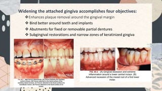
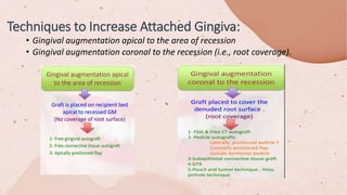
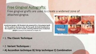
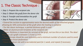
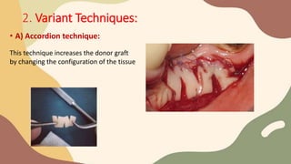
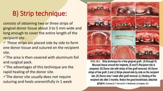
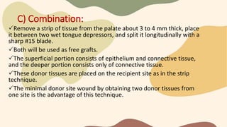
In the name of God
name: Maryam Adham Haghighipour
Guilan university of medical science/ Iran
Free Gingival Graft
(FGG)
History:
Friedman mucogingival surgery :
describes surgical procedures ,reference to three problem areas:
- Attached gingiva,
- Shallow vestibules,
- Frenum interfering with the marginal gingiva
Miller  periodontal plastic surgery:
Correct anatomic, developmental, or traumatic deformities of the gingiva or alveolar
mucosa.
finally:  the 1996 World Workshop
in Clinical Periodontics renamed mucogingival surgery as periodontal plastic surgery, a term originally
proposed by Miller in 1993 and broadened to include the following areas
Mucogingival therapy:
is a broader term that includes non-surgical procedures such as : papilla reconstruction by means of
orthodontic or restorative therapy.
1.Problems associated with attached gingiva
2. Problems associated with a shallow vestibule
3. Problems associated with an aberrant
frenum
4. Aesthetic surgical therapy
5. Tissue engineering
Widening the attached gingiva accomplishes four objectives:
Enhances plaque removal around the gingival margin
 Bind better around teeth and implants
 Abutments for fixed or removable partial dentures
 Subgingival restorations and narrow zones of keratinized gingiva
• Gingival augmentation apical to the area of recession
• Gingival augmentation coronal to the recession (i.e., root coverage).
Techniques to Increase Attached Gingiva:
Free Gingival Autografts:
Free gingival grafts are used to create a widened zone of
attached gingiva.
• 1. The Classic Technique
• 2. Variant Techniques :
• A) Accordion technique B) Strip technique C) Combination
1. The Classic Technique :
• Step 1: Prepare the recipient site
• Step 2: Obtain the graft from the donor site
• Step 3: Transfer and immobilize the graft
• Step 4: Protect the donor site
Extend the incision to approximately twice the desired width of the attached gingiva
allowing for 50% contraction of the graft when the healing is complete
For the classic technique, a partial-thickness graft is used
Make an aluminum foil template of the recipient site.
Proper thickness is important for survival of the graft, not too thin or too thick. The ideal
thickness of a graft is between 1.0 and 1,5 mm.
Tissue forceps should be used delicately and a minimum number of sutures used to avoid
unnecessary tissue perforation
Cover the donor site with a periodontal pack for 1 week, and repeat if necessary
2. Variant Techniques:
• A) Accordion technique:
This technique increases the donor graft
by changing the configuration of the tissue
B) Strip technique:
consists of obtaining two or three strips of
gingival donor tissue about 3 to 5 mm wide and
long enough to cover the entire length of the
recipient site .
 These strips are placed side by side to form
one donor tissue and sutured on the recipient
site.
The area is then covered with aluminum foil
and surgical pack.
The advantages of this technique are the
rapid healing of the donor site.
The donor site usually does not require
suturing and heals uneventfully in 1 week
C) Combination:
Remove a strip of tissue from the palate about 3 to 4 mm thick, place
it between two wet tongue depressors, and split it longitudinally with a
sharp #15 blade.
Both will be used as free grafts.
The superficial portion consists of epithelium and connective tissue,
and the deeper portion consists only of connective tissue.
These donor tissues are placed on the recipient site as in the strip
technique.
The minimal donor site wound by obtaining two donor tissues from
one site is the advantage of this technique.
HEALING OF THE GRAFT:
• The success of the graft depends on survival of the connective tissue.
• Fibrous organization of the interface between the graft and the recipient bed
occurs within 2 to several days.
• The graft is initially maintained by a diffusion of fluid from the host bed, adjacent
gingiva, and alveolar mucosa.
• The fluid is a transudate from the host vessels and provides nutrition and
hydration essential for the initial survival of the transplanted tissues.
• During the first day, the connective tissue becomes edematous and disorganized
and undergoes degeneration and lysis of some of its elements.
• As healing progresses, the edema is resolved, and degenerated connective tissue
is replaced by new granulation tissue.
• Revascularization of the graft starts by the second or third day.
• The graft is pale and the pallor changes to an ischemic grayish white during the
first 2 days until vascularization begins and a pink color appears.
• Loss of epithelium leaves the graft smooth and shiny.
• Functional integration of the graft occurs by the seventeenth day
• Capillaries from the recipient bed proliferate into the graft to form a network of
new capillaries and anastomose with preexisting vessels.
• The central section of the surface is the last to vascularize, but this is complete by
the tenth day.
• The epithelium undergoes degeneration and sloughing which is replaced by new
epithelium from the borders of the recipient site.
• A thin layer of new epithelium is present by the fourth day, with rete pegs
developing by the seventh day.
• As seen microscopically, healing of a graft of intermediate thickness (0.75 mm) is
complete by 10.5 weeks and thicker grafts (1.75 mm) may require 16 weeks or
longer.
Thank you


Free gingival graft

  • 1.
    In the nameof God name: Maryam Adham Haghighipour Guilan university of medical science/ Iran Free Gingival Graft (FGG)
  • 2.
    History: Friedman mucogingival surgery: describes surgical procedures ,reference to three problem areas: - Attached gingiva, - Shallow vestibules, - Frenum interfering with the marginal gingiva Miller  periodontal plastic surgery: Correct anatomic, developmental, or traumatic deformities of the gingiva or alveolar mucosa. finally:  the 1996 World Workshop in Clinical Periodontics renamed mucogingival surgery as periodontal plastic surgery, a term originally proposed by Miller in 1993 and broadened to include the following areas
  • 3.
    Mucogingival therapy: is abroader term that includes non-surgical procedures such as : papilla reconstruction by means of orthodontic or restorative therapy. 1.Problems associated with attached gingiva 2. Problems associated with a shallow vestibule 3. Problems associated with an aberrant frenum 4. Aesthetic surgical therapy 5. Tissue engineering
  • 4.
    Widening the attachedgingiva accomplishes four objectives: Enhances plaque removal around the gingival margin  Bind better around teeth and implants  Abutments for fixed or removable partial dentures  Subgingival restorations and narrow zones of keratinized gingiva
  • 5.
    • Gingival augmentationapical to the area of recession • Gingival augmentation coronal to the recession (i.e., root coverage). Techniques to Increase Attached Gingiva:
  • 6.
    Free Gingival Autografts: Freegingival grafts are used to create a widened zone of attached gingiva. • 1. The Classic Technique • 2. Variant Techniques : • A) Accordion technique B) Strip technique C) Combination
  • 7.
    1. The ClassicTechnique : • Step 1: Prepare the recipient site • Step 2: Obtain the graft from the donor site • Step 3: Transfer and immobilize the graft • Step 4: Protect the donor site Extend the incision to approximately twice the desired width of the attached gingiva allowing for 50% contraction of the graft when the healing is complete For the classic technique, a partial-thickness graft is used Make an aluminum foil template of the recipient site. Proper thickness is important for survival of the graft, not too thin or too thick. The ideal thickness of a graft is between 1.0 and 1,5 mm. Tissue forceps should be used delicately and a minimum number of sutures used to avoid unnecessary tissue perforation Cover the donor site with a periodontal pack for 1 week, and repeat if necessary
  • 8.
    2. Variant Techniques: •A) Accordion technique: This technique increases the donor graft by changing the configuration of the tissue
  • 9.
    B) Strip technique: consistsof obtaining two or three strips of gingival donor tissue about 3 to 5 mm wide and long enough to cover the entire length of the recipient site .  These strips are placed side by side to form one donor tissue and sutured on the recipient site. The area is then covered with aluminum foil and surgical pack. The advantages of this technique are the rapid healing of the donor site. The donor site usually does not require suturing and heals uneventfully in 1 week
  • 10.
    C) Combination: Remove astrip of tissue from the palate about 3 to 4 mm thick, place it between two wet tongue depressors, and split it longitudinally with a sharp #15 blade. Both will be used as free grafts. The superficial portion consists of epithelium and connective tissue, and the deeper portion consists only of connective tissue. These donor tissues are placed on the recipient site as in the strip technique. The minimal donor site wound by obtaining two donor tissues from one site is the advantage of this technique.
  • 11.
    HEALING OF THEGRAFT: • The success of the graft depends on survival of the connective tissue. • Fibrous organization of the interface between the graft and the recipient bed occurs within 2 to several days. • The graft is initially maintained by a diffusion of fluid from the host bed, adjacent gingiva, and alveolar mucosa. • The fluid is a transudate from the host vessels and provides nutrition and hydration essential for the initial survival of the transplanted tissues. • During the first day, the connective tissue becomes edematous and disorganized and undergoes degeneration and lysis of some of its elements. • As healing progresses, the edema is resolved, and degenerated connective tissue is replaced by new granulation tissue. • Revascularization of the graft starts by the second or third day. • The graft is pale and the pallor changes to an ischemic grayish white during the first 2 days until vascularization begins and a pink color appears. • Loss of epithelium leaves the graft smooth and shiny. • Functional integration of the graft occurs by the seventeenth day
  • 12.
    • Capillaries fromthe recipient bed proliferate into the graft to form a network of new capillaries and anastomose with preexisting vessels. • The central section of the surface is the last to vascularize, but this is complete by the tenth day. • The epithelium undergoes degeneration and sloughing which is replaced by new epithelium from the borders of the recipient site. • A thin layer of new epithelium is present by the fourth day, with rete pegs developing by the seventh day. • As seen microscopically, healing of a graft of intermediate thickness (0.75 mm) is complete by 10.5 weeks and thicker grafts (1.75 mm) may require 16 weeks or longer.
  • 13.