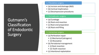
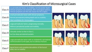
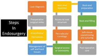
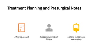
Objectives and rationale
Indications
Contraindications
False indications
Treatment planning and presurgical notes
Classification
Gutmann’s
Kim’s
Steps in endosurgery
Treatment planning & Presurgical notes
Mandatory investigations
Premedication
Local anaesthesia and hemostasis
Flap
Requirements of an ideal flap
Flap design
Semilunar flap
Vertical flaps
Horizontal flap
Ochsenbein-Luebke flap
Two-step or filling first technique
Disinfection immediately prior to filling
Preparation of surgical site
Soft tissue management
Opening the flap
Flap elevation
Flap retraction
Hard tissue considerations
Locating root apex
Osteotomy
Apical curettage
Apical rood end resection
Surgery from palatal access
Post-resection filling
Root end preparation
Root end filling materials
Reverse filling
Surgery for root fractures
Surgical management of internal resorption
Radisectomy and hemisection
Intentional replantation
Closure of surgical area
Repositioning of flap and compression
Needle selection
Suturing
Post surgical care
Electronic apex locator by dr.imran m.shaikhImran Shaikh
. Knowledge of apical anatomy, prudent use of radiographs and the correct use of an electronic apex locator will assist practitioners to achieve predictable results.
Electronic apex locator by dr.imran m.shaikhImran Shaikh
. Knowledge of apical anatomy, prudent use of radiographs and the correct use of an electronic apex locator will assist practitioners to achieve predictable results.
This short presentation discuss very important subject in endodontic field, which is the complications that most commonly occur during root canal treatment, like sodium hypochlorite accident and air emphysema and others. management of these complications is also discussed.
A well pictured presentation on Endodontic Instrumentation for UG students. Best for getting a good grip on the topic as a whole. Meant to supplement not substitute standard texts.
Surgical endodontics(Apicectomy) by Dr. Amit Suryawanshi .Oral & Maxillofac...All Good Things
Description:
Hi. This is Dr. Amit T. Suryawanshi. Oral & Maxillofacial surgeon from Pune, India. I am here on slideshare.com to share some of my own presentations presented at various levels in the field of OMFS. Hope this would somehow be helpful to you making your presentations. All the best & your replies are welcomed!
Surgical endodontics (Apicectomy) by Dr. Amit T. Suryawanshi, Oral Surgeon, ...All Good Things
Hi. This is Dr. Amit T. Suryawanshi. Oral & Maxillofacial surgeon from Pune, India. I am here on slideshare.com to share some of my own presentations presented at various levels in the field of OMFS. Hope this would somehow be helpful to you making your presentations. All the best.
Maxillectomy and craniofacial resection Mamoon Ameen
all maxillectomy types in detail and maxillofacial resection ,indications ,contraindications ,preoperative asssessment and detail techniques and rehabilitations
All furcation defects need to be classified and their possible prognosis should be defined. The treatment of the furcation defects should be carried out accordingly. Treatment include
Osteoplasty, Odontoplasty, Tunnel preparation, Root resection, Hemisection
Iatrogenic Perforation- A guide to fixing the hole in your patient's toothTaseef Hasan Farook
An overview of the possible types of perforation that may occur during endodontic treatment with their management. This slide presentation covers multiple management possibilities of said perforation proposed by various clinicians from around the world which can aid the readers in their treatment plan for the repair of a tooth perforation
Report Back from SGO 2024: What’s the Latest in Cervical Cancer?bkling
Are you curious about what’s new in cervical cancer research or unsure what the findings mean? Join Dr. Emily Ko, a gynecologic oncologist at Penn Medicine, to learn about the latest updates from the Society of Gynecologic Oncology (SGO) 2024 Annual Meeting on Women’s Cancer. Dr. Ko will discuss what the research presented at the conference means for you and answer your questions about the new developments.
Ethanol (CH3CH2OH), or beverage alcohol, is a two-carbon alcohol
that is rapidly distributed in the body and brain. Ethanol alters many
neurochemical systems and has rewarding and addictive properties. It
is the oldest recreational drug and likely contributes to more morbidity,
mortality, and public health costs than all illicit drugs combined. The
5th edition of the Diagnostic and Statistical Manual of Mental Disorders
(DSM-5) integrates alcohol abuse and alcohol dependence into a single
disorder called alcohol use disorder (AUD), with mild, moderate,
and severe subclassifications (American Psychiatric Association, 2013).
In the DSM-5, all types of substance abuse and dependence have been
combined into a single substance use disorder (SUD) on a continuum
from mild to severe. A diagnosis of AUD requires that at least two of
the 11 DSM-5 behaviors be present within a 12-month period (mild
AUD: 2–3 criteria; moderate AUD: 4–5 criteria; severe AUD: 6–11 criteria).
The four main behavioral effects of AUD are impaired control over
drinking, negative social consequences, risky use, and altered physiological
effects (tolerance, withdrawal). This chapter presents an overview
of the prevalence and harmful consequences of AUD in the U.S.,
the systemic nature of the disease, neurocircuitry and stages of AUD,
comorbidities, fetal alcohol spectrum disorders, genetic risk factors, and
pharmacotherapies for AUD.
ARTIFICIAL INTELLIGENCE IN HEALTHCARE.pdfAnujkumaranit
Artificial intelligence (AI) refers to the simulation of human intelligence processes by machines, especially computer systems. It encompasses tasks such as learning, reasoning, problem-solving, perception, and language understanding. AI technologies are revolutionizing various fields, from healthcare to finance, by enabling machines to perform tasks that typically require human intelligence.
- Video recording of this lecture in English language: https://youtu.be/lK81BzxMqdo
- Video recording of this lecture in Arabic language: https://youtu.be/Ve4P0COk9OI
- Link to download the book free: https://nephrotube.blogspot.com/p/nephrotube-nephrology-books.html
- Link to NephroTube website: www.NephroTube.com
- Link to NephroTube social media accounts: https://nephrotube.blogspot.com/p/join-nephrotube-on-social-media.html
Title: Sense of Taste
Presenter: Dr. Faiza, Assistant Professor of Physiology
Qualifications:
MBBS (Best Graduate, AIMC Lahore)
FCPS Physiology
ICMT, CHPE, DHPE (STMU)
MPH (GC University, Faisalabad)
MBA (Virtual University of Pakistan)
Learning Objectives:
Describe the structure and function of taste buds.
Describe the relationship between the taste threshold and taste index of common substances.
Explain the chemical basis and signal transduction of taste perception for each type of primary taste sensation.
Recognize different abnormalities of taste perception and their causes.
Key Topics:
Significance of Taste Sensation:
Differentiation between pleasant and harmful food
Influence on behavior
Selection of food based on metabolic needs
Receptors of Taste:
Taste buds on the tongue
Influence of sense of smell, texture of food, and pain stimulation (e.g., by pepper)
Primary and Secondary Taste Sensations:
Primary taste sensations: Sweet, Sour, Salty, Bitter, Umami
Chemical basis and signal transduction mechanisms for each taste
Taste Threshold and Index:
Taste threshold values for Sweet (sucrose), Salty (NaCl), Sour (HCl), and Bitter (Quinine)
Taste index relationship: Inversely proportional to taste threshold
Taste Blindness:
Inability to taste certain substances, particularly thiourea compounds
Example: Phenylthiocarbamide
Structure and Function of Taste Buds:
Composition: Epithelial cells, Sustentacular/Supporting cells, Taste cells, Basal cells
Features: Taste pores, Taste hairs/microvilli, and Taste nerve fibers
Location of Taste Buds:
Found in papillae of the tongue (Fungiform, Circumvallate, Foliate)
Also present on the palate, tonsillar pillars, epiglottis, and proximal esophagus
Mechanism of Taste Stimulation:
Interaction of taste substances with receptors on microvilli
Signal transduction pathways for Umami, Sweet, Bitter, Sour, and Salty tastes
Taste Sensitivity and Adaptation:
Decrease in sensitivity with age
Rapid adaptation of taste sensation
Role of Saliva in Taste:
Dissolution of tastants to reach receptors
Washing away the stimulus
Taste Preferences and Aversions:
Mechanisms behind taste preference and aversion
Influence of receptors and neural pathways
Impact of Sensory Nerve Damage:
Degeneration of taste buds if the sensory nerve fiber is cut
Abnormalities of Taste Detection:
Conditions: Ageusia, Hypogeusia, Dysgeusia (parageusia)
Causes: Nerve damage, neurological disorders, infections, poor oral hygiene, adverse drug effects, deficiencies, aging, tobacco use, altered neurotransmitter levels
Neurotransmitters and Taste Threshold:
Effects of serotonin (5-HT) and norepinephrine (NE) on taste sensitivity
Supertasters:
25% of the population with heightened sensitivity to taste, especially bitterness
Increased number of fungiform papillae
Factory Supply Best Quality Pmk Oil CAS 28578–16–7 PMK Powder in Stockrebeccabio
Factory Supply Best Quality Pmk Oil CAS 28578–16–7 PMK Powder in Stock
Telegram: bmksupplier
signal: +85264872720
threema: TUD4A6YC
You can contact me on Telegram or Threema
Communicate promptly and reply
Free of customs clearance, Double Clearance 100% pass delivery to USA, Canada, Spain, Germany, Netherland, Poland, Italy, Sweden, UK, Czech Republic, Australia, Mexico, Russia, Ukraine, Kazakhstan.Door to door service
Hot Selling Organic intermediates
TEST BANK for Operations Management, 14th Edition by William J. Stevenson, Ve...kevinkariuki227
TEST BANK for Operations Management, 14th Edition by William J. Stevenson, Verified Chapters 1 - 19, Complete Newest Version.pdf
Pulmonary Thromboembolism - etilogy, types, medical- Surgical and nursing man...VarunMahajani
Disruption of blood supply to lung alveoli due to blockage of one or more pulmonary blood vessels is called as Pulmonary thromboembolism. In this presentation we will discuss its causes, types and its management in depth.
HOT NEW PRODUCT! BIG SALES FAST SHIPPING NOW FROM CHINA!! EU KU DB BK substit...GL Anaacs
Contact us if you are interested:
Email / Skype : kefaya1771@gmail.com
Threema: PXHY5PDH
New BATCH Ku !!! MUCH IN DEMAND FAST SALE EVERY BATCH HAPPY GOOD EFFECT BIG BATCH !
Contact me on Threema or skype to start big business!!
Hot-sale products:
NEW HOT EUTYLONE WHITE CRYSTAL!!
5cl-adba precursor (semi finished )
5cl-adba raw materials
ADBB precursor (semi finished )
ADBB raw materials
APVP powder
5fadb/4f-adb
Jwh018 / Jwh210
Eutylone crystal
Protonitazene (hydrochloride) CAS: 119276-01-6
Flubrotizolam CAS: 57801-95-3
Metonitazene CAS: 14680-51-4
Payment terms: Western Union,MoneyGram,Bitcoin or USDT.
Deliver Time: Usually 7-15days
Shipping method: FedEx, TNT, DHL,UPS etc.Our deliveries are 100% safe, fast, reliable and discreet.
Samples will be sent for your evaluation!If you are interested in, please contact me, let's talk details.
We specializes in exporting high quality Research chemical, medical intermediate, Pharmaceutical chemicals and so on. Products are exported to USA, Canada, France, Korea, Japan,Russia, Southeast Asia and other countries.
2. FLOWCHART
• Objectives and rationale
• Indications
• Contraindications
• False indications
• Treatment planning and presurgical notes
• Classification
• Gutmann’s
• Kim’s
• Steps in endosurgery
3. • Treatment planning & Presurgical notes
• Mandatory investigations
• Premedication
• Local anaesthesia and hemostasis
• Flap
• Requirements of an ideal flap
• Flap design
• Semilunar flap
• Vertical flaps
• Horizontal flap
• Ochsenbein-Luebke flap
• Two-step or filling first technique
• Disinfection immediately prior to filling
• Preparation of surgical site
4. • Soft tissue management
• Opening the flap
• Flap elevation
• Flap retraction
• Hard tissue considerations
• Locating root apex
• Osteotomy
• Apical curettage
• Apical rood end resection
• Surgery from palatal access
• Post-resection filling
• Root end preparation
• Root end filling materials
• Reverse filling
5. • Surgery for root fractures
• Surgical management of internal
resorption
• Radisectomy and hemisection
• Intentional replantation
• Closure of surgical area
• Repositioning of flap and compression
• Needle selection
• Suturing
• Post surgical care
6. Objectives and
Rationale for
Surgery
• Curettage: Effective curettage of the pathologically
affected periradicular tissue which cannot be
accessed in an orthograde approach. This includes
therapy-resistant granuloma, true cysts, and foreign
body reactions.
• Resection: Surgical resection of root apex in cases
where the apical ramifications cannot be eliminated
in a nonsurgical endodontic treatment or surgical
resection of a root in cases of poor periodontal
support.
• Inspection: Inspection of the periradicular area to
ascertain causes of failure, inspection of isthmus,
and trace accessory canals in nonsurgical
endodontic cases that are clinically failing
8. Contraindications
• Inadequate periodontal support and active uncontrollable periodontal
disease
• Poor restorability with a postendodontic restoration
• Lesions situated very close to important anatomical structures such as the
inferior alveolar nerve, lingual nerve, mental foramen, maxillary sinus
when there is high risk of damage to these structures
• Systemic complications of patients such as bleeding disorders, severe
heart disease such as a patient recuperating from a myocardial infarction,
and immunocompromised patients
• Practitioner’s skill and experience with microsurgical treatment plays an
important role
9. False indications
Presence of an
incompletely formed
apex
Marked overextension
of canal filling
Persistent pain
Failure of previous
treatment
Extensive destruction
of periapical tissue and
bone involving one
third or more of the
root apex.
Root apex that appears
to be involved in a
cystic condition
Presence of crater-
shaped erosion of the
root apex
Internal resorption.
Extreme apical
curvature
Fracture of root apex
with pulpal death
10. SURGERY FOR CONVENIENCE OF
TREATMENT
Teeth with Radiolucencies and Brief Period of Time Available for Completion of
Therapy
Recurrent Acute Exacerbations
Root Configurations Presenting a Strong Possibility of Failure If Treated
Nonsurgically
Teeth with Most Convenient Access Available by Way of the Apex
11. SURGERY TO RE-TREAT A FAILURE OR
SYMPTOMATIC CASE
Failure of an Incompletely Formed Apex to Close
Marked Overextended Canal Filling Associated with Failure
Persistent Pain
Acute Exacerbation after Canal Filling
Lack of Apical Seal
Unfilled Portion of the Canal
Failures for Unknown Clinical Reason
12. SURGERY AFTER PROCEDURAL ACCIDENT
Broken Instruments
Broken Filling Materials
Ledging
Root Perforation
SURGERY TO GAIN INFORMATION FROM A
BIOPSY
Medical History of a Malignancy
Findings of a Periapical Lesion, Vital Pulp, and Extensive Apical Resorption
Lip Paresthesia
13. Gutmann’s
Classification
of Endodontic
Surgery
• (a) Incision and drainage (I&D)
• (b) Cortical trephination
• (c) Decompression procedures
1. Fistulative surgery
• (a) Curettage
• (b) Root-end resection
• (c) Root-end preparation
• (d) Root-end filling
2. Periradicular surgery
• (a) Perforation repair
• (i) Mechanical (iatrogenic)
• (ii) Resorptive
• (b) Periodontal management
• (i) Root resection
• (ii) Tooth resection
• (c) Intentional replantation
3. Corrective surgery
14. Kim’s Classification of Microsurgical Cases
no periradicular lesion, no mobility, normal pocket depth.
Clinical symptoms not resolved, after nonsurgical options.
Clinical symptoms are the only reason for the surgery.
Class A
small periradicular lesion with clinical symptoms.
Normal periodontal probing depth and no mobility.
Ideal candidates for microsurgery.
Class B
Large periradicular lesion, progressing coronally;
Without periodontal pocket and mobility.
Class C
Clinically similar to that in Class C,
but has deep periodontal pockets.
Class D
Large periradicular lesion with an endodontic–periodontal
communication to the apex, but no obvious fracture.Class E
Apical lesion and complete denudation of the buccal
plate, but no mobilityClass F
15. Case diagnosis
Preoperative
surgical notes
Anaesthesia/
Hemostasis
Management of
soft and hard
tissues
Surgical access
or osteotomy
Periradicular
curettage
Access to root
structure
Root-end
resection
Root-end
preparation
Root-end filling
Soft-tissue
repositioning
and suturing
Post-operative
care
Steps
In
Endosurgery
16.
17. Treatment Planning and Presurgical Notes
informed consent Preoperative medical
history
oral and radiographic
examination
19. Premedication
400 or 800 mg
ibuprofen per
day
5 mg valium on
the previous
night of surgery
and on the
morning of
surgery
chlorhexidine
gluconate
mouthwash 1
day prior and on
the day of
surgery and for
4–5 days after
the surgery.
Amoxicillin 500
mg t.i.d.
OR
Clindamycin 300
mg q.i.d. for a
week
20. Local
Anesthesia
and
Hemostasis
• Lidocaine (2%) with 1:50,000
epinephrine is administered.
• Liver/Renal dysfunction: amide
local anesthetic agents with
caution.
• Amide- absolute
contraindication ester
agents: procaine and
propoxycaine with
levonordefrin (procaine with
neo-cobefrin) are the only
choice
• Miliam et al., profound nerve
block anesthesia : local
anesthetic containing dilute
1:100,000 or 1:200,000
epinephrine.
• Local infiltration of local
anesthetic with a higher
concentration of
epinephrine (1:50,000)
• Safety limit: According to
the American Dental
Association, the maximal
permissible safety dosage of
epinephrine for a healthy
adult is 200 μg/day.
• 10 carpules of 1:50,000
epinephrine containing 2%
lidocaine to reach the
danger limit.
21. Clinical Management of
Hemorrhage in a Normal Patient
• Incision planning
• Use of hemostats
• Hemostasis through application of pressure
• Hemostatic agents
• Hypotensive anesthesia and vasoconstrictors
22. Soft-Tissue Management
Requirements
of an Ideal
Flap
Making Sure Base Is Widest Point of Flap
Avoiding Incision over a Bony Defect
Including the Full Extent of the Lesion
Avoiding Sharp Corners
Avoiding Incision across a Bony Eminence
Placing a Horizontal Incision in the Gingival Sulcus or Keeping It Away from the Gingival
Margin
Avoiding Incisions in the Mucogingival Junction
Avoiding Improper Treatment of Periosteum.
Taking Care during Retraction
24. Semilunar
Flap
• Suitable for incision and drainage
• esthetic crowns present
• trephination
Indications
• Limited access, poor visibility
• might tear at the edges during retraction
• minimal attached gingiva encroached on the
sulcus depth
• placed in the mucogingival junction
• Scar formation
Disadvantages
horizontal incision must be made a
minimum of 2 mm from the greatest
sulcus depth
25. Vertical Flaps
• single vertical releasing incision triangular flap
• two vertical releasing incisions rectangular flap
• optimal healing
• visualization of the surgical site
Disadvantage
possibility of opening a dehiscence is present
26. Triangular flap
• midroot perforation repair
• periapical surgery
• posterior area
• short roots
Indications
• easily modified
• small relaxing incision
• additional vertical incision
• extension of horizontal components
• easily repositioned
• maintains the integrity of blood supply
Advantages
• limited accessibilty
• tension creates on retraction gingival
attachment severed
Disadvantages
27. Rectangular
flap
• periapical surgery
• multiple teeth
• large lesions
• long or short roots
• lateral root repairs
Indications
• maximum access & visibility
• reduces retraction tension
• facilitates repositioning
Advantages
• reduced blood supply to flap
• increased incision & reflection time
• gingival attachment violated
• gingival recession
• crestal bone loss
• may uncover dehiscence
• suturing is more difficult
Disadvantages
28. Horizontal
flap
Indications
• cervical resorptive defects
• cervical area perforations
• periodontal procedures
Advantages
• no vertical incision
• ease of repositioning
Disadvantages
• limited access & visibiltiy difficult to reflect &
retract
• predisposed to streching & tearing
• gingival attachment violated
29. Ochsenbein-
Luebke flap
Indications
• esthetic crowns present
• periapical surgery
• anterior region
• long roots
• wide band of attached gingiva
Advantages
• ease in incision & reflection
• enhanced visibilty & access
• ease in repositioning
• Maintains integrity of gingival attachment
Disadvantages
• Horizontal component disrupts blood supply
• Vertical component crosses mucogingival junction
• difficult to alter if size of lesion misjudged
30. TWO-STEP OR FILLING-FIRST TECHNIQUE
• placement of the rubber dam and canal filling just as in the routine,
nonsurgical type of case. The surgery then becomes the second step
after the canal filling has been placed and the dam removed.
• The second method is the postresection filling technique, in which
the flap is opened, the apex of the tooth exposed, and the canal filled
• The canal filling method used is lateral condensation with
guttapercha, deep penetration of auxiliary cones particularly
important if the canal has been over-instrumented to gain an
overextension
31. Disinfection
Immediately
Prior to Filling
• Phenol: coagulant of protein halt periapical
bleeding
• 95% alcohol: hydrophilic canal quite free of water.
• treatment site is swabbed with a local disinfectant
• Rubber dam is applied
• Access reopened by a sterile bur and the
medicaments & other contents of the canal removed.
• If access cavity left open hypochlorite.
32. Preparation
of the
Surgical Site
Profound
anaesthesia: tooth,
soft tissues, hard
tissues
• Inferior alveolar,
mental, and
posterior superior
block injections
saliva ejector
two 2-by-2-inch
folded gauze sponges
are placed between
the patient's teeth,
patient asked to close
firmly until contact is
made.
surface disinfection
sterile gauze sponges
are placed on each
side of the site when
an anterior tooth is
being treated
anterior to the site
when a posterior
tooth is treated
Flap outlines: sterile
indelible pencil
33. Opening the flap
• scalpel with the size 15 blade mounted
• firm incision through the periosteum to the bone
• first along the vertical lines of incision and then
along the horizontal lines.
• Split-thickness flap is to be utilized: dissect the
periosteum from the overlying mucosa.
35. Flap Retraction
• Retractor placed beneath the raised tissue
with its edge against the bone.
• Pinching of any portion of the flap
maceration
• The raised tissue gently lies along the
length of the retractor, with its edges free
and blood supply unaffected
36. Locating the
Root Apex
• Obvious defect smallest surgical curette.
• Thin layer of bone sharp tip of the endodontic explorer
• no defect or no radiolucency premeasured file +
scalpel with a size 15 blade
• Lesion not exposed A no. 557 or 700 bur is placed in
the airotor, and by using a brush stroke with water spray
• Defect not found after bone removal Radiograph using
GP, silver point
• Most endodontists prefer to go in for Impact air 45°
handpiece and Lindeman bur or #6 or #8 round bur or
#57 fissure bur for an effective atraumatic osteotomy
37. Curettage
• area of pathosis is located
• Determination of extent sweeping motion
along the bone at the periphery using smallest
curette
• Gross removal of tissue
• Tip of root cleansed with surgical or periodontal
curette, excess GP/ sealer removed
• If no reverse fillingsmallest curette to
complete removal of tissue, leaving solid bone
to outline the cavity
38. Apical Root-
End Resection
• Apical 3 mm of the root tip is resected
perpendicular to the long axis of the root.
• microscope in a low-magnification mode
• Impact air 45° handpiece.
• The cut root end is examined with medium
magnification for the accuracy of cut and any
leftover cystic lining, cystic pathosis, or
granulation tissue.
• About 93% of the lateral canals and 98% of
apical ramifications are removed when 3 mm of
root apex is resected
39. Curettage vs
Apicoectomy
necrotic cementum may be removed during
apical surgery with a periodontal curette
without cutting off segments of the root
the crown-root ratio
Laterally, palatally or lingually extending
lesions tooth reduction for access
Unilateral lesion diagonal reduction
40. Surgery from
palatal access
• Palatal roots are extremely difficult to reverse
fill
• Wider palatal vault towards apex of palatal root,
curvature of palatal root buccally more bone
to penetrate
• Diagonal cut from palatal bone apical to gingival
margin directed apically and buccally
intersect with root few mm from tip
• Gp from tip visualised root tip removed from
buccal to palatal
41. Postresection Filling
Advantages
• total time involved for the canal
filling and curettage is lesser
• easier to locate a difficult-to-find
apex
• Broken instrument or filling
fragment may be surgically
removed before canal filling
• entire canal receives a filling
• original crown-root ratio is
maintained
Disadvantages
• time spent with the tissue reflected
and bone uncovered is greater
• presence of blood from the
periapical tissues interfering with
the condensation of the cones
• less dense filling because often
there is no solid dentinal matrix to
pack against.
• No radiograph taken no
additional information
• no rubber dam
42. Canal prepared in
previous appointment,
but not filled
Patient prepared for
surgery. Flap raised,
bone removal, apex
uncovering
Canal enlarged, GP one size
smaller placed into canal
should extend max 2mm
beyond apex, have retention
irrigated with 95%
alcohol and dried with
absorbent paper points
master cone is placed and
protruding portion of the cone
is seized with the locking cotton
pliers and pulled apically to gain
as tight a fit as possible within
the canal.
Lateral condensation
with auxiliary gutta-
percha cones
excess gutta-percha
removed with sharp
scalpel
Cold condensation of
apical portion of filling
• Hot condensation
improve seal on one
side; distort on other
Curettage of
inflammatory tissue,
radiograph
43. Removing
Broken
Instruments
and Filling
Materials
• postresection filling method is superior
• estimate radiographs with radiopaque materials in the
bone preparation.
• Fragment extends into periapical tissues avoid cutting
it off with a bur
• Once the apex and fragment are located, a cotton pellet
is placed into the lingual opening to prevent the
swallowing of the delivered piece.
• A sharp-tipped hemostat is used to seize the fragment,
and a firm incisal push is exerted in an attempt to
dislodge the instrument and return it down the canal
• Only if a tiny amount of the fragment is retained within
the canal will an apical pull remove it.
44. Fragment does not budge557
or 700 bur in the airotor is used
to trim off a portion of the root
Broken material still remains or
not extending into the periapical
tissues 1 mm of root apex is
removed endodontic explorer
is used to push into the canal in
an effort to gain dislodging
incisally.
If the material is not delivered
after this attempt, the method
may be repeated after removal
of an additional 1 mm of root
Undesirable crown-root ratio:
reverse filling in the apex and
pack the unfilled portion of the
canal from the access opening
with gutta-percha.
46. FLOWCHART
• Objectives and rationale
• Indications
• Contraindications
• False indications
• Treatment planning and presurgical notes
• Classification
• Gutmann’s
• Kim’s
• Steps in endosurgery
47. • Treatment planning & Presurgical notes
• Mandatory investigations
• Premedication
• Local anaesthesia and hemostasis
• Flap
• Requirements of an ideal flap
• Flap design
• Semilunar flap
• Vertical flaps
• Horizontal flap
• Ochsenbein-Luebke flap
• Two-step or filling first technique
• Disinfection immediately prior to filling
• Preparation of surgical site
48. • Soft tissue management
• Opening the flap
• Flap elevation
• Flap retraction
• Hard tissue considerations
• Locating root apex
• Osteotomy
• Apical curettage
• Apical rood end resection
• Surgery from palatal access
• Post-resection filling
• Root end preparation
• Root end filling materials
• Reverse filling
49. • Surgery for root fractures
• Surgical management of internal
resorption
• Radisectomy and hemisection
• Intentional replantation
• Closure of surgical area
• Repositioning of flap and compression
• Needle selection
• Suturing
• Post surgical care
50. Root-End Preparation
• KiS ultrasonic tips + ultrasonic units
• increased cutting efficiency,
• leaving the dentin surface smooth, yet
microscopically rough, which results in better
adaptation of filling materials, fewer
microfractures, and less leakage.
• Stainless steel, diamond-coated, or made of
zirconium nitride.
• Size in osteotomy preparation is reduced to less
than 5 mm. The ideal diameter needed is only 4
mm, thereby allowing a 3-mm ultrasonic tip to
move freely within the bone crypt.
• faster and better healing of the surgical wound.
• More precise and efficient retropreparation
• Reduced risk of lingual perforation
• cleaner and deeper root-end cavities
51. • Beveling: fissure bur in the airotor or straight
handpiece
• cutting from mesial to distal surface at 450 angle to the
long axis of tooth.
• Teeth with palatal or lingual inclination greater
angle
• Outline of root surface: slightly irregular oval, with
the canal having a smaller oval shape in the
approximate center Class I occlusal amalgam
preparation
• no. 33½ bur or ultrasonic tip down into the canal for a
minimum of 1 mm but preferably at least 2 to 3 mm
52. • figure-eight shape, with a long oval or slot canal in the center.
• the mesiobuccal root of a maxillary first molar, maxillary and
mandibular bicuspids, and mesial roots of mandibular molars and
mandibular anterior teeth.
• A no. 33½ bur or ultrasonic tip is used, and two round but touching
preparations
• onerooted tooth - two canals, even if only one canal was previously
filled
• Slot or Matsura type: bur used perpendicularly to the long axis
of the tooth and requires much less tooth and/or periapical
bone removal.
• Removal of root structure will lead to an inadequate crown-root ratio
• no. 700 bur in the straight handpiece or airotor. Starting at the apex of
the tooth, the bur is brought toward the cervical margin
approximately 2 mm, leaving a trough of tooth structure missing.
• no. 33½ or 35 bur or ultrasonic tipundercuts for the retention of the
filling material.
53. Root-End Filling Materials
Amalgam MTA
Intermediate
restorative
material (IRM)
SuperEBA
Glass ionomer
cement
Diaket
Composite
resins and resin
ionomer hybrids
54.
55. Reverse Filling of a Tooth with an
Incompletely Formed Apex
When removing gp undercut forms
If not, no. 35 inverted cone bur
Reverse Filling Incompletely Sealed Cases
failing silver point cases because much less reliable sealing ability than gutta-percha
after the root tip has been removed and the beveling performed.
short hook of the endodontic explorer is used to probe the margin between the canal wall
and the filling for any voids.
56. Reverse Filling of Significant Lateral Canals
radiolucency is present on the lateral portion of a root rather than at the apex
Class I preparation is used, with the modification that the preparation is placed at the point where
the lateral canal exits from the tooth substance. This is generally at a right angle to the long axis of
the tooth
multiplicity of small lateral canals appears at the apex best to trim the apical portion slightly with
a fissure bur in the airotor
57. Reverse Filling to Seal Perforation
Heavy condensation procedures allow the root canal sealer to locate the position of these
perforations, and the Class I type of preparation is used to retain the filling material
Very large lesions: large round bur, no. 4 or 6
Ultrasonic tips usually are ineffective for preparation against metal.
A no. 35 or 37 inverted cone bur - undercuts in sound dentin, and a no. 557 fissure bur removes
overhanging, badly unsupported tooth structure
When the lesion extends for some distance around the tooth, foil or bone wax may be placed against
the bone to prevent a forcing of the filling material into the bony crevices
58. Reverse Filling When the Most Convenient
Access Is from the Apex
well-fitting post and core, no apparent root canal, a sectioned silver point, or an
irretrievable broken instrument or filling Material
one-step procedure
the root apex is exposed and beveled.
Depending on the outline of the root face, either a Class I or a figure-eight preparation is
made and filled.
A radiograph is taken to verify that the apex has been properly sealed and the flap sutured
59. Filling When Enlargement Access Is
Obtained from the Apex
access for canal enlargement is from the apex: very sclerotic
canal
A 5 mm segment is cut from a small file, held in the beaks of a
hemostat, and pushed into the apical foramen to widen that
area
location of the canal,initial enlargement, location of apical
foramen, canal preparation
well packed gutta-percha filling is placed and no reverse filling is
needed.
60. Re-Treating Reverse Filling Failures
worth the effort if the canal is accessible
Unsuccessful re-treatment: vertical root fracture
Nonsurgical attempt: well fitted GP meets with apicoectomy
Sufficient root length available reverse filling is cut away down
to the site of the well-condensed gutta-percha, and the periapical
lesion is curetted.
61. SURGERY FOR
ROOT
FRACTURES
• Diagnosis
• Choices of Surgical Therapy
• fracture in apical third of root and sufficient root length
is present remove the apical fragment after canal
filling
• root length available is insufficient or fracture occurs in
middle third of the root removal of the apical
fragment and placement of a chrome-cobalt pin through
the prepared canal and into the tissues to restore the
previous crown-root ratio.
62. A, Preoperative view of
maxillary
central incisor.
B, Immediate postsurgical film
C, Three months after surgery,
D, One year after surgery, the
periapical bone has healed,
and the tooth is firm and
comfortable.
E, Nine years followup.
F, Nineteen years followup
G, Thirty-one years after original
treatment, still perfect
healing. Tooth is firm,
comfortable, and looks perfectly
normal!
63. Surgical Management of Internal
Resorption
Radisectomy: denotes the removal
of one or more roots of a molar.
Hemisection : sectioning of the
crown of a molar tooth, with either
the removal of half the crown and
its supporting root structure or the
retention of both halves, to be
used after reshaping and splinting
as two premolars.
64. Radisectomy
INDICATIONS
• Endodontic treatment of one root is
technically impossible or when such
treatment has failed
• Untreatable furcation involvement is
present and removal of the root will
facilitate oral hygiene in that area
• Extensive bone loss around one root
of an upper molar
• Fractured root of an upper molar
• Root has been perforated and cannot
be treated endodontically
CONTRAINDICATIONS
• When loss of bone involves more than
one root, and the remaining roots
would have inadequate support
• When the involved tooth is an
abutment tooth for a long span bridge
• When the roots are fused
65. Lesion in relation to the mesiobuccal
root
Osteotomy and radisection of the
mesiobuccal root completed
Ultrasonic retropreparation
completed in both the canals
MTA retrofilling done
Immediate
postoperative radiograph
Three-month follow-up
radiograph showing healing
66. Hemisection
INDICATIONS
• When periodontal involvement of one
root is severe
• When loss of bone is extensive in the
furcation area
• When caries involves much of one of
the roots
CONTRAINDICATIONS
• When loss of bone involves more than
one root, and the remaining roots
would have inadequate support
• When the involved tooth is an
abutment tooth for a long span bridge
• When the roots are fused
67. Hemisection done with
the help of a carbide bur
Mesial root surgically
extracted.
Postoperative healing
after 3 weeks
Full-coverage crown given
after 1 month.
68. Endodontic involvement in a
mandibular molar serving as an
abutment for a fixed partial
denture
Bridge removed, endodontic
therapy completed, and
hemisection done for improved
periodontal health and
maintenance
Healing after prosthodontic
rehabilitation
69. Intentional
Replantation
Indications
Difficulty of access for surgical
endodontics specially in lower
second and third molars
When the apex of the involved
tooth is in close proximity to key
anatomical structures such as
the mental nerve
According to Grossman, intentional replantation is the
purposeful removal of a tooth and its almost
immediate replacement, with the objective of
obturating the canals apically while the tooth is out of
the socket.
70. Atraumatic extraction of the tooth under
adequate asepsis and anesthesia.
frequently washed with HBSS solution during
the extraoral procedural time.
The root resection and retrofilling done with
ultrasonic tips and MTA.
Tooth reinserted into socket and the buccal and
lingual cortical plates are manually compressed.
Semirigid temporary splint
72. Repositioning
of Flap and
Compression
Reapproximation
Compress the tissues so that the flap does not
resist suturing.
kept moist with 2˝ × 2˝ moist gauze until
suturing has begun.
Compression thin fibrin clot initial
adhesion between the wound edges and
intravascular clotting
73.
74. Needle selection
• Oshenbein-Leubke Flap: taper point needle (TPN), 3/8
circle
• TPN >reverse cutting type needle (RCN) because there
isn’t the tendency to cut, or tear, the flap edges.
• TPN require less effort to exit at a point in the attached
tissue where the operator intends, not where the needle
wants to exit.
• Sulcular Flap: a reverse cutting needle (RCN), 3/8 circle
• larger size facilitates passing it through the contacts when
doing a sling suture.
• TPN is also used to suture the attached gingival area of
the flap at the coronal aspect of the releasing incision.
https://us.dental-tribune.com/clinical/apical-microsurgery-sutures-suturing-techniques-part-6/
75. Suturing
• Medical grade adhesives such as cyanoacrylates
• sutures remain the ultimate for closure of
periradicular surgical wounds.
• Synthetic fibers—nylon, polyester, polyglactin (PG), and
polyglycolic acid (PGA)
• Collagen, gut, and silk sutures
• Single interrupted sutures: closure and stabilization
of vertical releasing incisions.
• Interrupted Looped Sutures: for securing and
stabilizing the horizontal component of
mucoperiosteal flap
• The sling, or mattress type, suture is routinely used
to save time on closure, rather than doing individual
buccal to lingual sutures.
76. Principles of Suturing
Digitally press the flap before suturing
Never be skimpy with sutures
Take deep bites with the needle into the tissue
Do not pull the stitches too tightly
Avoid placing the knots over the lines of incision
Do not leave sutures in place for too long: maximum 7 days
Use a circumferential tie: around tooth for vertical flaps
77. TYPICAL
POSTOPERATIVE
INSTRUCTIONS
FOR THE
PATIENT
Rinse
Tomorrow, rinse your mouth with mouthwash solution
composed of ½ glass mouthwash to ½ glass warm
water. Try to do this for 5 minutes every hour
Brush
Brush your teeth in the normal manner, but be
particularly careful near the area that has been
surgically treated.
Do not lift Do not lift your lip to examine the stitches. This
may cause tearing of the tissues.
Apply Apply an ice bag, 10 minutes on, 20 minutes off,
during the remainder of the day.
Go After leaving the office, go directly home.