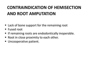
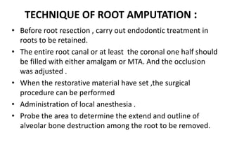
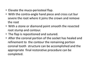
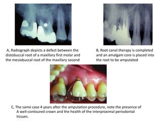
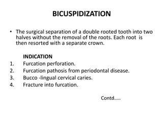
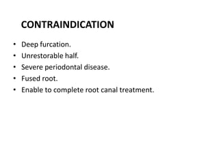
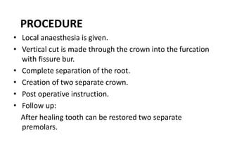
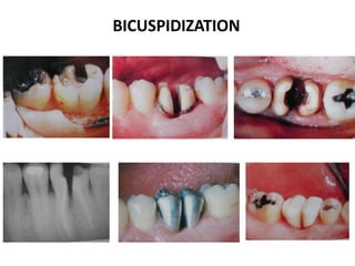
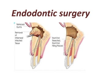
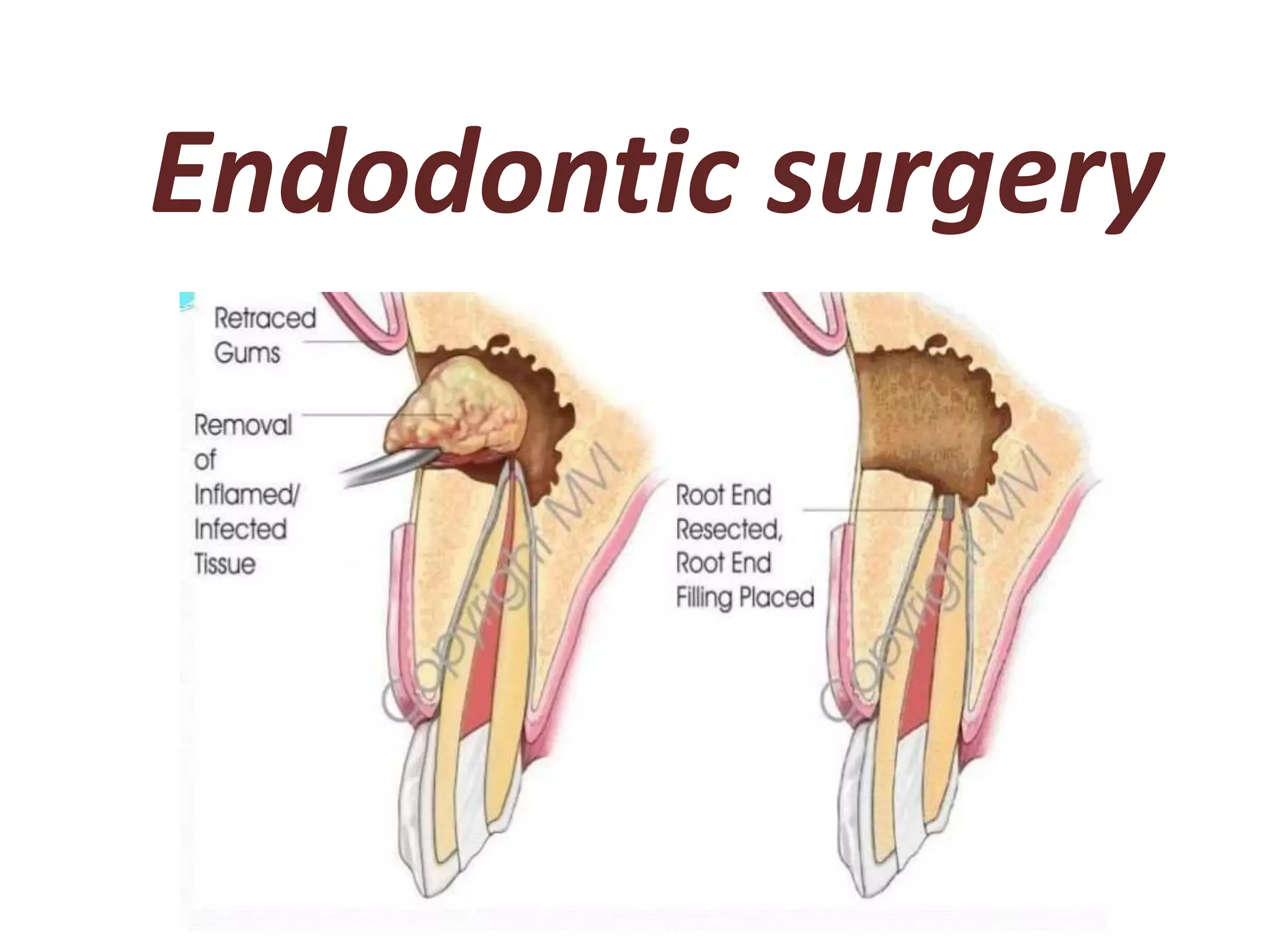
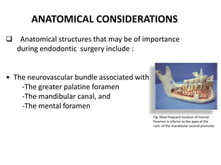
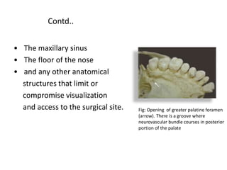
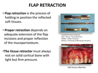
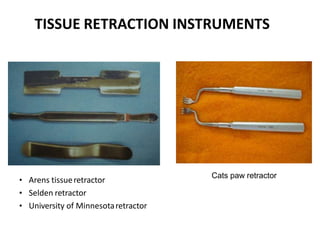
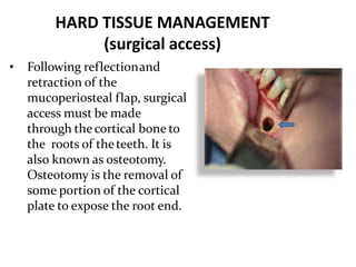
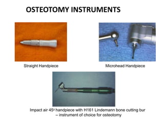
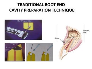
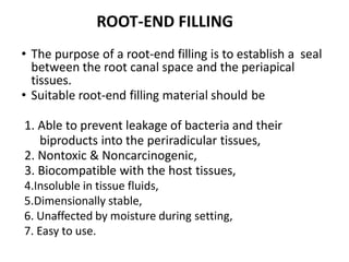
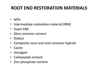
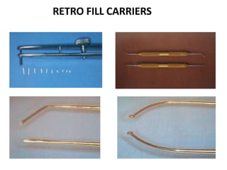
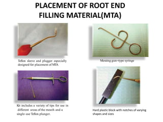
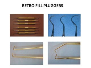
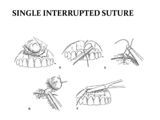
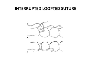
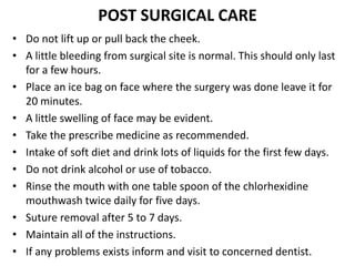
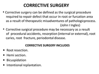
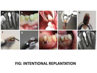
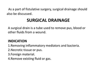
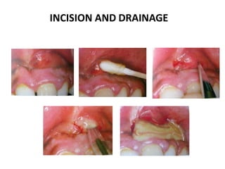
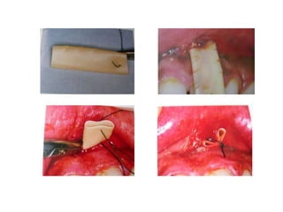
The document discusses endodontic surgery, including:
- Indications for endodontic surgery when non-surgical retreatment has failed or is not feasible.
- Classification, armamentarium, treatment planning considerations, and stages of surgical endodontics including flap design, osteotomy, periradicular curettage, root-end resection, and root-end preparation and filling.
- Key aspects are proper anesthesia, hemostasis, management of soft and hard tissues to access the surgical site and root structure for periradicular procedures.
One of the most dreaded nightmares of any clinician is broken instruments in the midst of an endodontic treatment. NiTi rotary instruments show a high incidence of instrument fracture despite their favourable qualities.
A concise and brief presentation on cleaning and shaping of root canals. Colorful and well pictured. Ideal for UG students and PG students to get a good understanding of BMP techniques.
In this lecture I explain the basic concept of root canal filling or what called obturation. The lectures discuss different techniques used in that matter in step-by-step fashion and explanatory pictures.
It is directed to the level of undergraduate mind.
Endodontic surgery / / rotary endodontic courses by indian dental academyIndian dental academy
Welcome to Indian Dental Academy
The Indian Dental Academy is the Leader in continuing dental education , training dentists in all aspects of dentistry and offering a wide range of dental certified courses in different formats.
Indian dental academy has a unique training program & curriculum that provides students with exceptional clinical skills and enabling them to return to their office with high level confidence and start treating patients
State of the art comprehensive training-Faculty of world wide repute &Very affordable.
Objectives and rationale
Indications
Contraindications
False indications
Treatment planning and presurgical notes
Classification
Gutmann’s
Kim’s
Steps in endosurgery
Treatment planning & Presurgical notes
Mandatory investigations
Premedication
Local anaesthesia and hemostasis
Flap
Requirements of an ideal flap
Flap design
Semilunar flap
Vertical flaps
Horizontal flap
Ochsenbein-Luebke flap
Two-step or filling first technique
Disinfection immediately prior to filling
Preparation of surgical site
Soft tissue management
Opening the flap
Flap elevation
Flap retraction
Hard tissue considerations
Locating root apex
Osteotomy
Apical curettage
Apical rood end resection
Surgery from palatal access
Post-resection filling
Root end preparation
Root end filling materials
Reverse filling
Surgery for root fractures
Surgical management of internal resorption
Radisectomy and hemisection
Intentional replantation
Closure of surgical area
Repositioning of flap and compression
Needle selection
Suturing
Post surgical care
Maxillectomy and craniofacial resection Mamoon Ameen
all maxillectomy types in detail and maxillofacial resection ,indications ,contraindications ,preoperative asssessment and detail techniques and rehabilitations
This PowerPoint presentation delivers a technical analysis of the midface orthognathic procedure. Explore surgical techniques, anatomical considerations, and treatment objectives.
Indian Dental Academy: will be one of the most relevant and exciting training center with best faculty and flexible training programs for dental professionals who wish to advance in their dental practice,Offers certified courses in Dental implants,Orthodontics,Endodontics,Cosmetic Dentistry, Prosthetic Dentistry, Periodontics and General Dentistry.
Surgical endodontics(Apicectomy) by Dr. Amit Suryawanshi .Oral & Maxillofac...All Good Things
Description:
Hi. This is Dr. Amit T. Suryawanshi. Oral & Maxillofacial surgeon from Pune, India. I am here on slideshare.com to share some of my own presentations presented at various levels in the field of OMFS. Hope this would somehow be helpful to you making your presentations. All the best & your replies are welcomed!
Surgical endodontics (Apicectomy) by Dr. Amit T. Suryawanshi, Oral Surgeon, ...All Good Things
Hi. This is Dr. Amit T. Suryawanshi. Oral & Maxillofacial surgeon from Pune, India. I am here on slideshare.com to share some of my own presentations presented at various levels in the field of OMFS. Hope this would somehow be helpful to you making your presentations. All the best.
Peri implantitis treatment with regenerative approachajayashreep
This study evaluates the clinical results and compare reentry hard tissue measurements following regenerative surgery after strict implant decontamination peri-implantitis cases.
Being a Periodontist, what necessary is to know what actually periodontal flaps are. So this presentation might provide you an insight into the field of periodontics as well as periodontal flaps.
endodontic Surgery /certified fixed orthodontic courses by Indian dental aca...Indian dental academy
Welcome to Indian Dental Academy
The Indian Dental Academy is the Leader in continuing dental education , training dentists in all aspects of dentistry and offering a wide range of dental certified courses in different formats.
Indian dental academy has a unique training program & curriculum that provides students with exceptional clinical skills and enabling them to return to their office with high level confidence and start treating patients
State of the art comprehensive training-Faculty of world wide repute &Very affordable.
Couples presenting to the infertility clinic- Do they really have infertility...Sujoy Dasgupta
Dr Sujoy Dasgupta presented the study on "Couples presenting to the infertility clinic- Do they really have infertility? – The unexplored stories of non-consummation" in the 13th Congress of the Asia Pacific Initiative on Reproduction (ASPIRE 2024) at Manila on 24 May, 2024.
Anti ulcer drugs and their Advance pharmacology ||
Anti-ulcer drugs are medications used to prevent and treat ulcers in the stomach and upper part of the small intestine (duodenal ulcers). These ulcers are often caused by an imbalance between stomach acid and the mucosal lining, which protects the stomach lining.
||Scope: Overview of various classes of anti-ulcer drugs, their mechanisms of action, indications, side effects, and clinical considerations.
These lecture slides, by Dr Sidra Arshad, offer a quick overview of physiological basis of a normal electrocardiogram.
Learning objectives:
1. Define an electrocardiogram (ECG) and electrocardiography
2. Describe how dipoles generated by the heart produce the waveforms of the ECG
3. Describe the components of a normal electrocardiogram of a typical bipolar leads (limb II)
4. Differentiate between intervals and segments
5. Enlist some common indications for obtaining an ECG
Study Resources:
1. Chapter 11, Guyton and Hall Textbook of Medical Physiology, 14th edition
2. Chapter 9, Human Physiology - From Cells to Systems, Lauralee Sherwood, 9th edition
3. Chapter 29, Ganong’s Review of Medical Physiology, 26th edition
4. Electrocardiogram, StatPearls - https://www.ncbi.nlm.nih.gov/books/NBK549803/
5. ECG in Medical Practice by ABM Abdullah, 4th edition
6. ECG Basics, http://www.nataliescasebook.com/tag/e-c-g-basics
Title: Sense of Smell
Presenter: Dr. Faiza, Assistant Professor of Physiology
Qualifications:
MBBS (Best Graduate, AIMC Lahore)
FCPS Physiology
ICMT, CHPE, DHPE (STMU)
MPH (GC University, Faisalabad)
MBA (Virtual University of Pakistan)
Learning Objectives:
Describe the primary categories of smells and the concept of odor blindness.
Explain the structure and location of the olfactory membrane and mucosa, including the types and roles of cells involved in olfaction.
Describe the pathway and mechanisms of olfactory signal transmission from the olfactory receptors to the brain.
Illustrate the biochemical cascade triggered by odorant binding to olfactory receptors, including the role of G-proteins and second messengers in generating an action potential.
Identify different types of olfactory disorders such as anosmia, hyposmia, hyperosmia, and dysosmia, including their potential causes.
Key Topics:
Olfactory Genes:
3% of the human genome accounts for olfactory genes.
400 genes for odorant receptors.
Olfactory Membrane:
Located in the superior part of the nasal cavity.
Medially: Folds downward along the superior septum.
Laterally: Folds over the superior turbinate and upper surface of the middle turbinate.
Total surface area: 5-10 square centimeters.
Olfactory Mucosa:
Olfactory Cells: Bipolar nerve cells derived from the CNS (100 million), with 4-25 olfactory cilia per cell.
Sustentacular Cells: Produce mucus and maintain ionic and molecular environment.
Basal Cells: Replace worn-out olfactory cells with an average lifespan of 1-2 months.
Bowman’s Gland: Secretes mucus.
Stimulation of Olfactory Cells:
Odorant dissolves in mucus and attaches to receptors on olfactory cilia.
Involves a cascade effect through G-proteins and second messengers, leading to depolarization and action potential generation in the olfactory nerve.
Quality of a Good Odorant:
Small (3-20 Carbon atoms), volatile, water-soluble, and lipid-soluble.
Facilitated by odorant-binding proteins in mucus.
Membrane Potential and Action Potential:
Resting membrane potential: -55mV.
Action potential frequency in the olfactory nerve increases with odorant strength.
Adaptation Towards the Sense of Smell:
Rapid adaptation within the first second, with further slow adaptation.
Psychological adaptation greater than receptor adaptation, involving feedback inhibition from the central nervous system.
Primary Sensations of Smell:
Camphoraceous, Musky, Floral, Pepperminty, Ethereal, Pungent, Putrid.
Odor Detection Threshold:
Examples: Hydrogen sulfide (0.0005 ppm), Methyl-mercaptan (0.002 ppm).
Some toxic substances are odorless at lethal concentrations.
Characteristics of Smell:
Odor blindness for single substances due to lack of appropriate receptor protein.
Behavioral and emotional influences of smell.
Transmission of Olfactory Signals:
From olfactory cells to glomeruli in the olfactory bulb, involving lateral inhibition.
Primitive, less old, and new olfactory systems with different path
HOT NEW PRODUCT! BIG SALES FAST SHIPPING NOW FROM CHINA!! EU KU DB BK substit...GL Anaacs
Contact us if you are interested:
Email / Skype : kefaya1771@gmail.com
Threema: PXHY5PDH
New BATCH Ku !!! MUCH IN DEMAND FAST SALE EVERY BATCH HAPPY GOOD EFFECT BIG BATCH !
Contact me on Threema or skype to start big business!!
Hot-sale products:
NEW HOT EUTYLONE WHITE CRYSTAL!!
5cl-adba precursor (semi finished )
5cl-adba raw materials
ADBB precursor (semi finished )
ADBB raw materials
APVP powder
5fadb/4f-adb
Jwh018 / Jwh210
Eutylone crystal
Protonitazene (hydrochloride) CAS: 119276-01-6
Flubrotizolam CAS: 57801-95-3
Metonitazene CAS: 14680-51-4
Payment terms: Western Union,MoneyGram,Bitcoin or USDT.
Deliver Time: Usually 7-15days
Shipping method: FedEx, TNT, DHL,UPS etc.Our deliveries are 100% safe, fast, reliable and discreet.
Samples will be sent for your evaluation!If you are interested in, please contact me, let's talk details.
We specializes in exporting high quality Research chemical, medical intermediate, Pharmaceutical chemicals and so on. Products are exported to USA, Canada, France, Korea, Japan,Russia, Southeast Asia and other countries.
Factory Supply Best Quality Pmk Oil CAS 28578–16–7 PMK Powder in Stockrebeccabio
Factory Supply Best Quality Pmk Oil CAS 28578–16–7 PMK Powder in Stock
Telegram: bmksupplier
signal: +85264872720
threema: TUD4A6YC
You can contact me on Telegram or Threema
Communicate promptly and reply
Free of customs clearance, Double Clearance 100% pass delivery to USA, Canada, Spain, Germany, Netherland, Poland, Italy, Sweden, UK, Czech Republic, Australia, Mexico, Russia, Ukraine, Kazakhstan.Door to door service
Hot Selling Organic intermediates
The prostate is an exocrine gland of the male mammalian reproductive system
It is a walnut-sized gland that forms part of the male reproductive system and is located in front of the rectum and just below the urinary bladder
Function is to store and secrete a clear, slightly alkaline fluid that constitutes 10-30% of the volume of the seminal fluid that along with the spermatozoa, constitutes semen
A healthy human prostate measures (4cm-vertical, by 3cm-horizontal, 2cm ant-post ).
It surrounds the urethra just below the urinary bladder. It has anterior, median, posterior and two lateral lobes
It’s work is regulated by androgens which are responsible for male sex characteristics
Generalised disease of the prostate due to hormonal derangement which leads to non malignant enlargement of the gland (increase in the number of epithelial cells and stromal tissue)to cause compression of the urethra leading to symptoms (LUTS
TEST BANK for Operations Management, 14th Edition by William J. Stevenson, Ve...kevinkariuki227
TEST BANK for Operations Management, 14th Edition by William J. Stevenson, Verified Chapters 1 - 19, Complete Newest Version.pdf
Pulmonary Thromboembolism - etilogy, types, medical- Surgical and nursing man...VarunMahajani
Disruption of blood supply to lung alveoli due to blockage of one or more pulmonary blood vessels is called as Pulmonary thromboembolism. In this presentation we will discuss its causes, types and its management in depth.
2. Weekly seminar on
Endodontic Surgery
Honorable Chair Person-
Prof. Dr. Umme Kulsum Rosy
Head of the department of Conservative Dentistry &
Endodontics, DDCH.
Supervised by- Presented by-
Dr. Raihana Nahar, Dr. Jannatul Ferdousy
Assistant Professor, Dr. Siffat ara,
Department of Conservative Dr. Rusvina Akter &
Dentistry & Endodontics , DDCH. Dr. Afrin Sultana.
(PGT trainee)
3. CONTENTS
Introduction
Definition
Rationale
Objectives
Indications
Contraindications
Classification
Armamentarium for periradicular surgery
Treatment planning for periradicular surgery
Stages in surgical endodontics
Post operative instruction
Corrective surgery
Conclusion
4. INTRODUCTION
Surgical intervention is required where endodontic
treatment has failed and tooth is to be retained rather than
extracted. The percentage of success of endodontic
treatment has been consistently high but failure may be arise
due to infection, poor access cavity preparation, inadequate
instrumentation, obturation , missed canals and coronal
leakage . So in this cases, Surgical endodontics is needed to
save the tooth.
5. DEFINITION:
A surgical procedure related to the problem of the pulp less
or periodontally involved tooth, requiring root amputation
and endodontic therapy.
(John I Ingles)
Removal of tissues other than the contents of root canal to
retain a tooth with pulpal or periapical involvement.
(Franklin weine)
6. RATIONALE:
• To remove the causative agents of periradicular
pathology.
• To restore the periodontium to a state of biologic and
functional health.
OBJECTIVES:
• To ensure placement of a proper seal between
periodontium and root canal foramina.
7. INDICATION
• Failure of non-surgical
retreatment (treatment has
been rendered at least two
times)
• If retreatment is not feasible.
• When a biopsy is necessary.
• Need for surgical drainage.
• Iatrogenic error
• Corrective surgery
Fig: Surgical drainage
Fig: Radiograph showing failure of
non surgical treatment.
8. CONTRAINDICATION
•Patient’s Medical status
Bleeding disorder
Recent heart surgery
Cancer
Old/ill patient
•Anatomic consideration
Proximity to nerve bundles
Second mandibular molar
Maxillary sinus
Fig: opening of greater palatine foramen(arrow).
There is a groove where neurovascular bundle
courses in posterior portion of the palate.
9. •Periodontal status : Inadequate
periodontal support and active
uncontrollable periodontal disease
•Poor restorability with a post
endodontic restoration.
10. Classification of Endodontic surgery
Ingle's classification
•Fistulative surgery
a.Incision and drainage
b. Cortical trephination
c. Decompression procedures
•Periradicular surgery (primary focus of this chapter)
a.Curettage
b.Root-end resection
c.Root-end preparation
d.Root-end filling
•Corrective surgery
a.Perforation repair
i.Mechanical (iatrogenic)
ii.Resorptive
b.Periodontal management
i.Root resection
ii.Tooth resection
c.Intentional replantation
12. Before going to any surgery we should have
knowledge about the classification of the
surgical flap.
13. CLASSIFICATION OF SURGICAL FLAP
1. Full thickness flaps:
(consists of epithelium ,connective tissue
and periosteum)
A. Triangular( one vertical releasing incision)
B. Rectangular( Two vertical releasing incision)
C. Trapezoidal (broad based rectangular )
D. Horizontal ( no vertical releasing incision)
E. Papilla-base flap
2. Partial thickness flaps:
(consists of epithelium and connective
tissue)
A. Sub marginal curved ( semilunar )
B. Sub marginal scalloped rectangular ( luebke-
ochsenbein)
14. TRIANGULAR FLAP
(One vertical releasing incision)
INDICATIONS
• It is recommended for maxillary
incisors and posterior teeth.
15. RECTANGULAR FLAP
(Two vertical releasing incision)
INDICATIONS
• Mandibular anteriors
• Multiple teeth
• Teeth with long roots like
maxillary canines
16. TRAPEZOIDAL FLAP
( Broad based rectangular)
• Similar to rectangular except the
2 vertical incisions intersect the
horizontal incision at an obtuse
angle .
• It is used to create a broad based
flap with the vestibular part wider
than the sulcular portion.
17. HORIZONTAL FLAP
(No vertical releasing incision)
Horizontal intrasulcular incision
with no vertical releasing incision.
INDICATIONS
• Repair of cervical defects (root perforations,
resorption , caries)
• Hemi sections and Root amputation
19. SUBMARGINAL SCALLOPED RECTANGULAR /
LUEBKEE OCHSENBEIN FLAP
• Modification of rectangular flap
• Horizontal incision is placed in buccal/labial
area, attached gingiva is scalloped – follows
the contour of marginal gingiva.
INDICATIONS
• Prosthetic crowns
• Periradicular surgery of anterior region
• Longer roots
20. 1. Anatomical Considerations
2. Pre-surgical patient management
3. Need for profound local anesthesia and hemostasis.
4. Management of soft tissue
5. Management of hard tissues
6. Surgical access, both visual and operative
7. Periradicular curettage
8. Access to root structure
9. Root-end resection
10.Root end preparation
11.Root-end restoration
12.Soft-tissue repositioning and suturing
13.Postsurgical care
TREATMENT PLANNING FOR PERIRADICULAR SURGERY
21. ANATOMICAL CONSIDERATIONS
Anatomical structures that may be of importance
during endodontic surgery include :
• The neurovascular bundle associated with
-The greater palatine foramen
-The mandibular canal, and
-The mental foramen
Fig: Most frequent location of mental
foramen is inferior to the apex of the
root of the mandibular second premolar
22. Contd..
• The maxillary sinus
• The floor of the nose
• and any other anatomical
structures that limit or
compromise visualization
and access to the surgical site. Fig: Opening of greater palatine foramen
(arrow). There is a groove where
neurovascular bundle courses in posterior
portion of the palate
23. • Proper history taking is the first key for success of any
surgical procedure.
• Patient should be evaluated for major systemic disorders
(cardiovascular, renal, hepatic, digestive, immune and
skeletal muscle) which may contraindicate or alter approach
to surgery.
• Periodontal evaluation
• Premedication for patient in normal or in presence of the
above medical conditions should be given priority and
consulted with physician.
PRESURGICAL PATIENT MANAGEMENT
24. • Premedication like sedatives or hypnotics, systemic
antibiotics etc for patient in order to improve
accessibility also postsurgical healing.
• Patient preparation start with patient communication
regarding reason for surgery, risk involved and factors
which improve prognosis for successful outcome of
surgical procedure over non-surgical treatment.
• Mouth rinse should be started a day before surgery.
Contd..
28. HEMOSTASIS
Hemostasis is the process of forming clot in the wall of
damaged blood vessels.
Some of the hemostatic agents are:
• Hemostatic collagen
(eg: CollaPlug, CollaTape, and Helistat)
• Gelatin (eg: Gelfoam)
30. SOFT TISSUE MANAGEMENT
PRINCIPLES OF FLAP DESIGN
1. Avoid horizontal and severely angled vertical incisions.
2. Avoid incisions over radicular eminences.
3. Incisions should be placed and flaps repositioned over
solid bone.
4. Avoid incisions across major muscle attachments.
5. Tissue retractor should rest on solid bone.
31. 6. Extent of the horizontal incision should be adequate to
provide visual and operative access with minimal soft
tissue trauma.
7. The junction of the horizontal and vertical
incisions should either include or exclude the involved
interdental papilla.
8. The flap should include the entire mucoperiosteum
(full thickness): marginal, interdental and attached
gingiva, alveolar mucosa, and periosteum
Contd..
32. INCISION
Surgical incision is a cut made through the skin and soft
tissue to facilitate an operation or procedure.
Types of incisions
• Vertical incision
• Sulcular incision
• Semilunar incision
• Modified semilunar incision
34. Incisions for the majority of mucoperiosteal flaps for
periradicular surgery can be accomplished by using one or
more of four scalpel blades No. 11, No. 12, No. 15, and
No. 15-C.
Figure: Horizontal sulcular incision
with a No.11 Bard Parker scapel
blade.
Figure: Vertical incision with a No.
15 Bard Parker scapel blade
Contd..
35. FLAP REFLECTION
• Flap reflection is the process of separating the soft tissue (
Gingiva Mucosa and Periosteum ) from the surface of the
alveolar bone.
Fig: Periosteal elevators for flap reflection;
A, Initial elevation of flap; B, Flap partially reflected; C, Flap completely reflected.
37. • Flap retraction is the process of
holding in position the reflected
soft tissues.
• Proper retraction depends on
adequate extension of the flap
incisions and proper reflection
of the mucoperiosteum.
•The tissue retractor must always
rest on solid cortical bone with
light but firm pressure.
Soft tissue reflection
Endodontic tissue retractors
(Top—Arenas Tissue Retractor ;
Middle—Selden retractor;
Bottom—University of Minnesota retractor).
FLAP RETRACTION
38. TISSUE RETRACTION INSTRUMENTS
• Arens tissueretractor
• Selden retractor
• University of Minnesotaretractor
Cats paw retractor
39. HARD TISSUE MANAGEMENT
(surgical access)
• Following reflectionand
retraction of the
mucoperiosteal flap, surgical
access must be made
through the cortical bone to
the roots of theteeth. It is
also known as osteotomy.
Osteotomy is the removal of
some portion of the cortical
plate to expose the root end.
42. ROOT-END LOCATION
A number of factors should be considered before
locating the root end:
• Determine the location of the bony window.
• The angle of the crown to the root should be assessed.
• When a root prominence or eminence is present in the
cortical plate , the root angulation and position can be
determined more easily.
• Measurement of the entire tooth length on well angled
radiograph and transferred to the surgical site by the use
of a sterile millimeter ruler.
43. • Once the root has been located and
identified, the bone covering the root
is slowly and carefully removed with
light brush strokes (using surgical hand
piece) and should continue this
process in an apical direction until the
root apex is identified.
Contd..
• When the cortical plate is intact, locate the
body of the root from coronal to the apex
because the bone covering the root is
thinner.
Steps
44. The root surface can be distinguished from the
surrounding osseous tissue( according to Barnes) by
following factors:
(1) Root structure generally has a yellowish color.
(2) Roots do not bleed when probed.
(3) Root texture is smooth and hard as compared to the
granular and porous nature of bone, and
(4) The root is surrounded by the periodontal ligament..
45. Hard tissue management in endodontic surgery involves 3
stages:
1. Trephination
2. Periradicular curretage
3. Periradicular surgery
(i) Root end resection.
(ii) Root end preparation & filling
STAGES OF HARD TISSUE
MANAGEMENT
46. TREPHINATION
• It is the perforation made through the cortical plate or
apical foramen to accomplish the release of pressure in the
periapical area from the accumulation of exudate within
the alveolar bone.
INDICATION
• This technique is employed in cases of periapical abscess in
which there is no swelling or drainage but much pain.
Types:
1. Apical trephination
2. Cortical trephination
47. APICAL TREPHINATION
• Penetration of the apical foramen with a small
endodontic file and enlarging the apical opening to a 20 or
number 25 file to allow drainage from the periradicular
lesion into the canal space.
• The treatment of choice for these patients is drainage
through the root canal system (apical trephination)
whenever possible.
48. CORTICAL TREPHINATION
It is a procedure involving the perforation of the
cortical plate to accomplish the release of pressure
from the accumulation of exudate within the alveolar
bone.
contd..
49. PROCEDURE
• Small incision is made over the periapical
region.
• Flap is reflected and bone is examined.
• Radiograph is taken with radiopaque marker
for confirmation. So that there is no chance of
penetration in the wrong area.
contd..
50. PERIRADICULAR CURETTAGE
Removal of a cyst, granuloma or periradicular inflammatory
tissue from its pathological bony crypt and fragments pieces
of death bone or debris from a tooth socket by using various
sizes and shapes of sharp surgical bone curettes and angled
periodontal curettes.
52. PERIAPICAL CURETTAGE TECHNIQUE
• When osteotomy has been
completed then pathosis present
in the periapical area has to be
curetted with curetters.
• The entire tissue mass is removed
by inserting the bone curette
between soft tissue mass and
lateral wall of the bony crypt.
Contd..
53. • Concave surface of the curette facing the bone.
• Ones the soft tissue lesion has been freed from the
periphery the bone curette should be turned with the
concave portion towards the soft tissue and used in a
scraping manner to free the tissue from the bony
crypt.
Contd..
54. PERIRADICULAR SURGERY
ROOT-END RESECTION (APICOECTOMY)
• Many authors have advocated periradicular curettage is
the definitive treatment in endodontic surgery without
root-end resection.
• Their rationale was to maintain cemental covering of the
root surface and root length as possible for tooth stability..
55. It is the ablasion of apical portion of root end and attached
soft tissue.
INDICATION
ROOT END RESECTION
1.Failed root canal treatment.
2.Large persistent lesions
after root canal treatment.
Large unresolved lesions after
root canal treatment :
58. • Factors to be considered before performing root
end resection:
(1) Instrumentation,
(2) Extent of the root end resection,
(3) Angle of the resection.
59. INSTRUMENTATION
• Ingle et al. recommended that root-end resection is best
accomplished by the use of tapered fissure bur or round
bur in a low-speed straight handpiece.
• Gutmann and Harrison, have suggested the use of a
high-speed handpiece and a surgical length plain fissure
bur.
60. EXTENT OF ROOT END RESECTION:
• Average length of root resection is 3mm which is
considered enough to eliminate the source of infection.
61. • It should be 0-10° or perpendicular to the
long axis of the tooth facing toward the
buccal or facial aspect of the root,this 10
bevel conserves the root structure,
maintains a better crown/root ratio and
increases the ability to visualize important
lingual anatomy.
( But 30 °-45 bevel removes more root
structure,increase the exposure of more
dentinal tubules and increases the
probability of overlooking important lingual
anatomy.)
ANGLE OF RESECTION
62. ROOT CONDITIONING
PURPOSE:
• Removes smear layer and improves the mechanical
adhesion of retrograde fillings.
• Exposes the dentine tetra acetic-acid.
CONDITIONERS USED:
• 15-24% EDTA-Ph 7.3 (best)
• Tetracycline Hcl-Ph 1%
• 50% Citric acid
63. • Factors to be considered during performing root end
resection:
1. Visual and operative access to the surgical site.
2. Anatomy of the root (shape, length, curvature).
3. Number of canals and their position in the root
4. Need to place a root-end filling surrounded by
solid dentin.
5. Presence and location of procedural error
6. Presence and extent of periodontal defects.
64. ROOT END PREPARATION:
PURPOSE
• To create a class-1 cavity(depth 2-3mm) to receive a root
end filling .
OBJECTIVE
• It must be placed paralal to the long axis of the root.
INSTRUMENT
• Small round bur or inverted cone bur.
• Ultrasonic tip.
• Straight low-speed hand piece.
66. • The purpose of a root-end filling is to establish a seal
between the root canal space and the periapical
tissues.
• Suitable root-end filling material should be
1. Able to prevent leakage of bacteria and their
biproducts into the periradicular tissues,
2. Nontoxic & Noncarcinogenic,
3. Biocompatible with the host tissues,
4.Insoluble in tissue fluids,
5.Dimensionally stable,
6. Unaffected by moisture during setting,
7. Easy to use.
ROOT-END FILLING
67. ROOT END RESTORATION MATERIALS
• MTA
• Intermediate restorative material (IRM)
• Super EBA
• Glass ionomer cement
• Dialect
• Composite resin and resin ionomer hybrids
• Cavite
• Amalgam
• Carboxylate cement
• Zinc phosphate cement
69. Teflon sleeve and plugger especially
designed for placement of MTA
Messing gun–type syringe
PLACEMENT OF ROOT END
FILLING MATERIAL(MTA)
Kit includes a variety of tips for use in
different areas of the mouth and a
single- use Teflon plunger.
Hard plastic block with notches of varying
shapes and sizes
71. SOFT TISSUE REPOSITIONING
• The elevated muco periosteum gently replaced to its
original position with the incision lines approximated as
closely as possible
• Tissue compression : Using a surgical gauze moistened
with sterile saline, gently apply firm pressure to the
flapped tissue for 2-3 mins before suturing
Enhances intra-vascular clotting in the severe blood
vessels.
75. POST SURGICAL CARE
• Do not lift up or pull back the cheek.
• A little bleeding from surgical site is normal. This should only last
for a few hours.
• Place an ice bag on face where the surgery was done leave it for
20 minutes.
• A little swelling of face may be evident.
• Take the prescribe medicine as recommended.
• Intake of soft diet and drink lots of liquids for the first few days.
• Do not drink alcohol or use of tobacco.
• Rinse the mouth with one table spoon of the chlorhexidine
mouthwash twice daily for five days.
• Suture removal after 5 to 7 days.
• Maintain all of the instructions.
• If any problems exists inform and visit to concerned dentist.
76. CORRECTIVE SURGERY
• Corrective surgery can be defined as the surgical procedure
required to repair defect that occur in root or furcation area
as a result of therapeutic misadventures of pathologicprocess.
(John I Ingles)
• Corrective surgical procedure may be necessary as a result
of procedural accidents, resorption (internal or external), root
caries, root fracture, periodontal disease.
CORRECTIVE SURGERY INCLUDES
Root resection.
Hemi section.
Bicuspidation
Intentional replantation.
77. • Root resection is the process by which one or more
roots of a tooth are removed at the level of the furcation
and leaves the crown and remaining roots functional.
• Root amputation procedure may be indicated when one
root of the multi rooted tooth that can not be retained and
the other roots have adequate periodontal support and the
remaining crown structure can be restored.
(John I Ingles)
ROOT AMPUTATION
78. • These procedure are most frequently indicated when one
root of a maxillary molar must be eliminated.
79. • Surgical removal of one root and overlying
crown.
When root removal is indicated in a mandibular
molar. Because of a vertical root fracture ,
procedural error or pathologic resorptive process .
Hemisection is usually treatment of choice.
HEMISECTION
80. INDICATION OF HEMISECTION AND ROOT
AMPUTATION:
• Extensive bone loss in relation to root where periodontal
therapy cannot correct it.
• Severely curved canal which cannot be treated.
• Extensive calcifications in root.
• Fracture of one root which does not involve other root.
• Resorption, caries , or perforation involving one root.
81. CONTRAINDICATION OF HEMISECTION
AND ROOT AMPUTATION
Lack of bone support for the remaining root
Fused root
If remaining roots are endodontically inoperable.
Root in close proximity to each other.
Uncooperative patient.
82. TECHNIQUE OF ROOT AMPUTATION :
• Before root resection , carry out endodontic treatment in
roots to be retained.
• The entire root canal or at least the coronal one half should
be filled with either amalgam or MTA. And the occlusion
was adjusted .
• When the restorative material have set ,the surgical
procedure can be performed
• Administration of local anesthesia .
• Probe the area to determine the extend and outline of
alveolar bone destruction among the root to be removed.
83. • Elevate the muco-periosteal flap.
• With the contra-angle hand piece and cross cut bur
severe the root where it joins the crown and remove
the root
• With a stone or diamond point smooth the resected
root stump and contour.
• The flap is repositioned and sutured.
• After the coronal portion of the socket has healed and
refinement to the contour the remaining portion
coronal tooth structure can be accomplished and the
appropriate final restorative procedure can be
completed.
84. A, Radiograph depicts a defect between the
distobuccal root of a maxillary first molar and
the mesiobuccal root of the maxillary second
B, Root canal therapy is completed
and an amalgam core is placed into
the root to be amputated
C, The same case 4 years after the amputation procedure, note the presence of
A well-contoured crown and the health of the interproximal periodontal
tissues.
85. • The surgical separation of a double rooted tooth into two
halves without the removal of the roots. Each root is
then resorted with a separate crown.
INDICATION
1. Furcation perforation.
2. Furcation pathosis from periodontal disease.
3. Bucco -lingual cervical caries.
4. Fracture into furcation.
Contd…..
BICUSPIDIZATION
87. PROCEDURE
• Local anaesthesia is given.
• Vertical cut is made through the crown into the furcation
with fissure bur.
• Complete separation of the root.
• Creation of two separate crown.
• Post operative instruction.
• Follow up:
After healing tooth can be restored two separate
premolars.
89. INTENTIONAL REPLANTATION
• It is the purposeful removal of a tooth to repair a defect
or cause of a treatment failure and then returning the
tooth to its original socket.
(John I Ingles)
INDICATIONS
• Difficult access.
• Anatomic limitations.
• Perforation in areas notaccessible surgically.
• Failed apical surgery.
• Apical surgery creatingdefect.
• Accidental avulsion( unintentional replantation).
contd…
91. PROCEDURE
• Anesthesia is given.
• Carefully loosen the tooth without minimizing
injuries to the soft tissue.
• After tooth is removed and soaked in a solution
during the entire extra oral time.
• The roots are thoroughly examined with
magnification.
• Gently curette the socket avoid to damage to the
socket and periodontal ligament.
• Root resection and root preparation.
92. • An Appropriate root end filling material will be
placed.
• The root is ready to be replaced in its socket.
• Before replacement the socket is gently ringe
with normal saline.
• Tooth is carefully and slowly replaced to the
socket in its proper orientation.
• Occlusion is checked and splinting is done.
• Splinting is removed after 2-3 weeks.
• Post operative instruction.
• Re-evaluate for removing stabilization.
• Follow up (1,3,6 and 12 month)
94. SURGICAL DRAINAGE
A surgical drain is a tube used to remove pus, blood or
other fluids from a wound.
INDICATION
1.Removing inflammatory mediators and bacteria.
2.Necrotic tissue or pus.
3.Foreign material.
4.Remove existing fluid or gas.
As a part of fistulative surgery, surgical drainage should
also be discussed.
97. POST SURGICAL COMPLICATION
INTRAOPERATIVE
• Bleeding – control with local application of adrenaline
pack, pressure pack.
• Damage to the neighboring root.
• Entry into sinus/ inferior alveolar canal.
POSTOPERATIVE
• Abscess formation.
• Fenestration, sinus tract formation. Increased
mobility of tooth
• Root resorption.
• Loss of bone.
• Ankylosis.
98. CONCLUSION
During the last 20 years, endodontics has encountered
dramatic shift in the use of periradicular surgery. Previously,
periradicular surgery was commonly considered as the
treatment of choice when nonsurgical treatment had failed
but now a days periradicular surgery has become very
selective in contemporary dental practice.
99. REFERENCE
Grossman:Grossmans Endodontic practice 13th .edn;new
Delhi;2014;wolters Kluwer pvt.ltd,Diseases of periradicular
tissues.p.112-143
Endodontics by John I. Ingle, Leif Bakland 5th Edition
Microsurgery in Endodontics: Syngkuc Kim
Surgical Endodontics: Guttman and Harrison: Mosby:1994.
Garg N Garg A :Textbook of endodontics
All picture and radiograph from textbooks and website.