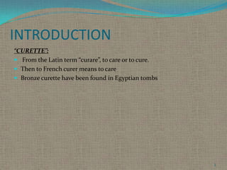
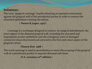
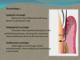
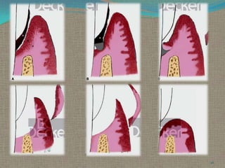
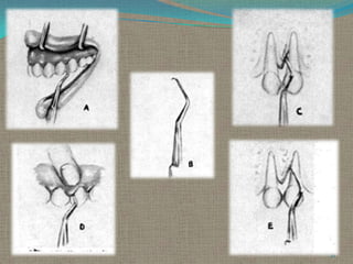
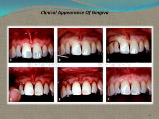
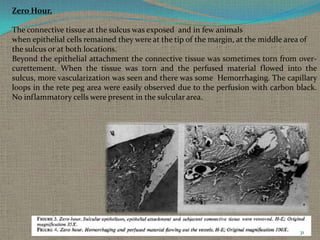
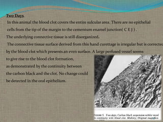
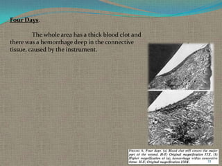
The document discusses gingival curettage, which involves using instruments to remove diseased soft tissue from periodontal pockets. It defines curettage and provides the history of the procedure. The basic technique is described as using curettes to scrape the inner lining of the pocket to remove ulcerated epithelium and damaged connective tissue. Indications include edematous pockets aiming to reduce inflammation and shrink tissue. Healing after curettage is examined through a study showing revascularization of the wound site over time.
Periodontitis is a complex infection initiated by bacteria –tissue destruction.
Host: the organism from which a parasite obtains its nourishment/ an individual who receives a graft
Modulation: the alteration of function or status of something in response to a stimulus or an altered physical or chemical environment
Every periodontal surgical procedure has its own indications. With proper knowledge of the etiology of the disease, correct diagnosis and treatment planning, the clinician is able to draw predictable success with periodontal flap surgery.
mucogingival surgery or plastic surgery of muco-gingival tissue is a surgical procedure targeted to correct and eliminate anatomic, developmental and traumatic alterations of gingiva.
A brief description of all topics to recent advances,SDD, host modulation and diabetes, host modulation in smokers, chemically modified tetracyclines, bisphosphonates
Gingival Curettage / /certified fixed orthodontic courses by Indian dental ac...Indian dental academy
Welcome to Indian Dental Academy
The Indian Dental Academy is the Leader in continuing dental education , training dentists in all aspects of dentistry and offering a wide range of dental certified courses in different formats.
Indian dental academy has a unique training program & curriculum that provides students with exceptional clinical skills and enabling them to return to their office with high level confidence and start treating patients
State of the art comprehensive training-Faculty of world wide repute &Very affordable.
Endodontic surgery / / rotary endodontic courses by indian dental academyIndian dental academy
Welcome to Indian Dental Academy
The Indian Dental Academy is the Leader in continuing dental education , training dentists in all aspects of dentistry and offering a wide range of dental certified courses in different formats.
Indian dental academy has a unique training program & curriculum that provides students with exceptional clinical skills and enabling them to return to their office with high level confidence and start treating patients
State of the art comprehensive training-Faculty of world wide repute &Very affordable.
Surgical endodontics(Apicectomy) by Dr. Amit Suryawanshi .Oral & Maxillofac...All Good Things
Description:
Hi. This is Dr. Amit T. Suryawanshi. Oral & Maxillofacial surgeon from Pune, India. I am here on slideshare.com to share some of my own presentations presented at various levels in the field of OMFS. Hope this would somehow be helpful to you making your presentations. All the best & your replies are welcomed!
Surgical endodontics (Apicectomy) by Dr. Amit T. Suryawanshi, Oral Surgeon, ...All Good Things
Hi. This is Dr. Amit T. Suryawanshi. Oral & Maxillofacial surgeon from Pune, India. I am here on slideshare.com to share some of my own presentations presented at various levels in the field of OMFS. Hope this would somehow be helpful to you making your presentations. All the best.
June 3, 2024 Anti-Semitism Letter Sent to MIT President Kornbluth and MIT Cor...Levi Shapiro
Letter from the Congress of the United States regarding Anti-Semitism sent June 3rd to MIT President Sally Kornbluth, MIT Corp Chair, Mark Gorenberg
Dear Dr. Kornbluth and Mr. Gorenberg,
The US House of Representatives is deeply concerned by ongoing and pervasive acts of antisemitic
harassment and intimidation at the Massachusetts Institute of Technology (MIT). Failing to act decisively to ensure a safe learning environment for all students would be a grave dereliction of your responsibilities as President of MIT and Chair of the MIT Corporation.
This Congress will not stand idly by and allow an environment hostile to Jewish students to persist. The House believes that your institution is in violation of Title VI of the Civil Rights Act, and the inability or
unwillingness to rectify this violation through action requires accountability.
Postsecondary education is a unique opportunity for students to learn and have their ideas and beliefs challenged. However, universities receiving hundreds of millions of federal funds annually have denied
students that opportunity and have been hijacked to become venues for the promotion of terrorism, antisemitic harassment and intimidation, unlawful encampments, and in some cases, assaults and riots.
The House of Representatives will not countenance the use of federal funds to indoctrinate students into hateful, antisemitic, anti-American supporters of terrorism. Investigations into campus antisemitism by the Committee on Education and the Workforce and the Committee on Ways and Means have been expanded into a Congress-wide probe across all relevant jurisdictions to address this national crisis. The undersigned Committees will conduct oversight into the use of federal funds at MIT and its learning environment under authorities granted to each Committee.
• The Committee on Education and the Workforce has been investigating your institution since December 7, 2023. The Committee has broad jurisdiction over postsecondary education, including its compliance with Title VI of the Civil Rights Act, campus safety concerns over disruptions to the learning environment, and the awarding of federal student aid under the Higher Education Act.
• The Committee on Oversight and Accountability is investigating the sources of funding and other support flowing to groups espousing pro-Hamas propaganda and engaged in antisemitic harassment and intimidation of students. The Committee on Oversight and Accountability is the principal oversight committee of the US House of Representatives and has broad authority to investigate “any matter” at “any time” under House Rule X.
• The Committee on Ways and Means has been investigating several universities since November 15, 2023, when the Committee held a hearing entitled From Ivory Towers to Dark Corners: Investigating the Nexus Between Antisemitism, Tax-Exempt Universities, and Terror Financing. The Committee followed the hearing with letters to those institutions on January 10, 202
Read| The latest issue of The Challenger is here! We are thrilled to announce that our school paper has qualified for the NATIONAL SCHOOLS PRESS CONFERENCE (NSPC) 2024. Thank you for your unwavering support and trust. Dive into the stories that made us stand out!
Biological screening of herbal drugs: Introduction and Need for
Phyto-Pharmacological Screening, New Strategies for evaluating
Natural Products, In vitro evaluation techniques for Antioxidants, Antimicrobial and Anticancer drugs. In vivo evaluation techniques
for Anti-inflammatory, Antiulcer, Anticancer, Wound healing, Antidiabetic, Hepatoprotective, Cardio protective, Diuretics and
Antifertility, Toxicity studies as per OECD guidelines
How to Make a Field invisible in Odoo 17Celine George
It is possible to hide or invisible some fields in odoo. Commonly using “invisible” attribute in the field definition to invisible the fields. This slide will show how to make a field invisible in odoo 17.
Introduction to AI for Nonprofits with Tapp NetworkTechSoup
Dive into the world of AI! Experts Jon Hill and Tareq Monaur will guide you through AI's role in enhancing nonprofit websites and basic marketing strategies, making it easy to understand and apply.
Honest Reviews of Tim Han LMA Course Program.pptxtimhan337
Personal development courses are widely available today, with each one promising life-changing outcomes. Tim Han’s Life Mastery Achievers (LMA) Course has drawn a lot of interest. In addition to offering my frank assessment of Success Insider’s LMA Course, this piece examines the course’s effects via a variety of Tim Han LMA course reviews and Success Insider comments.
Model Attribute Check Company Auto PropertyCeline George
In Odoo, the multi-company feature allows you to manage multiple companies within a single Odoo database instance. Each company can have its own configurations while still sharing common resources such as products, customers, and suppliers.
Acetabularia Information For Class 9 .docxvaibhavrinwa19
Acetabularia acetabulum is a single-celled green alga that in its vegetative state is morphologically differentiated into a basal rhizoid and an axially elongated stalk, which bears whorls of branching hairs. The single diploid nucleus resides in the rhizoid.
Operation “Blue Star” is the only event in the history of Independent India where the state went into war with its own people. Even after about 40 years it is not clear if it was culmination of states anger over people of the region, a political game of power or start of dictatorial chapter in the democratic setup.
The people of Punjab felt alienated from main stream due to denial of their just demands during a long democratic struggle since independence. As it happen all over the word, it led to militant struggle with great loss of lives of military, police and civilian personnel. Killing of Indira Gandhi and massacre of innocent Sikhs in Delhi and other India cities was also associated with this movement.
Synthetic Fiber Construction in lab .pptxPavel ( NSTU)
Synthetic fiber production is a fascinating and complex field that blends chemistry, engineering, and environmental science. By understanding these aspects, students can gain a comprehensive view of synthetic fiber production, its impact on society and the environment, and the potential for future innovations. Synthetic fibers play a crucial role in modern society, impacting various aspects of daily life, industry, and the environment. ynthetic fibers are integral to modern life, offering a range of benefits from cost-effectiveness and versatility to innovative applications and performance characteristics. While they pose environmental challenges, ongoing research and development aim to create more sustainable and eco-friendly alternatives. Understanding the importance of synthetic fibers helps in appreciating their role in the economy, industry, and daily life, while also emphasizing the need for sustainable practices and innovation.
Welcome to TechSoup New Member Orientation and Q&A (May 2024).pdfTechSoup
In this webinar you will learn how your organization can access TechSoup's wide variety of product discount and donation programs. From hardware to software, we'll give you a tour of the tools available to help your nonprofit with productivity, collaboration, financial management, donor tracking, security, and more.
3. INTRODUCTION
“CURETTE”:
From the Latin term “curare”, to care or to cure.
Then to French curer means to care
Bronze curette have been found in Egyptian tombs
3
4. Definitions :
The term "gingival curettage" implies directing an operative instrument
against the gingival wall of the periodontal pocket in order to remove the
ulcerated epithelium covering the sulcus.
( Nestor & Lopez ,1977)
Curettage is a technique designed to remove, by surgical debridement, the
inner aspect of the diseased gingival wall, including the ulcerated and
hyperplastic pocket epithelium and the contiguous zone of damaged
connective tissue downward and outward to the firm and intact aspect of the
gingival corium
(Simon Kon 1968 )
The word curettage is used in periodontics to mean the scraping of the gingival
wall of a periodontal pocket to separate diseased soft tissue
(F.A. carranza 10th edition )
4
5. Rationale:
Curettage is the conversion of chronic inflammatory ulcers to the gingival wall of
the pockets into surgical wound. Curettage can be used alone as the separate procedure
after root planing has to perform or as in combination with root planing to eliminate the
pocket.
The end result of the therapy is that the remaining gingival wall will shrink via
resolution of inflammatory infiltrates and the drainage of tissue fluid from the
wound, the exposed connective tissue from the contiguous oral epithelium will re
epithelialize , the new lamina propria subjacent to the new sulcular lining and epithelial
attachment forms, and the microvascularization of the tissue will return to the original
quality and distribution.
5
6. History :
1902 –Zanmensky published the classic paper ”Alveolar Pyorrhea – Its
pathological anatomy and its radiacal treatment. The infiltration with
white blood corpuscles is still not deep, it embraces almost exclusively
the papillary layer of gum ; the bone of socket is yet normal…”
He treated Pyorrhea with removal of calculus and also deep curettage
of the sockets, using cocaine anesthesia
1845-1928 -Salomon Robicsek He developed a surgical technique
consisting of a scalloped continuous gingivectomy excision, exposing
the marginal bone for subsequent curettage and remodeling
6
7. History :
1935-kronfeld proved that the bone in the peiodontal pockets was neither
necrotic nor infected but rather destroy by inflammatory process, and the
era of tissue curettage began as the attention was shifted to the soft tissue
surrounding the tooth as the source of infection.
The rationale for the radical treatment was supported by the authors
such as Neuman, Widman , Robicsek ,Zemsky , Ceszynki and Nodine
who popularized the surgical procedures for the elimination of periodontal
pocket.
7
8. 1948- Goldman discussed the rationale for curettage and differentiated
between those designed for elimination of gingival pocket. He stated that
the cure can be accomplished when the diseased epithelium lining and
attachment and the altered gingival corium are eliminated.
He later observed the soft tissue curettage with the use of ultrasonic
instruments.
1969- Morries,Moskow & Simao Kon,goldman
Carried out a study on the healing of periodontal pocket &
microvascularization of healing of periodontal wound.
8
9. Terminology : Gingival curettage
Removal of the inflammed soft tissue
lateral to the pocket wall.
Subgingival curettage
Procedure that is performed apical to the
epithelial attachment, severing the connective
tissue attachment down to the osseous crest.
Inadvertent curettage
Some degree of curettage is done
unintentionally when scaling and root planing
is performed.
9
11. Basic technique :
Curettage is a closed, definitive surgical procedure performed
under local anesthesia and aimed at pocket reduction, elimination,
reattachment, or new attachment.
It
is indicated primarily for edematous suprabony pockets, where
shrinkage and a reduction in inflammation will result in a shallow
sulcus, or prior to surgery for pocket elimination to reduce
inflammation (Hirschfeld, 1952).
It is performed with sharp curettes in an attempt to remove:
(1) The sulcular epithelium and the epithelial attachment and
(2) The inflammed connective issue of the pocket wall
11
12. Indications :
1. Edematous and inflammed tissues
2. Shallow pockets
3. Suprabony pockets
4. As part of initial preparation prior to open
surgical procedures in an attempt to achieve
tissue quality that can be handled easily
5. Progressive attachment or alveolar bone loss
6. Increased levels of pathogenic microorganisms
12
14. Basic Technique with a Curette
Instruments:
Gracey curettes
Universal curettes
Isolation & Anesthetize :
Local infiltration is given to anesthetize the
isolated selected site.
Insertion of Curettage :
Sharp Gracey or Universal curette is inserted with
the cutting edge against the tissue so as to engage the
inner lining of pocket wall & junctional epithelium.
14
15. Curette the soft tissue wall :
Curette is carried along the soft tissue in a horizontal stroke
The pocket wall is supported by gentle finger pressure on
the external surface
Several overlapping strokes are used to completely remove
the epithelium & underlying granulation tissue .
In subgingival curettage, the tissue attached to the bottom
of the pocket & alveolar crest are removed with a scooping
motion of the curette to the tooth surface.
Irrigation :
Irrigate the area to remove debris & press the tissue to the
tooth surface gently which enables the arrest of bleeding &
adaptation of soft tissue to the root surface
Suturing :
Indicated if the clot area has been disrupted & the papilla
have been separated.
Healing :
It will result in shrunken, firm well adapted & well
contoured tissue.
15
17. Excisional New Attachment Procedure (ENAP)
1931 Kirkland described a modified flap operation for treating periodontal
disease
1939 Barkann described a conservative surgical approach to treat
periodontal pockets.
Barkann's procedure closely approximates the ENAP technique reported on
by Yukna et al. 1976
The ENAP is essentially subgingival curettage performed with a knife.
Objectives :
•permit thorough soft tissue preparation
•secure better access to the root surface
•advantages over traditional subgingival curettage is the definitive, clean
excision of the junctional epithelium and the subjacent tissue with a greater
probability of new clinical attachment
17
18. Indications
1. Suprabony pockets
2. Adequate keratinized tissue
3. When esthetics are unimportant
Contraindictions
1. Pockets exceed mucogingival junction
2. Edematous tissue
3. Lack of keratinized tissue
4. Osseous defects must be treated
5. Hyperplastic tissue
6. Close root proximity
7. Furcation involvement
8. Probing depths of 3 mm or less
18
19. Advantages
1. Improved root visualization
2. Complete removal of sulcular epithelium and epithelial
attachment
3. Minimal gingival trauma
4. No loss of keratinized gingiva
Disadvantages
1. Difficult to determine apical extent of epithelial
attachment
2. Does not result in new attachment
19
20. Procedure :
Instruments
Surgical handle
(Bard parker no. 3)
surgical blades no. 11, 12, 15
Curettes
Technique
Scaling and root planing are performed at least 1 week before
the ENAP, which increases the healing potential
Anesthesia
Adequate anesthesia is given, after which pockets are checked
to ensure that the zone of keratinized tissue is adequate and that the
pockets do not exceed the mucogingival junction
20
21. Incision :
With a no. 11 or no. 15 scalpel blade, a scalloped, partialthickness, inverse-beveled incision is made from the crest of the gingiva to the
base of the sulcus.
The incisions are carried facially, lingually, and
interproximally as far as possible. The papilla is thinned interproximally to
remove any inflammed connective tissue and the triangular wedge of
interproximal tissue. This tissue is difficult to remove once the flap is free.
Removal of the tissue :
With scalers and curettes, the inflammed granulated and
excised tissues are removed. All tissue tags are carefully removed. The root is
scaled until it is hard and smooth and is free of calculus and softened
cementum.
21
22. Irrigation
The area is flushed with normal saline to remove debris, blood
clots, and tissue tags.
Suturing
Interproximal sutures are used to position the tissue as closely as
possible to the presurgical height and to adapt the papillae and tissue
tightly about the necks of the teeth. Primary closure is desirable.
Dressing
A periodontal dressing is now placed interproximally, without
being forced.
22
23. Clinical improvement was reported at 1 and 3 year
evaluations following the procedure but probing depths
increased slightly and the amount of newly gained
attachment decreased slightly at each postoperative
evaluation from 1 to 5 years. However, an overall gain of 1.5
mm in clinical attachment was still evident 5 years after the
treatment.
Studies by Yukna (1980) and associates suggest a trend
towards a relapse somewhere around the 5-year mark.
Since no clinically significant tissue loss occurs with this
technique, retreatment by the ENAP procedure every 5
years or so may well preserve the maximum amount of
attachment for the longest possible time.
Both subgingival curettage and ENAP have the advantage
of minimizing postsurgical recession and root sensitivity
because the free gingival margin is kept virtually intact and
tissue elevation is not a part of these procedures.
23
25. ENAP Modification
In 1977, Fredi and Rosenfeld modified the technique by
advocating a partial-thickness inverse beveled incision down to the crest of
bone to completely remove tissue about the periodontal ligament. The flaps
were then sutured at the presurgical height . The technique is basically the
same in all other aspects
A. Initial incision made
to the crest of bone of
the pocket
B. Inner wall removed
down to the crest of bone
& periodontal ligament
C. Healed tissue
25
26. Caustic Drugs.
Since early in the development of periodontal procedures, the use
of caustic drugs has been recommended to induce a chemical curettage of the
lateral wall of the pocket or even the selective elimination
of the epithelium.
Drugs such as :
sodium sulfide
alkaline sodium hypochlorite solution (Antiformin),
phenol
These drugs were discarded after studies showed their ineffectiveness.
The extent of tissue destruction with these drugs cannot be controlled,
and they may increase rather than reduce the amount of tissue to be
removed by enzymes and phagocytes (Kenneth – 1981) (Lorraine et al-1986)
26
27. Ultrasonic Curettage.
The use of ultrasonic devices has been recommended for gingival curettage.
When applied to the gingiva of experimental animals, ultrasonic vibrations
disrupt tissue continuity, lift off epithelium, dismember collagen bundles,
and alter the morphologic features of fibroblast nuclei.
Ultrasound is effective for debriding the epithelial lining of periodontal
pockets. It results in a narrow band of necrotic tissue (microcauterization),
which strips off the inner lining of the pocket
The Morse scaler-shaped and rod-shaped ultrasonic instruments
are used for this purpose.
Nadler H-1962 found ultrasonic instruments to be as effective as manual
instruments for curettage but resulted in less inflammation
and less removal of underlying connective tissue.
27
28. CLINICAL APPEARANCE AFTER SCALING AND CURETTAGE
Immediately after scaling and curettage, the gingiva appears
hemorrhagic and bright red.
After 1 week,
The gingiva appears reduced in height
owing to an apical shift in the position of the gingival
margin. The gingiva is also slightly redder than normal,
but much less so than on previous days.
After 2 weeks
The normal color, consistency, surface texture, and
contour of the gingiva are attained, and the gingival
margin is well adapted to the tooth
28
30. HEALING FOLLOWED BY CURETTAGE
Visualization Of Microvascularization Of The Healing Periodontal
Wound Followed By Curettage
(Simao kon , H. Goldman et al 1969)
A study was carried out for the purpose of observing
the wound healing process and the behaviour of the blood
vessels when curettage was performed.
Pelikan carbon black suspension (Gunther Wagner) was filtered and
injected into the common carotid arteries of young
adult mongrel dogs.
The perfusion technique was performed 2, 4, 6, 7, 12, 16, 23, 31,
38, 55 and 85 days after the curettage.
The macroscopic aspect of the wound was evaluated
clinically by means of kodachromes. Sections (8
microns in thickness) were stained by the Mallory technique
(Goldman-Bloom modification) and HematoxylinEosin.
30
31. Zero Hour.
The connective tissue at the sulcus was exposed and in few animals
when epithelial cells remained they were at the tip of the margin, at the middle area of
the sulcus or at both locations.
Beyond the epithelial attachment the connective tissue was sometimes torn from overcurettement. When the tissue was torn and the perfused material flowed into the
sulcus, more vascularization was seen and there was some Hemorrhaging. The capillary
loops in the rete peg area were easily observed due to the perfusion with carbon black.
No inflammatory cells were present in the sulcular area.
31
32. Two Days.
In this animal the blood clot covers the entire sulcular area. There are no epithelial
cells from the tip of the margin to the cementum enamel junction( C E J ) .
The underlying connective tissue is still disorganized.
The connective tissue surface derived from this hand curettage is irregular but is corrected
by the blood clot which presents an even surface. A large perfused vessel seems
to give rise to the blood clot formation,
as demonstrated by the continuity between
the carbon black and the clot. No change could
be detected in the oral epithelium.
32
33. Four Days.
The whole area has a thick blood clot and
there was a hemorrhage deep in the connective
tissue, caused by the instrument.
33
34. The sulcular epithelium is being formed by a proliferation of
the oral epithelium which migrates underneath the clot.
The subsulcular connective tissue is still disorganized.
34
35. Six Days.
The epithelium already covers the cut surface of the connective tissue. It is very
thin, being formed by an average of five layers of flat epithelial cells. The
connective tissue is healed and there is no
apparent inflammatory infiltrate. A large
number of loaded small capillaries is present
directly beneath the sulcular epithelium.
At the cemento-enamel junction the bottom of
the long epithelial attachment and the insertion
of Group III fibers can be seen forming a well
collagenated area. Group I is still disorganized..
35
37. Seven Days.
It is also completely epithelized as far as the
cementum enamel junction. The epithelium is
thin, but irregular and there are some epithelial
projections.
Many desquamated cells and debris are present in the
sulcus, in which area there is an inflammatory
response.
37
38. 12 Days.
The sulcular epithelium remains thin and is located at the cementum enamel
junction. Keratinization is present at the margin where the sulcular and oral
epithelium join. There is no inflammatory infiltrate
in the connective tissue which underlies the newly
formed sulcular epithelium. It is well collagenated,
and Group I fibers as well as the subsulcular capillaries
show some semblance of organization.
The tip of the margin is well vascularized
38
40. 16 Days.
The sulcular epithelium remains at the
cementum enamel junction. It is thin and there are no
rete pegs. There is a flattening of cells against the
enamel, forming the cuticle which identifies the
bottom of the new sulcus. The connective tissue is well
organized, with little vascularization and without
inflammatory infiltrate. Group I fibers are not as
distinguishable at this stage.
40
41. 23 Days.
No significant change in the epithelium and vascularization can be observed;
however, Group I fibers seem to be better organized and there is more
delineation.
There are more dark spots of carbon black at the tip of the margin. Flat
cells, separated from the sulcular epithelium, form a cuticle that remains free at
the enamel space.
41
42. 31 Days.
There is no significant difference
between this specimen and that of the 23-day
specimen. The sulcular epithelium appears to be
somewhat thicker and there is a noticeable
increase in vascularization, predominantly at the
gingival margin. The epithelial attachment
remains at the cementum enamel junction
42
43. 38 Days.
The sulcus remains normal, with no inflammation, and the
connective tissue is well collagenated. Fewer vessels can be
observed here than in the control, since there is no inflammatory
reaction.
43
44. Studies
1949 Henry M. Goldman conducted a study for a treatment of
intrabony pockets with subgingival curettage procedure and he
concluded that new bone formation has been achieved in all the
6 cases
1971 Herbert Oshrain, Albart Salkind conducted a study in
which , a contoured mylar strips were inserted into periodontal
pockets immediately after curettage, removed after ten minutes
and examined for microbial population.
A viable flora was present subgingivally which probably
represents remnants of a pre curettage population.
1977 Nestor Lopez , Mario conducted a comparative study
to check effects upon gingival inflammation by subgingival
scaling with root planing and curettage.
They concluded , the clinical appearance of gingiva after 14
days was similar in both groups where as the depth of gingival
sulcus recorded prior the treatment & 4 and 6 weeks following
treatment suggest that curettage accomplish a greater retraction
of the gingiva than subgingival scaling and root planing.
44
45. Dennis et. Al. 1980 conducted his study for testing
short term clinical differences in periodontal status
after treatment with osseous recontouring and flap
curettage in humans.
Twelve systemically healthy patients with
bilaterally similar marginal periodontal destruction
received a standardized presurgical therapy.
The posterior segments of these patients were
then treated with osseous recontouring and flap
curettage.
45
46. Following results are obtain in his study
(1) Osseous recontouring and open flap curettage equally reduced
plaque and gingival inflammation;
(2) Each surgical procedure equally increased attached gingiva;
(3) Pocket reduction achieved with osseous recontouring was
maintained over 6 months, pockets recurring after open curettage;
(4) Open curettage did not induce bone regeneration;
(5) Osseous recontouring did not result in irreversible tooth
mobility;
(6) Osseous recontouring resulted in a net loss of attachment; open
curettage producing a net gain, especially in deeper pockets;
(7) Both procedures improved periodontal health.
46
47. 1981 Kenneth, tussing et al conducted a study in which a
histological evaluation of gingival curettage was performed
by sodium hypochlorite solution.
According to this study chemical curettage gives better
results than routine gingival curettage therapy
Predictable , uniform removal of pocket lining
Local anesthesia is not required
Reduce hemorrhage due to the styptic action of sodium
hypochlorite solution allowing better vision and easier
tissue management.
47
48. 1984 Harold Meador , James et al were conducted a study
to evaluate long term effectiveness of periodontal therapy
in clinical practice.
In this study various periodontal therapies were assessed
such as non surgical treatment , close curettage, open
curettage, modified widman flap and osseous surgery.
The major conclusions were that the modified widman flap
and open curettage were more effective that flap and
osseous surgery and much more effective than close
curettage
48
49. 1986 Lorraine B Forgas , sherry Gound were conducted a study to
investigate the effect of Antiformin-citric Acid chemical curettage
Combine with SRP would significantly reduce pathogenic bacteria
of the periodontal pocket when compared to SRP alone.
No significant result were found
1988 G. Biagini , L. checchi et al were conducted a study to evaluate
Gingival repair in periodontitis were treated with scaling and root planing
Performed with ultrasonic and hand scalers.
In this study no differences were found in the histological features of repair
Process triggered by either type of scaler
49
50. AAP Statement Regarding Gingival curettage
(2002)
The American Dental Association has deleted that code from the fourth edition
Of current Dental Terminology(CDT-4). In addition the American Academy
of Periodontology in its Guidelines for peiodontal therapy, did not include
Gingival curettage as a method of treatment
50
51. Lindhe J, Nyman S. The effect of plaque control and surgical pocket
elimination on the establishment and maintenance of periondontal
health. A longitudinal study of periodontal therapy in cases of
advanced disease. J Clin Periodontol 1975;2:67–79.
Stern T, Everett F, Robicsek K. S. Robicsek a pioneer in the surgical
treatment of periodontal disease. J Periodontol 1965;36:265–268.
The American Academy of Periodontology; Glossary of Periodontal
Terms, 3rd ed. Chicago; The American Academy of Periodontology;
1992
Nabers, CL. Repositioning the attached gingiva. J Periodontol
1954;25:38–39.
Newman M, Sanz M, Nachnani S, Saltini C, Anderson L. Effect of a
0.12% chlorhexidine on bacterial colonization following periodontal
surgery. J Periodontol 1989;60:577–581.
Becker W, Becker BE, Ochsenbein C, et al. A longitudinal study
comparing scaling osseous surgery and modified Widman procedures.
Results after 1 year. J Periodontol 1988;59:351–365.
Trylovich D, Cobb C, Pippin D, Spencer, P, Killoy W. The effects of
Nd:YAG laser on in-vitro fibroblast attachment to endotoxin treated
root surfaces. J Periodontol 1992;63:626–632
Chace, R.: Subgingival curettage in periodontal therapy. J Periodontol
45:107, 1974
51
52. Raust, G.: What is the value of gingival curettage in
periodontal therapy. Periodont Abst 17:142, 1969.
Beube, F.: A rationale approach to periodontal surgery.
Dent Clin North Am 4:425, 1960
Caton, J. and Zander, H.: The attachment between
tooth and gingival tissues after periodic root planning
and soft tissue curettage. J Periodontol 50:462, 1979
52
Editor's Notes
The pocket wall is supported by gentle finger pressure on the external surface