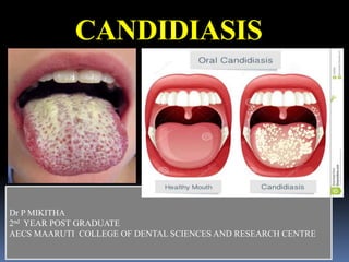
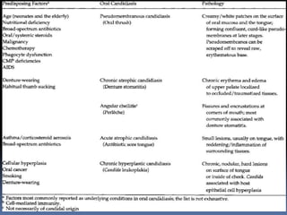
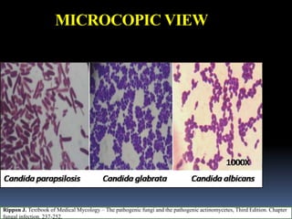
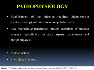
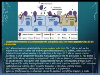
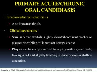
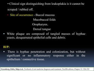
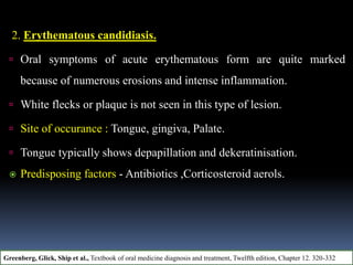
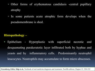
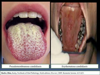
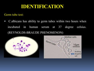
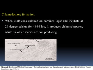
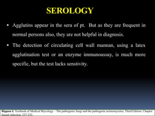
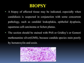
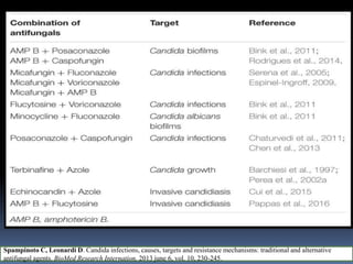
This document discusses candidiasis, caused by the yeast Candida. It begins with an introduction to mycology and the morphology of fungi. It then discusses candidiasis specifically, including its history, epidemiology, predisposing factors, pathogens involved, and pathophysiology. The document classifies the different types of candidiasis and discusses Candida in HIV patients. It covers the diagnosis and treatment of candidiasis and concludes with references.
Haemophilus influenzae type b (Hib) is a gram-negative bacteria that was formerly a leading cause of bacterial meningitis and other invasive diseases in children under 5 years old. Before Hib vaccines were introduced in 1988, approximately one in 200 children would develop invasive Hib disease and it was responsible for 50% of bacterial meningitis cases. Since widespread use of Hib conjugate vaccines, the incidence of invasive Hib disease has declined over 99%. Public health actions focus on rapid reporting, isolation, prophylaxis of contacts, and ensuring vaccination to prevent additional cases.
Candida albicans is a yeast that can cause infections in humans. It normally lives harmlessly in the body but can overgrow and cause disease when the environment changes. It has several forms ranging from yeast to hyphal filaments. Over 75% of women experience at least one vaginal candidiasis infection. Risk factors include antibiotics use, pregnancy, diabetes, and weakened immune system. Symptoms depend on the infected area and include rashes, vaginal discharge, and oral thrush. Diagnosis involves microscopic examination of samples. Treatment involves topical or oral antifungal drugs like fluconazole that work by disrupting the fungal cell membrane or inhibiting ergosterol production. Prevention focuses on good
This document summarizes common fungal infections caused by dimorphic and opportunistic fungi. It lists endemic mycoses including paracoccidioidomycosis, coccidioidomycosis, histoplasmosis, and blastomycosis caused by fungi that exist in both mold and yeast forms. It also describes opportunistic mycoses including candidiasis, cryptococcosis, and aspergillosis caused by fungi that exist only in yeast or mold forms and primarily infect immunocompromised individuals. Key characteristics of each fungal species such as morphology, habitat, disease manifestations, diagnosis and treatment are provided.
Parvoviruses are the smallest DNA viruses, including human parvovirus B19. B19 is pathogenic in humans, infecting erythroid progenitor cells and causing fifth disease in children characterized by a rash. It can also cause aplastic crisis, hydrops fetalis in fetuses, and chronic anemia in immunocompromised patients. Diagnosis involves detecting IgG and IgM antibodies by ELISA or PCR to detect the virus. There is no treatment, though a vaccine is in clinical trials.
Candida is a common fungus that can cause infections in humans. It normally lives on the skin and mucosal surfaces without causing problems, but under certain conditions it can overgrow and lead to candidiasis. Candidiasis includes infections of the mouth (thrush), skin, esophagus, and vagina. Risk factors include diabetes, pregnancy, HIV/AIDS, and broad-spectrum antibiotic use. Symptoms vary by location but may include redness, cracks, sores, white patches or lesions, itching, and pain. Treatment involves topical or oral antifungal medications.
The document discusses candidiasis, a fungal infection commonly caused by Candida albicans that usually occurs in the mouth and pharynx but can spread to the esophagus, describing its causes as immunosuppression or altered oral environments, symptoms of oral discomfort and white plaques, diagnosis through smears or cultures, treatment with topical or oral antifungals like nystatin or fluconazole, and nursing management including ensuring proper use of medications and attaining adequate nutrition and therapy.
Candidiasis refers to infections caused by Candida species, most commonly C. albicans. These fungi typically infect skin, nails, mucous membranes, and the GI tract, but can also cause systemic disease. Predisposing factors include mechanical trauma, moisture, nutrition deficiencies, extremes of age, illnesses, and medications. Clinical manifestations vary by site of infection and include oral thrush, vaginal candidiasis, skin infections like intertrigo. Diagnosis involves microscopic examination of samples and culture. Treatment depends on site and severity but commonly involves topical or oral antifungal medications.
This document provides an overview of Capnocytophaga, including its classification, characteristics, species, culture methods, incidence, detection, identification, interactions, virulence factors, antibiotic therapy and resistance. Capnocytophaga is an anaerobic, gram-negative bacterium that is facultative and exhibits gliding movement. It includes species such as C. ochracea, C. gingivalis, C. sputigena, and C. canimorsus. The document discusses methods for culturing and identifying Capnocytophaga, its role in periodontal diseases, potential virulence factors, and antibiotic susceptibility.
This document provides an overview of systemic and opportunistic mycoses. It defines systemic mycoses as deep fungal infections caused by soil-dwelling dimorphic fungi that are accidentally inhaled. The main causative agents described are Blastomyces dermatitidis, Paracoccidioides brasiliensis, Coccidioides immitis, and Histoplasma capsulatum. Opportunistic mycoses occur in immunocompromised individuals and the most common causes are Candida species, Aspergillus species, and Cryptococcus neoformans. Key clinical features, laboratory diagnostics including microscopy, culture, and serology, treatment approaches, and important epidemiological details are summarized
Burkholderia species are found in diverse habitats including soil, plants, animals, and clinical specimens. B. pseudomallei and B. mallei cause melioidosis and glanders, respectively, in humans and animals. B. cepacia is an opportunistic pathogen in cystic fibrosis patients. Characteristics of B. cepacia strains from CF infections include production of extracellular enzymes and ability to reduce nitrate. B. cepacia and B. gladioli LPS exhibits high endotoxic activity. B. cepacia contains cable pili associated with highly infectious epidemic strains infecting CF patients.
Candida albicans is a common fungal pathogen that normally lives in the human gastrointestinal tract. It can cause infections when there is an overgrowth, especially in immunocompromised individuals. C. albicans exists in both yeast and hyphal forms and is able to cause a variety of diseases depending on where in the body it infects, such as oropharyngeal candidiasis (oral thrush), vulvovaginal candidiasis (yeast infection), and invasive candidiasis. Transmission primarily occurs endogenously when there is a disruption to the normal microbiota. Treatment involves antifungal medications depending on the specific infection.
1. Actinomyces and Nocardia are filamentous, Gram-positive bacteria found in the environment and as normal flora in humans.
2. Actinomyces causes cervicofacial and abdominal actinomycosis in humans through tissue invasion following trauma or medical procedures. Diagnosis involves identifying sulfur granules in biopsy specimens.
3. Nocardia is an opportunistic pathogen that can cause pulmonary, cutaneous, or disseminated nocardiosis through inhalation or skin inoculation. It is an important cause of infection in immunocompromised individuals.
Epidemiological characterisation of Burkholderia cepacia complex (Bcc) from c...Bhoj Raj Singh
The presentation is extracted from the thesis talking about
1. The presence of Bcc organisms in the clinical infections of animals.
2. Ultrasound gels as a potential source of pathogens, especially Bcc.
3. Multidrug resistance in BCCs.
4. Lack of regulatory guidelines in Indian Pharmacopeia as existing in USP.
Influenza viruses are a major cause of respiratory disease and are responsible for periodic epidemics and pandemics worldwide. There are three main types of influenza viruses (A, B, and C) with type A being the most variable and causing the largest epidemics. Antigenic drift allows the virus to evade immunity between epidemics, while antigenic shift of surface proteins can cause pandemics by introducing novel subtypes into humans. Transmission occurs via respiratory droplets or contact. Clinical signs include fever, cough, and muscle aches. Complications can include pneumonia or Reye's syndrome in children. Diagnosis is through antigen detection, virus isolation, or serology. Treatment focuses on antivirals and symptom relief
Histoplasmosis is caused by the dimorphic fungus Histoplasma capsulatum. It exists in the mycelial phase in the environment and the yeast phase in tissues. Infection occurs via inhalation of microconidia from contaminated soil. Most infections are asymptomatic, but some may cause flu-like symptoms. Diagnosis involves microscopy of clinical samples or cultures to identify the yeast cells. Serological tests and skin tests also assist in diagnosis. Amphotericin B and itraconazole are used to treat severe or disseminated cases.
Candida are a genus of yeasts that are normally present on human skin and mucous membranes. There are over 150 Candida species, with Candida albicans being the most common cause of infection. Candida can cause infections through reproduction via budding. Major diseases include oropharyngeal candidiasis, intestinal candidiasis, vulvovaginal candidiasis, and candidemia. Risk factors for infection include antibiotic or steroid use, diabetes, and a diet high in sugar. Prevention strategies incorporate dietary modifications, probiotic supplementation, and antifungal medications.
Campylobacter is a common cause of foodborne illness worldwide and Campylobacter jejuni is a typical organism, which is a gram-negative, motile bacterium that causes both intestinal and systemic infections. Key virulence factors of Campylobacter include lipopolysaccharides and enterotoxins, and pathogenesis involves oral transmission from contaminated food or animals followed by colonization of the small intestine. Symptoms include acute diarrhea and abdominal pain that are usually self-limiting but antibiotics may be used for severe or prolonged cases.
Haemophilus influenzae is a Gram-negative coccobacillus first isolated in 1892 during an influenza outbreak. It is classified within the phylum Proteobacteria and can cause several diseases in humans like meningitis, pneumonia, epiglottitis and cellulitis. H. influenzae possesses several virulence factors like a capsule, lipopolysaccharides and IgA1 protease that help it evade the immune system and cause disease. Diagnosis involves culture and identification of the bacteria from clinical specimens as well as antigen and molecular detection techniques. Vaccination against H. influenzae type b is available to prevent disease.
Candidiasis – clinical manifestations and lab diagnosis of oral candidiasisShiv Joshi
This document discusses clinical manifestations and lab diagnosis of oral candidiasis. It outlines the various types of candidiasis infections including mucocutaneous, cutaneous, and systemic manifestations. For lab diagnosis of oral candidiasis, samples are collected via sterile swabs from oral mucous membranes and examined microscopically after KOH and Gram staining. Fungal cultures aid identification through colony morphology on agar plates. Biochemical tests and typing methods further characterize Candida isolates.
Yersinia includes human pathogens Y. pestis, Y. pseudotuberculosis, and Y. enterocolitica. Y. pestis causes plague, a zoonotic disease transmitted by fleas that has caused several pandemics throughout history. Currently it remains endemic in parts of Asia, Africa, and Americas, causing thousands of cases annually. Yersiniosis is caused by Y. pseudotuberculosis and Y. enterocolitica through consumption of contaminated food or water, commonly presenting as self-limiting gastroenteritis. Both diseases are diagnosed through culture, biochemical tests, and serology to identify the Yersinia species. Prevention focuses on flea control for plague and food safety for yers
This document summarizes common viral infections including measles, varicella, mumps, and viral hepatitis. Measles is caused by a paramyxovirus and causes a rash and respiratory symptoms. Varicella (chickenpox) is caused by varicella zoster virus and presents with a pruritic vesicular rash that spreads. Mumps is caused by a paramyxovirus and presents with painful swelling of the salivary glands. Hepatitis A and B viruses are described as common causes of viral hepatitis transmitted through fecal-oral and blood-borne routes respectively.
1. The document describes four systemic mycoses caused by dimorphic fungi: histoplasmosis, blastomycosis, coccidioidomycosis, and paracoccidioidomycosis.
2. It provides details on the causative agents, pathogenesis, clinical manifestations, laboratory diagnosis including histopathology, culture, and serology, and treatment recommendations for each fungal infection.
3. Key diagnostic methods include histopathological staining of tissue samples to identify characteristic fungal structures, culture of samples to demonstrate the dimorphic nature of the fungi, and serological tests to detect antibodies.
This document discusses Burkholderia cepacia, a gram-negative bacterium that can cause serious infections in immunocompromised individuals such as those with cystic fibrosis. It was originally known as Pseudomonas cepacia and is commonly found in soil and water. For cystic fibrosis patients, B. cepacia infections can lead to a rapidly fatal necrotizing pneumonia called cepacia syndrome. The bacteria are highly resistant to antibiotics and can form biofilms, contributing to its ability to cause infections. Proper diagnosis requires use of selective media such as Burkholderia cepacia Selective Agar to detect the bacteria.
There are over 100,000 known fungal species that inhabit different environments. Only around 600 species can cause disease in humans. Historically, invasive fungal infections were rare in immunocompromised patients but have increased in recent decades due to factors like improved diagnostics and more immunosuppressed individuals. Fungi use various virulence factors like thermal tolerance, dimorphism, and production of toxins or extracellular enzymes to establish infections by evading or weakening the host's immune response.
This document provides information about histoplasmosis, including its characteristics, pathogenesis, types, clinical presentation, and laboratory diagnosis. It can be summarized as follows:
1. Histoplasmosis is caused by the dimorphic fungus Histoplasma capsulatum, which exists in both a mycelial and yeast form. It is found worldwide in soil contaminated with bird or bat droppings.
2. Infection typically occurs via inhalation of yeast cells into the lungs. It can cause pulmonary or disseminated disease, spreading to organs in immunocompromised individuals.
3. Laboratory diagnosis involves direct examination of samples for yeast cells, culture of the fungus, and serological tests like complement fixation
Mycetoma is a chronic subcutaneous infection caused by fungi or bacteria that enters through trauma. It is characterized by painless swelling containing grains of the causative organism, which can discharge through sinuses. Diagnosis involves demonstrating grains in infected tissue. Treatment involves surgery and antifungal therapy. Sporotrichosis is also a subcutaneous infection caused by Sporothrix schenckii fungus through skin penetration. It causes skin nodules and lymph node involvement. Diagnosis uses microscopy and culture. Treatment uses potassium iodide, itraconazole or amphotericin B. Rhinosporidiosis is caused by Rhinosporidium seeberi and presents as friable polyps in the
Yersinia and Pasteurella are important bacterial pathogens. Yersinia pestis causes plague via transmission from rodents and their fleas to humans. It can cause bubonic, septicemic or pneumonic plague. Yersinia enterocolitica and Y. pseudotuberculosis can cause gastroenteritis and mesenteric lymphadenitis via the fecal-oral route. Pasteurella multocida is a zoonotic pathogen that can cause abscesses and meningitis in rare cases of human infection. Laboratory diagnosis involves culture, staining and serology of specimens depending on the suspected infection. Control relies on surveillance, treatment of cases, and measures to reduce rodent and flea
This document discusses opportunistic mycoses that occur in individuals with compromised immune systems. It focuses on several common fungal causes including Aspergillus, Mucor, and Penicillium species. Aspergillosis is described in detail, outlining the different clinical manifestations depending on infection site and immune status. Diagnosis involves microscopic examination and culture of specimens. Treatment involves antifungal therapies like amphotericin B and azoles.
Fortunately we are not alone and we provide residence to numerous microbial
communities comprising of bacterial species. The human body is made up of 10 14 cells of which
only 10% are mammalian and the remainder is contributed by the microorganisms that make up
the resident micro flora of the host. Normal microbial flora is a term that denotes the population
of microorganism that inhabit the skin and mucous membranes of healthy normal persons.
Numerous bacterial species colonize the mouth, upper airways, skin, vagina and intestinal tract
of humans where each one have a unique site specific fingerprint made of distinct microbe with
high level of diversity which is still unexplained.These microorganism are inhabit in the oral
cavity and their unavoidable interrelationships are essential component in maintaining
homeostasis between health and disease moreover the skin and mucous membrane always harbor
variety of organisms and they can be further categorized into two groups. One is the resident
flora which consists of relatively fixed type of microorganisms regularly found in given area at a
given age, and promptly gathers and re-establishes itself even if it is disturbed. The other one is
the transient flora, consist of non-pathogenic or potentially pathogenic microorganisms that
inhabit skin or mucous membrane for hours, days or week and it is derived from the environment
that does not produce disease and does not have the capacity to reestablish permanently on the
surface. However if the resident flora is disturbed, transient microorganisms may get colonized,
proliferate and produce disease. This indigenous microbiota plays an important role in health and
diseases of humans and contributing to the development of the immune system and provides
resistance to colonization by pathogenic microorganisms. Thereby the presentation is intended to
review on the importance and thrust areas of oral microbiome in health and disease.
This document discusses the microbiology of periodontal diseases. It describes the typical microbiota found in healthy sites, gingivitis sites, and chronic or aggressive periodontitis sites. The microbiota shifts from mostly gram-positive facultative bacteria in health to include more gram-negative anaerobic bacteria in disease. Key pathogenic bacteria associated with periodontitis include Porphyromonas gingivalis, Tannerella forsythia, Treponema denticola, Aggregatibacter actinomycetemcomitans, and Prevotella intermedia. These bacteria produce virulence factors like proteases, lipopolysaccharides, and leukotoxins that promote tissue destruction.
Streptococcus anginosus group (formerly known as Streptococcus milleri) refers to a group of streptococcal species that commonly cause abscesses and infections. They were initially discovered in oral infections and named after a microbiologist. While usually considered harmless oral commensals, they can cause serious infections, especially in immunocompromised individuals or those with underlying conditions. Identification and treatment of infections involves bacterial culture and antibiotic therapy along with drainage of abscesses.
Porphyromonas gingivalis is a gram-negative, anaerobic bacterium implicated as a primary pathogen in periodontal disease. It produces several virulence factors that allow it to invade tissues, evade the host immune system, and cause damage. These include proteolytic enzymes, lipopolysaccharide, capsular polysaccharides, fimbriae, and outer membrane proteins. Fimbriae aid in adhesion to host cells, while enzymes degrade host proteins and tissues. Capsular polysaccharides and outer membrane structures help resist phagocytosis. P. gingivalis is strongly associated with periodontitis and its virulence factors contribute directly to tissue destruction and immune evasion during infection.
This document provides an overview of the microbiological and immunological aspects of the microbial-host interaction in periodontal disease. It discusses the various bacterial species involved, including the "red complex" bacteria Porphyromonas gingivalis, Treponema denticola, and Tannerella forsythia. It describes the virulence factors of these bacteria and how they evade host defenses. It also summarizes the innate and adaptive immune response phases, focusing on the acute inflammatory response and roles of neutrophils in controlling bacterial challenge through opsonization and phagocytosis.
This document provides an overview of the microbiology of periodontal disease. It discusses the various bacteria associated with periodontal diseases, including the "red complex" bacteria Porphyromonas gingivalis, Treponema denticola, and Tannerella forsythia. It also examines the virulence factors of these pathogens and how they contribute to periodontal tissue destruction and bone loss. The document establishes the role of these microorganisms in periodontal diseases based on criteria such as their association with disease, elimination during treatment, ability to induce disease in animal models, and production of virulence factors that can damage host tissues.
This document discusses oral fungal infections. It begins by introducing mycology, the study of fungi, and describes the characteristics of fungi including that they are eukaryotic, lack chlorophyll, and have cell walls containing chitin. It then discusses the various types of oral fungal infections including candidiasis, the most common oral fungal infection caused by Candida albicans. The document describes the risk factors, pathogenesis, clinical features of different forms of oral candidiasis, and concludes with information on other oral fungal infections such as histoplasmosis and cryptococcosis.
Candidiasis is a fungal infection caused by Candida species. Since the 1940s when antibiotics became widespread, cases of candidiasis have risen significantly. Candida is now the fourth most common cause of bloodstream infections in hospitalized patients in the US. The burden of candidiasis in terms of morbidity, mortality, and costs is considerable. Common manifestations include vaginal candidiasis, chronic mucocutaneous candidiasis, and bloodstream infections. Host defenses against Candida include physical barriers and immune cells such as macrophages and neutrophils. Candida virulence factors that enhance its pathogenicity include adhesins, polymorphism, biofilms, invasins, secreted hydrolases, and metabolic adaptation.
Candidiasis is a fungal infection caused by Candida species. Since the 1940s when antibiotics became widespread, cases of candidiasis have risen sharply. Candida is the fourth most common cause of bloodstream infections in hospitalized patients in the US. Morbidity, mortality and costs associated with candidiasis are significant. Predisposing factors include immunosuppression, prolonged antibiotic use, and medical procedures. Candida normally inhabits the skin and GI tract but can cause infections like vaginitis, bloodstream infections, and chronic mucocutaneous candidiasis in immunocompromised individuals.
This document discusses fungal infections of the oral cavity, specifically those caused by Candida species. It covers topics like the introduction, terminology, morphology and carriage vs infection of Candida. It also discusses the etiology and pathogenesis of Candidal infections, including factors like adherence, invasion, dimorphism and evasion of the host immune system. Predisposing factors to Candidal infections include local changes to the oral mucosa as well as systemic factors that compromise the host's defenses. The document outlines various clinical presentations of oral Candidal infections.
The document summarizes various fungal infections that can affect the oral cavity. It discusses candidiasis, the most common fungal infection caused by Candida albicans. Candidiasis ranges from mild to severe and can manifest as white plaques (pseudomembranous candidiasis), erythematous lesions, or cause denture stomatitis. Risk factors include immunocompromise, antibiotics, and dentures. Diagnosis involves microscopy and culture. Treatment involves antifungal medications like amphotericin B and fluconazole.
ORAL MICROBIOME.pptx by UMNA FATIMA- BIOMEDumnajmi123
This PowerPoint presentation provides a thorough exploration of the oral microbiome and its significance in both maintaining health and contributing to disease. Beginning with an introduction to the oral microbiome, the presentation outlines its diverse composition and its crucial role in oral health. It further examines the concept of dysbiosis within the oral microbiome, highlighting the factors contributing to imbalance and its implications for oral and systemic health. The presentation also delves into emerging research linking oral microbiome dysbiosis to systemic diseases, shedding light on potential mechanisms and clinical implications. Methods for studying the oral microbiome are discussed, along with recent advancements in research methodologies and therapeutic strategies targeting microbial dysbiosis. Additionally, the presentation explores the evolving field of precision dentistry and its integration with oral microbiome analysis for personalized treatment approaches. Through case studies and examples, the audience gains insight into the practical applications of oral microbiome research. The presentation concludes with a summary of key points and an invitation for questions and discussion, emphasizing the importance of ongoing research in understanding and harnessing the potential of the oral microbiome for improving health outcomes.
The document discusses immunity to fungal infections. It notes that fungi can cause diseases through either a lack of immune recognition or overactivation of the inflammatory response. The immune system uses pattern recognition receptors and innate immune cells like phagocytes to recognize and respond to fungal pathogens. Both resistance mechanisms that limit fungal growth and tolerance mechanisms that limit host damage are important for maintaining immune homeostasis during fungal infections.
Candidiasis infections are caused by an overgrowth of the yeast Candida, which normally inhabits human mucosal surfaces. There are several types of Candidiasis depending on the infected site, including oral thrush, diaper rash, and invasive bloodstream infections. Candida albicans is the most common cause of fungal infections worldwide. It was first discovered in the 1800s and its ability to cause disease opportunistically depends on virulence factors like adhesion proteins and morphological changes between yeast and filamentous forms. Diagnosis involves microscopic examination or culturing of infected sites to identify characteristic Candida features, and treatment depends on the type and severity of infection.
The etiology of a disease refers to the causative trigger(s), whereas pathogenesis refers to the mechanism(s) by which the disease progresses.
In other words, while the microbial biofilm developing on the tooth surface constitutes a necessary etiological factor, its mere presence is insufficient for the initiation of the disease.
Further risk factors, such as host genetics, lifestyle, stress, and systemic conditions, that dictate the immunopathogenesis are crucial for the transition from a healthy to a diseased state.
ggregatibacter actinomycetemcomitans (previously Actinobacillus actinomycetemcomitans) is a Gram-negative, facultative nonmotile, rod-shaped oral commensal often found in association with localized aggressive periodontitis, a severe infection of the periodontium, although it is also associated with nonoral infections. Its role in periodontitis was first discovered by Danish-born periodontist Jørgen Slots, a professor of dentistry and microbiology at the University of Southern California School of Dentistry.
'Bacterium actinomycetem comitans' was described by Klinger (1912) as coccobacillary bacteria isolated together with Actinomyces from actinomycotic lesions of man. It was reclassified as Actinobacillus actinomycetemcomitans by Topley & Wilson (1929) and as Haemophilus actinomycetemcomitans by Potts et al. (1985). The species has attracted attention because of its association with localized aggressive periodontitis. is explained here by Dr Harshavardhan Patwal
This document provides an overview of endodontic microbiology. It begins with an introduction to how microorganisms cause pulp and periradicular infections. It then discusses the mechanisms of microbial pathogenicity and virulence factors. It describes the microbial ecology of the root canal ecosystem and how the environment changes over time. It outlines the various routes of root canal infection and the typical microbes involved in primary, secondary, and persistent intraradicular infections. It also discusses extraradicular infections. The document covers biofilm formation and bacterial interactions. It concludes with discussing treatment measures and references.
The etiology of a disease refers to the causative trigger(s), whereas pathogenesis refers to the mechanism(s) by which the disease progresses.
In other words, while the microbial biofilm developing on the tooth surface constitutes a necessary etiological factor, its mere presence is insufficient for the initiation of the disease.
Further risk factors, such as host genetics, lifestyle, stress, and systemic conditions, that dictate the immunopathogenesis are crucial for the transition from a healthy to a diseased state.
The document discusses periodontal flaps, including their definition, historical background, objectives, indications and contraindications. It describes the advantages and disadvantages of flap surgery, as well as principles of flap design such as ensuring adequate blood supply. The document outlines different flap techniques and factors that can affect surgical outcomes. In summary:
- A periodontal flap involves surgically separating gingiva/mucosa to access bone and roots. Objectives include enabling root instrumentation and re-establishing periodontal health.
- Indications include deep pockets inaccessible to non-surgical treatment, while contraindications involve poor patient health/cooperation.
- Principles of flap design focus on blood supply to reduce
The document discusses the junctional epithelium (JE), providing definitions, historical concepts, and details on its structure and function. Some key points:
- JE is the non-keratinized stratified squamous epithelium that forms a collar around the cervical portion of the tooth below the cementoenamel junction.
- There has been debate around its attachment to the tooth surface, but transmission electron microscopy showed it is attached via hemidesmosomes to the internal basal lamina on the tooth surface.
- JE develops as the tooth erupts, with the reduced enamel epithelium transforming into JE over 1-2 years in a coronal to apical direction via cell changes.
- It plays a
This document discusses Porphyromonas gingivalis, a bacterium associated with periodontal disease. It covers the taxonomy, biochemical characteristics, structure, methods of detection including culture and molecular techniques, oral ecology and transmission. Regarding transmission, it notes that vertical transmission from parents to children is possible but rare, while horizontal transmission between siblings or spouses sharing the same strain is more common. Person-to-person transmission can occur through saliva, direct mucosal contact or sharing toothbrushes.
This document discusses the epidemiology of periodontal diseases. It defines epidemiology and describes its aims and principles. It discusses epidemiological measures like incidence and prevalence. It describes different epidemiological study designs including observational studies like case-control and cohort studies, and experimental studies like randomized controlled trials. It also discusses risk factors, indices, and epidemiological studies of chronic and aggressive periodontitis conducted in India.
This document discusses trauma from occlusion (TFO), defined as pathological alterations or adaptive changes that develop in the periodontium due to excessive occlusal forces. It provides historical context on TFO research dating back to 1901, classifications of TFO, stages of tissue response to TFO including injury and repair, and factors that can increase occlusal forces or decrease the periodontium's resistance to forces. TFO can be acute or chronic and primary (due to occlusal factors) or secondary (due to reduced periodontal support). Excessive forces can cause tissue injury through thrombosis, hemorrhage or necrosis while the body attempts repair through new tissue formation and bone remodeling.
Desquamative gingivitis is a clinical manifestation characterized by erythema, desquamation and ulceration of the gingiva that can be indicative of an underlying condition. It is not a specific disease but rather a gingival response associated with various disorders. The document discusses the definition, pathogenesis, clinical presentation and diagnosis of desquamative gingivitis. It also describes three disorders that are commonly associated with desquamative gingivitis: lichen planus, bullous pemphigoid, and pemphigus.
The document discusses bleeding and coagulation. It defines blood and its components like plasma, red blood cells, white blood cells and platelets. It describes the processes of erythropoiesis, the formation of red blood cells in the bone marrow, and hemostasis, the prevention of blood loss through vascular constriction, platelet plug formation and blood clotting. It also discusses the clotting factors involved in the coagulation cascade and the mechanisms of platelet activation, shape change, secretion and aggregation that form the platelet plug during hemostasis.
This document discusses aggressive periodontitis, including its history, classification, localized aggressive periodontitis, and generalized aggressive periodontitis. It defines aggressive periodontitis as a specific type of periodontitis featuring rapid attachment loss, bone destruction, and familial aggregation, with inconsistent microbial deposits and severe tissue destruction. Localized aggressive periodontitis is characterized by circumpubertal onset and localized first molar/incisor involvement. Generalized aggressive periodontitis affects at least three teeth other than first molars and incisors and has an episodic nature. Both forms are associated with elevated levels of certain bacteria and potential immunological and genetic factors.
Calcium and phosphorus are essential minerals that have important functions in the body. Calcium is important for bone and teeth formation, muscle contraction, nerve impulse transmission and other processes, while phosphorus is essential for energy storage and transfer, cell membrane structure, and acid-base balance. The absorption and regulation of calcium and phosphorus levels is controlled by hormones like vitamin D, parathyroid hormone, and calcitonin, which act to maintain appropriate levels in the blood and bones. Imbalances in calcium and phosphorus can lead to disorders that impact bone health and other bodily functions.
Cell Therapy Expansion and Challenges in Autoimmune DiseaseHealth Advances
There is increasing confidence that cell therapies will soon play a role in the treatment of autoimmune disorders, but the extent of this impact remains to be seen. Early readouts on autologous CAR-Ts in lupus are encouraging, but manufacturing and cost limitations are likely to restrict access to highly refractory patients. Allogeneic CAR-Ts have the potential to broaden access to earlier lines of treatment due to their inherent cost benefits, however they will need to demonstrate comparable or improved efficacy to established modalities.
In addition to infrastructure and capacity constraints, CAR-Ts face a very different risk-benefit dynamic in autoimmune compared to oncology, highlighting the need for tolerable therapies with low adverse event risk. CAR-NK and Treg-based therapies are also being developed in certain autoimmune disorders and may demonstrate favorable safety profiles. Several novel non-cell therapies such as bispecific antibodies, nanobodies, and RNAi drugs, may also offer future alternative competitive solutions with variable value propositions.
Widespread adoption of cell therapies will not only require strong efficacy and safety data, but also adapted pricing and access strategies. At oncology-based price points, CAR-Ts are unlikely to achieve broad market access in autoimmune disorders, with eligible patient populations that are potentially orders of magnitude greater than the number of currently addressable cancer patients. Developers have made strides towards reducing cell therapy COGS while improving manufacturing efficiency, but payors will inevitably restrict access until more sustainable pricing is achieved.
Despite these headwinds, industry leaders and investors remain confident that cell therapies are poised to address significant unmet need in patients suffering from autoimmune disorders. However, the extent of this impact on the treatment landscape remains to be seen, as the industry rapidly approaches an inflection point.
Rasamanikya is a excellent preparation in the field of Rasashastra, it is used in various Kushtha Roga, Shwasa, Vicharchika, Bhagandara, Vatarakta, and Phiranga Roga. In this article Preparation& Comparative analytical profile for both Formulationon i.e Rasamanikya prepared by Kushmanda swarasa & Churnodhaka Shodita Haratala. The study aims to provide insights into the comparative efficacy and analytical aspects of these formulations for enhanced therapeutic outcomes.
TEST BANK For An Introduction to Brain and Behavior, 7th Edition by Bryan Kol...rightmanforbloodline
TEST BANK For An Introduction to Brain and Behavior, 7th Edition by Bryan Kolb, Ian Q. Whishaw, Verified Chapters 1 - 16, Complete Newest Versio
TEST BANK For An Introduction to Brain and Behavior, 7th Edition by Bryan Kolb, Ian Q. Whishaw, Verified Chapters 1 - 16, Complete Newest Version
TEST BANK For Community Health Nursing A Canadian Perspective, 5th Edition by...Donc Test
TEST BANK For Community Health Nursing A Canadian Perspective, 5th Edition by Stamler, Verified Chapters 1 - 33, Complete Newest Version Community Health Nursing A Canadian Perspective, 5th Edition by Stamler, Verified Chapters 1 - 33, Complete Newest Version Community Health Nursing A Canadian Perspective, 5th Edition by Stamler Community Health Nursing A Canadian Perspective, 5th Edition TEST BANK by Stamler Test Bank For Community Health Nursing A Canadian Perspective, 5th Edition Pdf Chapters Download Test Bank For Community Health Nursing A Canadian Perspective, 5th Edition Pdf Download Stuvia Test Bank For Community Health Nursing A Canadian Perspective, 5th Edition Study Guide Test Bank For Community Health Nursing A Canadian Perspective, 5th Edition Ebook Download Stuvia Test Bank For Community Health Nursing A Canadian Perspective, 5th Edition Questions and Answers Quizlet Test Bank For Community Health Nursing A Canadian Perspective, 5th Edition Studocu Test Bank For Community Health Nursing A Canadian Perspective, 5th Edition Quizlet Test Bank For Community Health Nursing A Canadian Perspective, 5th Edition Stuvia Community Health Nursing A Canadian Perspective, 5th Edition Pdf Chapters Download Community Health Nursing A Canadian Perspective, 5th Edition Pdf Download Course Hero Community Health Nursing A Canadian Perspective, 5th Edition Answers Quizlet Community Health Nursing A Canadian Perspective, 5th Edition Ebook Download Course hero Community Health Nursing A Canadian Perspective, 5th Edition Questions and Answers Community Health Nursing A Canadian Perspective, 5th Edition Studocu Community Health Nursing A Canadian Perspective, 5th Edition Quizlet Community Health Nursing A Canadian Perspective, 5th Edition Stuvia Community Health Nursing A Canadian Perspective, 5th Edition Test Bank Pdf Chapters Download Community Health Nursing A Canadian Perspective, 5th Edition Test Bank Pdf Download Stuvia Community Health Nursing A Canadian Perspective, 5th Edition Test Bank Study Guide Questions and Answers Community Health Nursing A Canadian Perspective, 5th Edition Test Bank Ebook Download Stuvia Community Health Nursing A Canadian Perspective, 5th Edition Test Bank Questions Quizlet Community Health Nursing A Canadian Perspective, 5th Edition Test Bank Studocu Community Health Nursing A Canadian Perspective, 5th Edition Test Bank Quizlet Community Health Nursing A Canadian Perspective, 5th Edition Test Bank Stuvia
Clinic ^%[+27633867063*Abortion Pills For Sale In Tembisa Central19various
Clinic ^%[+27633867063*Abortion Pills For Sale In Tembisa Central Clinic ^%[+27633867063*Abortion Pills For Sale In Tembisa CentralClinic ^%[+27633867063*Abortion Pills For Sale In Tembisa CentralClinic ^%[+27633867063*Abortion Pills For Sale In Tembisa CentralClinic ^%[+27633867063*Abortion Pills For Sale In Tembisa Central
These lecture slides, by Dr Sidra Arshad, offer a quick overview of the physiological basis of a normal electrocardiogram.
Learning objectives:
1. Define an electrocardiogram (ECG) and electrocardiography
2. Describe how dipoles generated by the heart produce the waveforms of the ECG
3. Describe the components of a normal electrocardiogram of a typical bipolar lead (limb II)
4. Differentiate between intervals and segments
5. Enlist some common indications for obtaining an ECG
6. Describe the flow of current around the heart during the cardiac cycle
7. Discuss the placement and polarity of the leads of electrocardiograph
8. Describe the normal electrocardiograms recorded from the limb leads and explain the physiological basis of the different records that are obtained
9. Define mean electrical vector (axis) of the heart and give the normal range
10. Define the mean QRS vector
11. Describe the axes of leads (hexagonal reference system)
12. Comprehend the vectorial analysis of the normal ECG
13. Determine the mean electrical axis of the ventricular QRS and appreciate the mean axis deviation
14. Explain the concepts of current of injury, J point, and their significance
Study Resources:
1. Chapter 11, Guyton and Hall Textbook of Medical Physiology, 14th edition
2. Chapter 9, Human Physiology - From Cells to Systems, Lauralee Sherwood, 9th edition
3. Chapter 29, Ganong’s Review of Medical Physiology, 26th edition
4. Electrocardiogram, StatPearls - https://www.ncbi.nlm.nih.gov/books/NBK549803/
5. ECG in Medical Practice by ABM Abdullah, 4th edition
6. Chapter 3, Cardiology Explained, https://www.ncbi.nlm.nih.gov/books/NBK2214/
7. ECG Basics, http://www.nataliescasebook.com/tag/e-c-g-basics
3. CONTENTS
Introduction to mycology
Candidiasis
History of candidiasis
Epidemiology
Predisposing factors
Pathogens
Pathophysiology
Classification of candidiasis
Candida in HIV immuno-compromised patients
Diagnosis
Treatment
Conclusion
References
4. INTRODUCTION TO MYCOLOGY
Fungi are eukaryotic microorganisms (eukaryotic -true
nucleus) i.e. has a membrane bound nucleus seen in fungi,
algae, protozoa, plants and animals.
Prokaryotes---- (Primitive nucleus) --- does not have a
membrane bound nucleus - e.g. true bacteria.
Study of fungi is called mycology. In Greek Mykos- meaning
mushroom.
Ananthanarayan, Panicker. Textbook of microbiology, fourth edition. Chapter The mycology. 432-455.
5. MORPHOLOGY OF FUNGI
Rippon J. Textbook of Medical Mycology – The pathogenic fungi and the pathogenic actinomycetes, Third Edition. Chapter
fungal infection. 237-252.
6. CANDIDIASIS
Oral candidiasis is a collective term given to a group of disorders
caused by the yeast candida and its species.
An older name for this disease is moniliasis.
Like many other pathogenic fungi, candida albicans may
exist in two forms, a trait known as dimorphism.
The yeast form of the organism is believed to be relatively
innocuous, but the hyphal form is usually associated with
the invasion of host tissue.
Rippon J. Textbook of Medical Mycology – The pathogenic fungi and the pathogenic actinomycetes, Third Edition. Chapter
fungal infection. 237-252.
7. HISTORY OF CANDIDIASIS
The descriptions oral thrush go back to the time of hippocrates.
Vulvovaginal candidiasis was first described in 1849 by Wilkinson.
In 1875, Hausmann demonstrated the causative organism in both
vulvovaginal and oral candidiasis is the same
With the advent of antibiotics following World War II, the rates of
candidiasis increased.
The rates then decreased in 1950s following the development of
nystatin.
Rippon J. Textbook of Medical Mycology – The pathogenic fungi and the pathogenic actinomycetes, Third Edition. Chapter
fungal infection. 237-252.
8. EPIDEMIOLOGY
Incidence of candida albicans isolated from the oral cavity
has been reported to be neonates - 45%
Healthy children - 45% - 65%
Healthy adults - 30% - 45%
People wearing removable dentures -50% - 65%
Acute leukemia patients on chemotherapy – 90%
HIV patients ->95%
Effects of oral candidiasis may range form localized
infections to acute systemic disseminated disease.
Rippon J. Textbook of Medical Mycology – The pathogenic fungi and the pathogenic actinomycetes, Third Edition. Chapter
fungal infection. 237-252.
9. PREDISPOSING FACTORS
Candida species are normal oral commensals
It is generally accepted that candidiasis as an opportunistic
infection affecting individuals who were debilitated by
another disease. It is also known that they develop in normal
healthy individuals. The transition of this innocuous
commensal to the disease causing parasite may be associated
with the virulence attributes of the organism.
Greenberg, Glick, Ship et al., Textbook of oral medicine diagnosis and treatment, Twelfth edition, Chapter 12. 320-332
10.
11. Neville et al, have identified three general factors
that may lead to clinically evident oral candidiasis.
These factors are
The immune status of the host.
The oral mucosal environment.
The particular strain of C.albicans (the hyphae
form is usually associated with pathogenic
infection).
Shafer, Hine, Levy, Textbook of Oral Pathology, Sixth edition, Elsevier; 2009. Keratotic lesions. 613-623.
12. Factors that alter the immune status of the host.
Blood dyscrasis / advanced malignancy.
Old age / infancy.
Radiation therapy / chemotherapy
HIV inf. / other immuno deficiency disorders.
Endocrine abnormalities like
Diabetes mellitus
Hypothyroidism or Hypoparathyrodisim
Pregnancy
Corticosteroid therapy / hypoadrenalism
Greenberg, Glick, Ship et al., Textbook of oral medicine diagnosis and treatment, Twelfth edition, Chapter 12. 320-332
13. Factors that alter the oral mucosal environment
Xerostomia
Prolong antibiotic therapy.
Poor oral or denture hygiene
Mal nutrition / gastro intestinal absorption.
Iron, folic acid or vitamin def.
Acidic saliva / carbohydrate rich diets
Heavy smoking.
Oral epithelial dysplasia.
Chronic local irritants.
Greenberg, Glick, Ship et al., Textbook of oral medicine diagnosis and treatment, Twelfth edition, Chapter 12. 320-332
14. PATHOGENS
Candida spp are asexual yeast of genus ascomycetes and
genetically diploid with presence of 8 chromosomes.
They are small( 4-6 um) thin walled ovoid cells(
blastospores) that reproduce by budding.
The important candidal species are C.albicans (commonest)
C.tropicalis, C.glabrata, C. pseudotrophicalis, C.
guilleierimondii, C. krusei.
C.albicans, C. glabrata and C .tropoicalis represent more
than 80% of isolates from clinical infection.
Rippon J. Textbook of Medical Mycology – The pathogenic fungi and the pathogenic actinomycetes, Third Edition. Chapter
fungal infection. 237-252.
15.
16. MICROCOPIC VIEW
Rippon J. Textbook of Medical Mycology – The pathogenic fungi and the pathogenic actinomycetes, Third Edition. Chapter
fungal infection. 237-252.
17. PATHOPHYSIOLOGY
Establishment of the infection requires thigmotropism
(contact sensing) and attachment to epithelial cells.
Also intercellular penetration through secretions of protease
enzymes, specifically secretory aspartyl proteinases and
phospholipase B.
A. host factors.
B. virulence factors.
Janaina C. O. Sardi et. al. candida spp. In periodontal disease – a review. J Oral Sci 52, 177-185, 2010
18.
19. HOST FACTORS
To colonize and infect the oral tissues candida requirs specific local and
systemic defects in host immune responses.
Among healthy people, several immunological and non-immunological
defense mechanism are collectively responsible for antifungal properties
of the oral enviornment.
The host factors include:
A. properties of saliva
B. oral keratinocytes
C. T-cell mediated responses.
20. 1. Properties of saliva:
Mechanical debridement, facilitated by salivary mucins and
proteoglycans.
A close to neutral pH that reduces fungal adherence to
epithelial surfaces and also reduces the expression of C
albicans virulence genes.
Antibacterial proteins such as lysozyme, lactoferrin, histatins,
calprotectin, and secretory leukocyte protease inhibitor.
Janaina C. O. Sardi et. al. candida spp. In periodontal disease – a review. J Oral Sci 52, 177-185, 2010
21. 2. Oral keratinocytes:
Provides a physical barrier to candida
Secrete a number of growth factors and cytokines thus playing
a crucial role in the inflammatory response on the epithelial
cells.
3. T-Cell mediated response:
An intact T-helper cell 1(Th1) CD4+ response to c. albicans
and a CD4+ T-cell augmentation of monocyte/macrophage
and PMN functions appear to be necessary for the clearance
of oropharyngeal candidiasis.
Janaina C. O. Sardi et. al. candida spp. In periodontal disease – a review. J Oral Sci 52, 177-185, 2010
22.
23. VIRULENCE FACTORS
Adhesins
Polymorphism
Biofilm
Secreted hydrolases
Invasins
Metabolic adaptation
Janaina C. O. Sardi et. al. candida spp. In periodontal disease – a review. J Oral Sci 52, 177-185, 2010
24. Adhesins
They have special sets of (GPI) linked cell surface glycoproteins that
allow it to adhere to the surface of microorganisms.
These glycoproteins are encoded by 8 sets of agglutinin-like
sequence (ALS) genes, ranging from Als1-7 and Als9.
For adhesion, the Als3 gene appears to be the most important as it is
upregulated during an infection of oral and vaginal epithelial cells.
Also, it helps with biofilm formation by helping with adhesion to
each other.
Janaina C. O. Sardi et. al. candida spp. In periodontal disease – a review. J Oral Sci 52, 177-185, 2010
25. Polymorphism
Polymorphic fungus that can grow in several different forms, primarily yeast,
pseudohyphae, and hyphae.
For its pathogenicity, its ovoid-shaped budding yeast and parallel walled true
hyphae forms are the most important.
The hyphae form is more prevalent for an infection, while the yeast form is
believed to be important in the spread.
The role of pseudohyphae is not very well understood, other than being an
intermediate form between yeast and hyphae.
Several factors can cause a change in morphology, such as pH differences,
temperature changes, CO2 levels, starvation and quorum-sensing molecules.
Janaina C. O. Sardi et. al. candida spp. In periodontal disease – a review. J Oral Sci 52, 177-185, 2010
26. Biofilm
Candida albicans have the ability to form biofilms on living and
non-living surfaces, such as mucosal membranes and catheters,
respectively.
After the adherance of yeast cells to the surface, there is
development of hyphae cells in the upper part of the biofilm.
Eventually, this leads to a more resistant, mature biofilm and
dispersion of yeast cells- both contributing to the pathogens
virulence.
Janaina C. O. Sardi et. al. candida spp. In periodontal disease – a review. J Oral Sci 52, 177-185, 2010
27. In the process of biofilm formation, Bcr1, Tec1 and Efg1
function as important transcriptional factors.
Recent studies show that biofilms protect C.albicans
colonization from neutrophil attack and deter the formation of
ROS.0
Janaina C. O. Sardi et. al. candida spp. In periodontal disease – a review. J Oral Sci 52, 177-185, 2010
28. Invasins
Along with adhesion, Als3 proteins can function as invasins
that help with the invasion of C.ablicans into host epithelial and
endothelial cells.
Another important invasin gene is Ssa1, which normally codes
for heat shock proteins.
Janaina C. O. Sardi et. al. candida spp. In periodontal disease – a review. J Oral Sci 52, 177-185, 2010
29. Basically, these specialised proteins on the pathogens surface
mediate binding to host ligands, such as E-cadherin on
epithelial cells and N-cadherin on endothelial cells, and it
induces host cells to engulf the fungal pathogen (induced
endocytosis).
Another method of invasion is the active penetration of
C.albicans into host cells by an unknown mechanism
involving hyphae (active penetration)
Janaina C. O. Sardi et. al. candida spp. In periodontal disease – a review. J Oral Sci 52, 177-185, 2010
30. Secreted hydrolases
Secrete 3 main classes of hydrolases: proteases, phospholipase, and
lipases.
It is proposed that these hydrolases help facilitate the pathogens active
penetration into host cells and the uptake of extracellular nutrients from
the enviornment.
There are about 10 known secreted aspartic proteases ( Sap 1-10).
Their exact contribution to the pathogenicity is controversial.
For phospholipases, there are 4 major classes (A,B,C,D) and all 5
members of the B class are involved with the disruption of the host cell
surface.
Janaina C. O. Sardi et. al. candida spp. In periodontal disease – a review. J Oral Sci 52, 177-185, 2010
31. Metabolic adaptation
Are usually found in the gastrointestinal microbiome of healthy
individuals and in this enviornment nutrient levels are relatively
high.
During niche changes in the course of infection, available nutrient
levels will also change.
Consequently the fungus can quickly undergo metabolic adaptation,
such as their glycolysis, gluconeogenesis, and starvation responses.
For example, in the case of candidemia, C. albicans infect the blood
stream which is typically rich in glucose.
Janaina C. O. Sardi et. al. candida spp. In periodontal disease – a review. J Oral Sci 52, 177-185, 2010
32. Neverthless, it might be phagocytosed into a macrophage or
neutrophil, where its surrounded by ROS, AMP’s.
In response C. albicans quickly switch the glycolysis to
starvation response with the activation of glyoxylate cycle.
Due to this flexibility, C. albicans can infect almost every organ
in a human host through bloodstream, providing candidemia’s
higher mortality rate.
Janaina C. O. Sardi et. al. candida spp. In periodontal disease – a review. J Oral Sci 52, 177-185, 2010
33. CLASSIFICATION OF
CANDIDIASIS
Rippon J. Textbook of Medical Mycology – The pathogenic fungi and the pathogenic actinomycetes, Third Edition. Chapter
fungal infection. 237-252.
34. Tony Axell, Samaranayaka and Reichart (1997) have proposed a re classification.
One of reasons being unusual clinical variants have appeared with the pandemic
progression of the HIV infection.
I. Primary oral candidiasis
Acute forms - Pseudomembranous
Erythematous
Chronic types -hyperplastic - Nodular.
- Plaque Like
-Erythematous
-Pseudomembranous.
Greenberg, Glick, Ship et al., Textbook of oral medicine diagnosis and treatment, Twelfth edition, Chapter 12. 320-332
35. II. Secondary oral candidiasis - Oral manifestations of systemic
mucocutaneous candidiasis as a result of diseases such as thymic
aplasia and candidiasis endocrinopathy syndrome.
III. Candida associated lesions - Angular cheilitis
Denture stomatitis
Median rhomboid glossitis
IV. Keratinised primary lesions super infected with candida:
Leukoplakia
Lichen planus
Lupus erythematosus
Greenberg, Glick, Ship et al., Textbook of oral medicine diagnosis and treatment, Twelfth edition, Chapter 12. 320-332
36. PRIMARYACUTE/CHRONIC
ORALCANDIDIASIS
1.Pseudomembranous candidiasis:
Also known as thrush.
Clinical appearance
Semi adherent, whitish, slightly elevated confluent patches or
plaques resembling milk curds or cottage cheese.
Plaques can be easily removed by wiping with a gauze swab,
leaving a red and slightly bleeding surface or even a shallow
ulceration.
Greenberg, Glick, Ship et al., Textbook of oral medicine diagnosis and treatment, Twelfth edition, Chapter 12. 320-332
37. Site of occurrence - Buccal mucosa
Mucobuccal folds
Oropharynx.
Dorsal tongue
White plaque are composed of tangled masses of hyphae
yeasts, desquamated epithelial cells and debris.
H/P:
There is hyphae penetration and colonization, but without
significant or no inflammatory response either in the
epithelium / connective tissue.
Greenberg, Glick, Ship et al., Textbook of oral medicine diagnosis and treatment, Twelfth edition, Chapter 12. 320-332
• Clinical sign distinguishing from leukoplakia is it cannot be
scraped / rubbed off.
38. 2. Erythematous candidiasis.
Oral symptoms of acute erythematous form are quite marked
because of numerous erosions and intense inflammation.
White flecks or plaque is not seen in this type of lesion.
Site of occurance : Tongue, gingiva, Palate.
Tongue typically shows depapillation and dekeratinisation.
Predisposing factors - Antibiotics ,Corticosteroid aerols.
Greenberg, Glick, Ship et al., Textbook of oral medicine diagnosis and treatment, Twelfth edition, Chapter 12. 320-332
39. Other forms of erythematous candidasis -central papillary
atrophy
In some patients acute atrophic form develops when the
pseudomembrane is shed.
Histopathology –
Epithelium - Hyperplastic with superficial necrotic and
desquamating parakeratotic layer infiltrated both by hyphae and
yeasts and by inflammatory cells. Predominantly neutrophil
leucocytes. Neutrophils may accumulate to form micro abscesses.
Greenberg, Glick, Ship et al., Textbook of oral medicine diagnosis and treatment, Twelfth edition, Chapter 12. 320-332
41. PRIMARY CHRONIC
CANDIDIASIS
1. Hyperplastic candidiasis
Also referred as candidal leukoplakia.
Clinical appearance :
Persistent white patch on the oral mucosa indistinguishable from
leukoplakia.
Characteristically the lesions present as dense, opaque white
patches of irregular thickness and density with a rough or nodular
surfaces.
Greenberg, Glick, Ship et al., Textbook of oral medicine diagnosis and treatment, Twelfth edition, Chapter 12. 320-332
42. Site of occurrence
Buccal mucosa adjacent to the commmissure of the lips and
present as roughly triangular, often bilateral white plaques
tapering posteriorly.
Often associated with angular cheilitis.
In many patients there is a strong association with tobacco
smoking.
Greenberg, Glick, Ship et al., Textbook of oral medicine diagnosis and treatment, Twelfth edition, Chapter 12. 320-332
43. Histo pathology :
Epithelium is parakeratinized and is generally marked and
hyperplastic and acanthotic.
Many of the cell in the para keratinized surface of the
epithelium are separated by oedema and numerous neutrophil
leucocytes, the neutrophils together collecting together as
micro abscesses.
Chronic hyper plastic candidosis is considered to be a pre
malignant lesion and epithelial dysplasia is seen in about 50%
of cases.
Greenberg, Glick, Ship et al., Textbook of oral medicine diagnosis and treatment, Twelfth edition, Chapter 12. 320-332
44. CANDIDIASISASSOCIATED
LESIONS
1.Candida associated lesions denture stomatitis.
Chronic inflammatory changes of the denture bearing mucosa.
Occurs in the confined space between the mucosa and the
upper denture
Inadequate cleaning of the fitting surface.
Wearing the denture throughout the night all appear to
favor the over growth of candida resulting in a local
imbalance of the host parasite relationship.
Shafer, Hine, Levy, Textbook of Oral Pathology, Sixth edition, Elsevier; 2009. Keratotic lesions. 613-623.
45. Histo pathology :
The epithelium shows hyperplasia / atrophy.
Surface being either parakeratinized or non keratinised.
Leucocytes infiltrate the epithelium forming micro
abscesses.
Shafer, Hine, Levy, Textbook of Oral Pathology, Sixth edition, Elsevier; 2009. Keratotic lesions. 613-623.
46. 2. Angular Chelitis / Perleche / Angular stomatitis.
Multifactorial disease of infectious origin.
Occurs predominantly in denture wearers.
Commonly caused by >yeasts, staphylococci, β-Streptococci
Clinical appearance
Soreness.
Erythema
Crust formation
Fissuring at the corners of the mouth.
Shafer, Hine, Levy, Textbook of Oral Pathology, Sixth edition, Elsevier; 2009. Keratotic lesions. 613-623.
47. Seen on deep folds of skin at the angles of the mouth, which
may be associated with loss of occlusal height in old age or
due to decrease vertical height in dentures.
Nutritional deficiencies -Thiamine. ,Riboflavin ,Folic acid,
Iron deficiency anemia
Shafer, Hine, Levy, Textbook of Oral Pathology, Sixth edition, Elsevier; 2009. Keratotic lesions. 613-623.
48. 3. Median Rhomboid glossitis
In the past, this was thought to be a developmental defect of
the tongue. Investigators have noted a consistent relationship
between the lesion and candida albicans.
Clinical appearance
Diamond shaped depapillated erythematous patch on the
midline of the tongue dorsum.
Erythema is due in part of the loss of the filiform papillae in
this area.
Mixed microbiological etiology
Shafer, Hine, Levy, Textbook of Oral Pathology, Sixth edition, Elsevier; 2009. Keratotic lesions. 613-623.
50. MUCO CUTANEOUS CANDIDIASIS
(CHRONIC MUCOCUTANEOUS
CANDIDIASIS -CMC)
Several oral candidiasis may also be seen as a component of a
relatively rare group of immunologic disorders known as
mucocutaneous candidiasis.
Result of a defect in cell mediated immunity or structure of the
epidermis. Several distinct immunologic dysfunctions have been
identified and severity of the candidal infection correlates with the
immunologic defect.
Shafer, Hine, Levy, Textbook of Oral Pathology, Sixth edition, Elsevier; 2009. Keratotic lesions. 613-623.
51. Types
Familial CMC.
Diffuse CMC
Candidiasis endocrinopathy syndrome.
CMC of late onset > 35 yrs.
Clinical appearance
Thick white plaque that cannot be scraped / rubbed off.
Usually associated with endocrine abnormalities
Nutritional def. >Iron deficiencies.
Shafer, Hine, Levy, Textbook of Oral Pathology, Sixth edition, Elsevier; 2009. Keratotic lesions. 613-623.
53. ID REACTION
Some persons with chronic candida infections develop a secondary
skin response characterized by a localized or generalized sterile
vesiculopapular rash that is believed to be an allergic response to
candida antigens.
These lesions referred to as monilids, id reaction, usually resolve
with treatment of the candida infection.
Shafer, Hine, Levy, Textbook of Oral Pathology, Sixth edition, Elsevier; 2009. Keratotic lesions. 613-623.
54. AIDS related oral candidiasis
Primary HIV infection is associated with
Severe aphthous stomatitis
Oropharyngeal and oesophageal candidiasis.
Clinical variants of oral candidiasis seen are
Pseudomembranous
Erythematous.
Hyperplastic
Angular chelitis.
Shafer, Hine, Levy, Textbook of Oral Pathology, Sixth edition, Elsevier; 2009. Keratotic lesions. 613-623.
55. The pseudomembranous and erythematous variants forms the
most common clinical presentations of the mucosal candidiasis
associated with HIV-infection.
Symptoms include burning pain, altered taste sensation, difficulty
swallowing liquids and solids.
Immunodeficiency T-helper cells predisposes to secondary
infections (fungi being fairly common)
C. albicans serotype B is prevalent.
Spampinoto C, Leonardi D. Candida infections, causes, targets and resistance mechanisms: traditional and alternative
antifungal agents. BioMed Research Internation, 2013 june 6, vol. 10, 230-245.
56. The pseudomembranous form can be easily diagnosed by
demonstrating the presence of candidal yeast and pseudohyphae
on wet mounts or stained smears of material obtained by
swabbing the lesions.
Confirmed by the isolation of candida species on culture.
In erythematous form, the sparse presence of candida at the
mucosal surface frequently requires a biopsy and PAS ataining
to establish a formal diagnosis.
Candidiasis can occur at any time during the course of HIV
infection.
Spampinoto C, Leonardi D. Candida infections, causes, targets and resistance mechanisms: traditional and alternative
antifungal agents. BioMed Research Internation, 2013 june 6, vol. 10, 230-245.
57. Oral burdens of C. albicans are augmented in HIV-infected
patients even prior to the first episode of candidiasis and the
intensity of carriage increases significantly in the progression
from asymptomatic candida carrier to an episode of candidiasis.
Pseudomembranous form of OPC and oesophageal candidiasis
increases dramatically in advanced HIV disease associated with
CD4+ cell counts of <200/mm3, while erythematous candidiasis
and angular chelitis are less strongly associated with late disease.
Spampinoto C, Leonardi D. Candida infections, causes, targets and resistance mechanisms: traditional and alternative
antifungal agents. BioMed Research Internation, 2013 june 6, vol. 10, 230-245.
58. Histopathology
Candida infection in HIV infected persons show weak
inflammatory reaction.
Epithelium invaded by numerous hyphae or Pseudo hyphae
without the characteristic massive infiltrate of
polymorphonuclear leucocytes.
Areas of sub epithelial inflammation often contain few or no
leucocytes.
Spampinoto C, Leonardi D. Candida infections, causes, targets and resistance mechanisms: traditional and alternative
antifungal agents. BioMed Research Internation, 2013 june 6, vol. 10, 230-245.
59. CLINICAL EXPRESSION OFCANDIDA
INFECTION IN HIV – IMMUNO
COMPROMISED PATIENTS.
Reichart et al (1994) reported on seven dentate homosexual
AIDS patients who developed inflammatory papillary hyperplasia
of the palate in association with candida infections.
A characteristic form of periodontal disease: linear gingival
erythema, is clinically expressed as a gingival erythematous band
of at least 2mm extending to adjacent papilla.
Spampinoto C, Leonardi D. Candida infections, causes, targets and resistance mechanisms: traditional and alternative
antifungal agents. BioMed Research Internation, 2013 june 6, vol. 10, 230-245.
60. Grbic et al (1995) have studied the presence of linear gingival
erythema in two groups of patients. One group was composed of
homosexual men and other group was composed of parenteral
drug users.
Both groups, included HIV sero positive and HIV sero negative
patients. Linear gingival erythema was present in greater numbers
in the drug user group. (Statistically significant) irrespective of
serostatus
Spampinoto C, Leonardi D. Candida infections, causes, targets and resistance mechanisms: traditional and alternative
antifungal agents. BioMed Research Internation, 2013 june 6, vol. 10, 230-245.
61. The authors suggest that this finding may be related to the
extremely poor oral hygiene observed in the parenteral drug user
group.
The presence of candidiasis in homo sexual men was related with
true HIV associated linear gingival erythema. The authors
hypothesis that HIV positive patients may be more sensitive to
certain bacterial pathogens that, in the presence of candida would
notice them prone to developing linear gingival erythema.
Spampinoto C, Leonardi D. Candida infections, causes, targets and resistance mechanisms: traditional and alternative
antifungal agents. BioMed Research Internation, 2013 june 6, vol. 10, 230-245.
63. Direct microscopy:
gram stained smears & KOH mounts from lesions of skin, nail
or mucous membrane shows budding gram positive yeast
cells.
Other stains: PAS and Gomori’s methenamine silver stain.
Ananthanarayan, Panicker. Textbook of microbiology, fourth edition. Chapter The mycology. 432-455.
64. CULTURE
Cultures are obtained on sabourad’s dextrose agar (SDA) and
on ordinary bacteriological culture media, eg: blood agar at
room temperature or at 37 degrees celsius.
Colonies are creamy white, smooth with a yeast odour.
Gram stained smears from colonies shows gram positive
budding yeast cells.
Ananthanarayan, Panicker. Textbook of microbiology, fourth edition. Chapter The mycology. 432-455.
65. Cornmeal agar:
for identification of characteristic terminal chlamydospores,
incubation at 25 degree celsius.
Blood agar:
non hemolytic, smooth, moderate colonies, resemble staph.
Colonies.
Ananthanarayan, Panicker. Textbook of microbiology, fourth edition. Chapter The mycology. 432-455.
66. Sabouraud’s agar
Cornmeal agar Blood agar
Ananthanarayan, Panicker. Textbook of microbiology, fourth edition. Chapter The mycology. 432-455.
67. Chromagar:
this is special media for candida, use to identify the different
species, each species colonies have a characteristic color.
C.albicans- light green to bluish green
C.glabrata-rose
C.krusei-rose pale
C.tropicalis-blue-violet
Ananthanarayan, Panicker. Textbook of microbiology, fourth edition. Chapter The mycology. 432-455.
68.
69. IDENTIFICATION
Germ tube test:
C.albicans has ability to germ tubes within two hours when
incubated in human serum at 37 degree celsius.
(REYNOLDS-BRAUDE PHENOMENON)
70. Chlamydospore formation:
When C.albicans cultured on cornmeal agar and incubate at
26 degree celsius for 48-96 hrs, it produces chlamydospores,
while the other species are non producing.
Rippon J. Textbook of Medical Mycology – The pathogenic fungi and the pathogenic actinomycetes, Third Edition. Chapter
fungal infection. 237-252.
71. SEROLOGY
Agglutins appear in the sera of pt. But as they are frequent in
normal persons also, they are not helpful in diagnosis.
The detection of circulating cell wall mannan, using a latex
agglutination test or an enzyme immunoassay, is much more
specific, but the test lacks senstivity.
Rippon J. Textbook of Medical Mycology – The pathogenic fungi and the pathogenic actinomycetes, Third Edition. Chapter
fungal infection. 237-252.
72. BIOPSY
A biopsy of affected tissue may be indicated, especially when
candidiasis is suspected in conjunction with some concurrent
pathology, such as candidal leukoplakia, epithelial dysplasia,
squamous cell carcinoma or lichen planus.
The section should be stained with PAS or Gridley’s or Gomori
methenamine silver(GMS), because candida species stain poorly
by hematoxylin and eosin.
73. SKIN TEST
Delayed hypersenstivity to candida is so universal that skin
testing with candida extracts is used as an indicator of the
functional integrity of cell mediated immunity
Rippon J. Textbook of Medical Mycology – The pathogenic fungi and the pathogenic actinomycetes, Third Edition. Chapter
fungal infection. 237-252.
74. IMPRINT CULTURE TECHNIQUE
This technique uses a sterile plastic foam pad of known size
(2.5*2.5) dipped in sabourard’s broth and placed on the
suspect mucosal surface for 60 sec.
Then this plastic foam is placed directly on sabouraud’s or
Pagano-Levin agar.
Rippon J. Textbook of Medical Mycology – The pathogenic fungi and the pathogenic actinomycetes, Third Edition. Chapter
fungal infection. 237-252.
75. Candida density at each site is determined by a GallenKamp
colony contcat and expressed as CFU/mm2.
≥30 CFU/mm2 of mucosa in dentate.
≥ 49 CFU/mm2 in denture wearers.
Rippon J. Textbook of Medical Mycology – The pathogenic fungi and the pathogenic actinomycetes, Third Edition. Chapter
fungal infection. 237-252.
76. ORALRINSE TECHNIQUE
Here the pt. is asked to rinse the mouth for 60 sec. with 10ml of
sterile phosphate buffered saline or sterile water. The oral rinse is
centrifuged at 1700g for 10min and the deposit resuspended in
1ml of sterile PBS.
The concetrated oral rinse is now inoculated on apropriated media
to assess CFU/mm of rinse sample using a spiral plate prior to
incubation.
Rippon J. Textbook of Medical Mycology – The pathogenic fungi and the pathogenic actinomycetes, Third Edition. Chapter
fungal infection. 237-252.
77. SALIVARY CULTURE TECHNIQUE
This involves pt. 2ml of mixed unstimulated saliva into a
sterile, universal container.
The number of canida expressed as CFU/mm of saliva is
estimated by counting the resultant growth of sabouraud’s
agar.
Rippon J. Textbook of Medical Mycology – The pathogenic fungi and the pathogenic actinomycetes, Third Edition. Chapter
fungal infection. 237-252.
78. TREATMENT
Cutaneous candidiasis: most localized cutaneous candidiasis
infections may be treated with any number of topical
antifungal agents.
If the infection is paronychia, the most important aspect of
therapy is drainage of abscess, followed by oral antifungal
therapy with either fluconazole or itraconazole.
Spampinoto C, Leonardi D. Candida infections, causes, targets and resistance mechanisms: traditional and alternative
antifungal agents. BioMed Research Internation, 2013 june 6, vol. 10, 230-245.
79. Spampinoto C, Leonardi D. Candida infections, causes, targets and resistance mechanisms: traditional and alternative
antifungal agents. BioMed Research Internation, 2013 june 6, vol. 10, 230-245.
80. Spampinoto C, Leonardi D. Candida infections, causes, targets and resistance mechanisms: traditional and alternative
antifungal agents. BioMed Research Internation, 2013 june 6, vol. 10, 230-245.
81. Chronic mucocutaneous candidiasis - I.V. Fluconazole
Amphotericin B.
Acute oral candida infection - topical nystatin. Oral Susp
USP. 100,000 units/ml.
More effective treatment of acute and chronic candidasis
can usually obtained by a once daily dose of 200 mg
ketoconazole or 100 mg fluconazole for 2 weeks.
Spampinoto C, Leonardi D. Candida infections, causes, targets and resistance mechanisms: traditional and alternative
antifungal agents. BioMed Research Internation, 2013 june 6, vol. 10, 230-245.
82. Treatment of denture base mouth and angular chelitis must
include elimination of candida from the denture surface either by
making a new denture or by relining an existing denture.
Nystatin applied as a cream under a denture or to the lesions of
angular chelitis will eliminate redness and promote shrinkage of
mucosa and healing of fissures.
Spampinoto C, Leonardi D. Candida infections, causes, targets and resistance mechanisms: traditional and alternative
antifungal agents. BioMed Research Internation, 2013 june 6, vol. 10, 230-245.
83. Spampinoto C, Leonardi D. Candida infections, causes, targets and resistance mechanisms: traditional and alternative
antifungal agents. BioMed Research Internation, 2013 june 6, vol. 10, 230-245.
84. CONCLUSION
Fungal infections aptly recognized as “Diseases of the
diseased” is increasingly alarmingly due to the pandenic of
AIDS. Advances in medical and pharmacological treatment
have resulted in greater life expectancy for patients with
malignant and metabolic disease and other systemic,
conditions, which can predispose patients to oral and systemic
fungal infections.
85. REFERENCES
Greenberg, Glick, Ship et al., Textbook of oral medicine
diagnosis and treatment, Twelfth edition, Chapter 12. 320-
332.
Joklik, Willett, Amos et al., Textbook of Zinsser
Microbiology, Ninth edition, Jaypee brothers medical
publishers; Mycoplasma and ureaplasma. 210-237.
86. •Shafer, Hine, Levy, Textbook of Oral Pathology, Sixth
edition, Elsevier; 2009. Keratotic lesions. 613-623.
•Harsh Mohan, Textbook of Pathology, fourth edition,
Jaypee Brothers Medical Publishers; 2003. Chapter 8. 227-
232.
•Rippon J. Textbook of Medical Mycology – The pathogenic
fungi and the pathogenic actinomycetes, Third Edition. Chapter
fungal infection. 237-252.
87. Ananthanarayan, Panicker. Textbook of microbiology, fourth
edition. Chapter The mycology. 432-455.
Spampinoto C, Leonardi D. Candida infections, causes, targets
and resistance mechanisms: traditional and alternative antifungal
agents. BioMed Research Internation, 2013 june 6, vol. 10, 230-
245.
Janaina C. O. Sardi et. al. candida spp. In periodontal disease –
a review. J Oral Sci 52, 2010, 177-185.