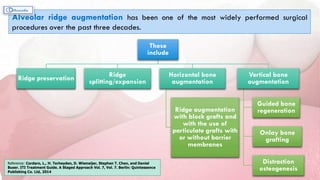
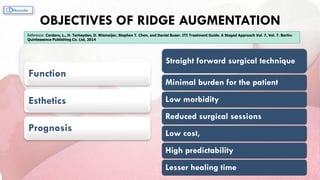
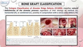
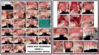
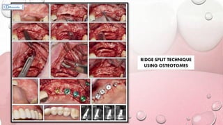
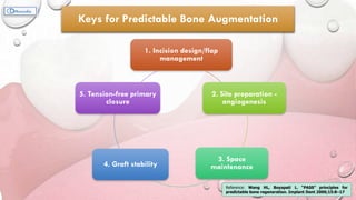
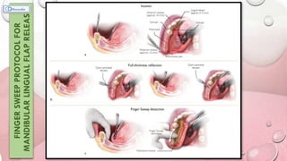
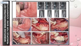
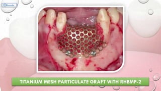
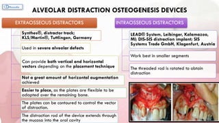
This document provides information on various ridge augmentation techniques. It begins with an introduction describing how tooth loss leads to bone resorption and impaired function. It then discusses the history of using autogenous bone grafts for ridge augmentation. The objectives of ridge augmentation are also outlined. Key techniques discussed include ridge preservation, ridge splitting, use of autogenous bone blocks, and distraction osteogenesis. Advantages and disadvantages of different graft sources and incision designs are compared. The document emphasizes the importance of adequate bone volume for successful implant placement and summarizes various methods to augment bone.
Vertical ridge augmentation is sometimes required for dental implant placement. The presentation looks at various conventional and newer techniques for ridge augmentation in the oral cavity.
The biological fixation determines the longevity of dental implant treatment. It ensures the long term survival of dental implant. Better the osseointegration,higher will be the survival rate
The content covers majority of the aspect of immediate implant placement - why immediate implants?, case selection, decision making, classifications, surgical technique, healing following immediate implant placement, immediate implants in infected sockets/periapical infections, literature reviews and recommendations for clinical practice.
Fundamentals of Soft Tissue Grafting Principles for Dental Clinicians
by Dr. Jin Y. Kim
Board-Certified Periodontist
Lecturer, UCLA School of Dentistry
“Program on Ridge Split and Ridge Augmentation for Implant Placement”- Two lectures on “Concepts of Ridge Augmentation” and “Novel and Simpler Approaches to Ridge Augmentation”. Event organized by the Dental Experts and held at Paneenya Mahavidyalaya Institute of Dental Sciences, Hyderabad, India on 18/11/2016.
Platelet Rich Fibrin (PRF) in Dentistry, What is PRF ? , What are the difference between PRP,PRGF and PRF ?, Preparation of PRF , shapes of PRF, Role of PRF in wound healing, APPLICATIONS OF PRF, Applications of PRF In Oral and Maxillofacial Surgery, Applications of PRF In Periodontics, Applications of PRF In Endodontics, Applications of PRF In Tissue Engineering
Coronal advanced flap in combination with a connective tissue graft. Is the t...MD Abdul Haleem
Coronal advanced flap in combination with a connective tissue graft. Is the thickness of the flap a predictor for root coverage? - A prospective clinical study.
Department of Periodontology and Oral Implantology.
"A Journal Club Presentation"
Furcation involvement is a common sequela of severe chronic periodontal disease. Its effective management has a profound influence on the outcome of periodontal therapy.
“Perio-Implant Synergy”- Two lectures on “Lost Buccal Plate- Complications and Management” and “Failing to Plan is Planning to Fail”. Organized by the Society of Periodontists and Implantologists of Kerala” at PMS Dental College, Trivandrum, India on 17/9/2018.
The denture-wearing history should provide information on the age of existing dentures, the frequency of denture replacement, the patient's experiences and expectations. It is important to identify whether any previous dentures have been successful as it may be suitable to copy features from a previously successful set. It will be important to manage expectations for those patients with a history of denture intolerance, yet technically satisfactory prostheses.
Clinical examination
Clinical examination should fully evaluate both the patient's anatomy and previous dentures to anticipate challenges and the potential to improve upon retention, stability, support, appearance and/or other factors. This should be undertaken in a systematic manner and would typically involve assessment of anatomy followed by an assessment of any existing dentures. This should follow a diagnostic process to determine if the patient presents with:
Technically adequate dentures on a favourable tissue base
Technically adequate dentures on an unfavourable tissue base
Technically inadequate dentures on a favourable tissue base
Technically inadequate dentures on an unfavourable tissue base.
Orthodontics has been developing greatly in achieving the desired results both clinically and technically.
Today, it is still very challenging to reduce the duration of orthodontic treatments.
It is one of the common deterents that the orthodontist faces and it causes irritation among adults plus increasing risks of caries, gingival recession, and root resorption.
A number of attempts have been made to create different approaches both preclinically and clinically in order to achieve quicker results, but still there are a lot of uncertainties and unanswered questions towards most of these techniques.
Atrophied Edentulous Mandible with Implant-Supported Overdenture; A 10-year f...Abu-Hussein Muhamad
Abstract: Severe atrophy of the inferior alveolar process and underlying basal bone often results in problems with a lower denture. These problems include insufficient retention of the lower denture, intolerance to loading by the mucosa, pain, difficulties with eating and speech, loss of soft-tissue support, and altered facial appearance. These problems are a challenge for the prosthodontist and surgeon. In this case report, patient with resorbed edentulous mandible was successfully rehabilitated using two dental implants placed in the interforaminal region with ball abutments opposing conventional maxillary complete denture. Key Words: dental implants; dental prosthesis, implant-supported; resorption,
Abstract: Corticotomy-assisted orthodontic treatment is an established and efficient orthodontic technique that has recently been studied in a number of publications. Corticotomy facilitated orthodontics have been employed in various forms over speed up orthodontic treatment It involves selective alveolar decortication in the form of decortication lines and dots performed around the teeth that are to be moved. It is done to induce a state of increased tissue turnover and a transient osteopenia, which is followed by a faster rate of orthodontic tooth movement. This technique has several advantages, including faster tooth movement, shorter treatment time, safer expansion of constricted arches, enhanced post-orthodontic treatment stability and extended envelope of tooth movement. The aim of this article is to present a comprehensive review of the literature, including historical background, contemporary clinical techniques, indications, contraindications, complications and side effects. Keywords: Corticotomy, decortication, review, orthodontic treatment
IOSR Journal of Dental and Medical Sciences is one of the speciality Journal in Dental Science and Medical Science published by International Organization of Scientific Research (IOSR). The Journal publishes papers of the highest scientific merit and widest possible scope work in all areas related to medical and dental science. The Journal welcome review articles, leading medical and clinical research articles, technical notes, case reports and others.
Being a Periodontist, what necessary is to know what actually periodontal flaps are. So this presentation might provide you an insight into the field of periodontics as well as periodontal flaps.
Local anesthesia, all in one place with all the references and all the important points.
It contains some videos and animations, for which feel free to contact. As such animations are not compatible with Slideshare. Enjoy and please hit the like button if you liked the presentation.
A complete presentation made on the topic of blood with everything needed together in one place.
This presentation contains some animations and videos that might not be that compatible with Slideshare, so feel free to contact me and I shall share it.
Everything a dentist needs to know about a periodontal abscess is here.
Along with all the relevant facts, references, definitions, classifications, and each and every statement is given with proper detail
Here's all that you need to know about dental calculus.
With proper references and all the recent advances, along with the detailed facts and description.
Each and every statement is provided with an accurate reference and all the things to know are very well summarised in one place.
Aberrant Frenum !!
No worries... When Frenectomy is here.
Hello Periodontists,
Here's the entire process of Frenectomy in a nutshell and various ways to encounter the same.
Lets Shoot ...
Report Back from SGO 2024: What’s the Latest in Cervical Cancer?bkling
Are you curious about what’s new in cervical cancer research or unsure what the findings mean? Join Dr. Emily Ko, a gynecologic oncologist at Penn Medicine, to learn about the latest updates from the Society of Gynecologic Oncology (SGO) 2024 Annual Meeting on Women’s Cancer. Dr. Ko will discuss what the research presented at the conference means for you and answer your questions about the new developments.
ARTIFICIAL INTELLIGENCE IN HEALTHCARE.pdfAnujkumaranit
Artificial intelligence (AI) refers to the simulation of human intelligence processes by machines, especially computer systems. It encompasses tasks such as learning, reasoning, problem-solving, perception, and language understanding. AI technologies are revolutionizing various fields, from healthcare to finance, by enabling machines to perform tasks that typically require human intelligence.
New Directions in Targeted Therapeutic Approaches for Older Adults With Mantl...i3 Health
i3 Health is pleased to make the speaker slides from this activity available for use as a non-accredited self-study or teaching resource.
This slide deck presented by Dr. Kami Maddocks, Professor-Clinical in the Division of Hematology and
Associate Division Director for Ambulatory Operations
The Ohio State University Comprehensive Cancer Center, will provide insight into new directions in targeted therapeutic approaches for older adults with mantle cell lymphoma.
STATEMENT OF NEED
Mantle cell lymphoma (MCL) is a rare, aggressive B-cell non-Hodgkin lymphoma (NHL) accounting for 5% to 7% of all lymphomas. Its prognosis ranges from indolent disease that does not require treatment for years to very aggressive disease, which is associated with poor survival (Silkenstedt et al, 2021). Typically, MCL is diagnosed at advanced stage and in older patients who cannot tolerate intensive therapy (NCCN, 2022). Although recent advances have slightly increased remission rates, recurrence and relapse remain very common, leading to a median overall survival between 3 and 6 years (LLS, 2021). Though there are several effective options, progress is still needed towards establishing an accepted frontline approach for MCL (Castellino et al, 2022). Treatment selection and management of MCL are complicated by the heterogeneity of prognosis, advanced age and comorbidities of patients, and lack of an established standard approach for treatment, making it vital that clinicians be familiar with the latest research and advances in this area. In this activity chaired by Michael Wang, MD, Professor in the Department of Lymphoma & Myeloma at MD Anderson Cancer Center, expert faculty will discuss prognostic factors informing treatment, the promising results of recent trials in new therapeutic approaches, and the implications of treatment resistance in therapeutic selection for MCL.
Target Audience
Hematology/oncology fellows, attending faculty, and other health care professionals involved in the treatment of patients with mantle cell lymphoma (MCL).
Learning Objectives
1.) Identify clinical and biological prognostic factors that can guide treatment decision making for older adults with MCL
2.) Evaluate emerging data on targeted therapeutic approaches for treatment-naive and relapsed/refractory MCL and their applicability to older adults
3.) Assess mechanisms of resistance to targeted therapies for MCL and their implications for treatment selection
micro teaching on communication m.sc nursing.pdfAnurag Sharma
Microteaching is a unique model of practice teaching. It is a viable instrument for the. desired change in the teaching behavior or the behavior potential which, in specified types of real. classroom situations, tends to facilitate the achievement of specified types of objectives.
Couples presenting to the infertility clinic- Do they really have infertility...Sujoy Dasgupta
Dr Sujoy Dasgupta presented the study on "Couples presenting to the infertility clinic- Do they really have infertility? – The unexplored stories of non-consummation" in the 13th Congress of the Asia Pacific Initiative on Reproduction (ASPIRE 2024) at Manila on 24 May, 2024.
Prix Galien International 2024 Forum ProgramLevi Shapiro
June 20, 2024, Prix Galien International and Jerusalem Ethics Forum in ROME. Detailed agenda including panels:
- ADVANCES IN CARDIOLOGY: A NEW PARADIGM IS COMING
- WOMEN’S HEALTH: FERTILITY PRESERVATION
- WHAT’S NEW IN THE TREATMENT OF INFECTIOUS,
ONCOLOGICAL AND INFLAMMATORY SKIN DISEASES?
- ARTIFICIAL INTELLIGENCE AND ETHICS
- GENE THERAPY
- BEYOND BORDERS: GLOBAL INITIATIVES FOR DEMOCRATIZING LIFE SCIENCE TECHNOLOGIES AND PROMOTING ACCESS TO HEALTHCARE
- ETHICAL CHALLENGES IN LIFE SCIENCES
- Prix Galien International Awards Ceremony
Title: Sense of Smell
Presenter: Dr. Faiza, Assistant Professor of Physiology
Qualifications:
MBBS (Best Graduate, AIMC Lahore)
FCPS Physiology
ICMT, CHPE, DHPE (STMU)
MPH (GC University, Faisalabad)
MBA (Virtual University of Pakistan)
Learning Objectives:
Describe the primary categories of smells and the concept of odor blindness.
Explain the structure and location of the olfactory membrane and mucosa, including the types and roles of cells involved in olfaction.
Describe the pathway and mechanisms of olfactory signal transmission from the olfactory receptors to the brain.
Illustrate the biochemical cascade triggered by odorant binding to olfactory receptors, including the role of G-proteins and second messengers in generating an action potential.
Identify different types of olfactory disorders such as anosmia, hyposmia, hyperosmia, and dysosmia, including their potential causes.
Key Topics:
Olfactory Genes:
3% of the human genome accounts for olfactory genes.
400 genes for odorant receptors.
Olfactory Membrane:
Located in the superior part of the nasal cavity.
Medially: Folds downward along the superior septum.
Laterally: Folds over the superior turbinate and upper surface of the middle turbinate.
Total surface area: 5-10 square centimeters.
Olfactory Mucosa:
Olfactory Cells: Bipolar nerve cells derived from the CNS (100 million), with 4-25 olfactory cilia per cell.
Sustentacular Cells: Produce mucus and maintain ionic and molecular environment.
Basal Cells: Replace worn-out olfactory cells with an average lifespan of 1-2 months.
Bowman’s Gland: Secretes mucus.
Stimulation of Olfactory Cells:
Odorant dissolves in mucus and attaches to receptors on olfactory cilia.
Involves a cascade effect through G-proteins and second messengers, leading to depolarization and action potential generation in the olfactory nerve.
Quality of a Good Odorant:
Small (3-20 Carbon atoms), volatile, water-soluble, and lipid-soluble.
Facilitated by odorant-binding proteins in mucus.
Membrane Potential and Action Potential:
Resting membrane potential: -55mV.
Action potential frequency in the olfactory nerve increases with odorant strength.
Adaptation Towards the Sense of Smell:
Rapid adaptation within the first second, with further slow adaptation.
Psychological adaptation greater than receptor adaptation, involving feedback inhibition from the central nervous system.
Primary Sensations of Smell:
Camphoraceous, Musky, Floral, Pepperminty, Ethereal, Pungent, Putrid.
Odor Detection Threshold:
Examples: Hydrogen sulfide (0.0005 ppm), Methyl-mercaptan (0.002 ppm).
Some toxic substances are odorless at lethal concentrations.
Characteristics of Smell:
Odor blindness for single substances due to lack of appropriate receptor protein.
Behavioral and emotional influences of smell.
Transmission of Olfactory Signals:
From olfactory cells to glomeruli in the olfactory bulb, involving lateral inhibition.
Primitive, less old, and new olfactory systems with different path
HOT NEW PRODUCT! BIG SALES FAST SHIPPING NOW FROM CHINA!! EU KU DB BK substit...GL Anaacs
Contact us if you are interested:
Email / Skype : kefaya1771@gmail.com
Threema: PXHY5PDH
New BATCH Ku !!! MUCH IN DEMAND FAST SALE EVERY BATCH HAPPY GOOD EFFECT BIG BATCH !
Contact me on Threema or skype to start big business!!
Hot-sale products:
NEW HOT EUTYLONE WHITE CRYSTAL!!
5cl-adba precursor (semi finished )
5cl-adba raw materials
ADBB precursor (semi finished )
ADBB raw materials
APVP powder
5fadb/4f-adb
Jwh018 / Jwh210
Eutylone crystal
Protonitazene (hydrochloride) CAS: 119276-01-6
Flubrotizolam CAS: 57801-95-3
Metonitazene CAS: 14680-51-4
Payment terms: Western Union,MoneyGram,Bitcoin or USDT.
Deliver Time: Usually 7-15days
Shipping method: FedEx, TNT, DHL,UPS etc.Our deliveries are 100% safe, fast, reliable and discreet.
Samples will be sent for your evaluation!If you are interested in, please contact me, let's talk details.
We specializes in exporting high quality Research chemical, medical intermediate, Pharmaceutical chemicals and so on. Products are exported to USA, Canada, France, Korea, Japan,Russia, Southeast Asia and other countries.
3. INTRODUCTION
Teeth loss Continuous
resorption
Diminished volume and
strength of residual bone
Impaired masticatory function
Loss of facial vertical
dimension
Speech difficulty
Facial soft tissue changes
Reference: Cordaro, L., H. Terheyden, D. Wismeijer, Stephen T. Chen, and
Daniel Buser. ITI Treatment Guide. A Staged Approach Vol. 7, Vol. 7. Berlin:
Quintessence Publishing Co. Ltd, 2014
4. SUCCESSFUL IMPLANT THERAPY depends upon “ADEQUATE VOLUME OF BONE”
To increase the
rate of bone
formation and to
augment bone
volume:
Osteoinduction Appropriate growth
factors
Osteoconduction
A grafting material
serving as a
scaffold for new
bone growth
Distraction
osteogenesis
A fracture is
surgically induced
and the two
fragments are then
slowly pulled apart
Guided Tissue
Regeneration
Allows spaces
maintained by
barrier membranes
to be filled with
new bone
Reference: Reddi 1981; Urist 1965
Reference: Buch et al. 1986; Reddi
et al. 1987
Reference: Ilizarov 1989a,b
Reference: Dahlin et al. 1988, 1991a; Kostopoulos
& Karring 1994; Nyman & Lang 1994
5. The use of autogenous bone
grafts with osseointegrated implants
was originally discussed by
Brånemark et al, who used the
iliac crest as a donor site.
This early Swedish study looked
at completely edentulous cases
and proposed the
autogenous
retransplantation of bone
from the iliac crest to add
bulk to thin cortex bone.
Naturally a learning curve
was expressed whereby the
authors attempted to
simultaneously graft and
place implants. However,
this resulted in low
implant survival.
HISTORY
Reference: Breine U, Brånemark PI. Reconstruction of alveolar
jaw bone. An experimental and clinical study of immediate
and preformed autologous bone grafts in combination with
osseointegrated implants. Scand J Plast Reconstr Surg
1980;14:23–48.
Brånemark PI, Lindström J, Hallén O, Breine U, Jeppson PH,
Öhman A. Reconstruction of the defective mandible. Scand J
Plast Reconstr Surg 1975;9:116–128.
6. Alveolar ridge augmentation has been one of the most widely performed surgical
procedures over the past three decades.
These
include
Ridge preservation Ridge
splitting/expansion
Horizontal bone
augmentation
Ridge augmentation
with block grafts and
with the use of
particulate grafts with
or without barrier
membranes
Vertical bone
augmentation
Guided bone
regeneration
Onlay bone
grafting
Distraction
osteogenesis
Reference: Cordaro, L., H. Terheyden, D. Wismeijer, Stephen T. Chen, and Daniel
Buser. ITI Treatment Guide. A Staged Approach Vol. 7, Vol. 7. Berlin: Quintessence
Publishing Co. Ltd, 2014
7. OBJECTIVES OF RIDGE AUGMENTATION
Function
Esthetics
Prognosis
Straight forward surgical technique
Minimal burden for the patient
Low morbidity
Reduced surgical sessions
Low cost,
High predictability
Lesser healing time
Reference: Cordaro, L., H. Terheyden, D. Wismeijer, Stephen T. Chen, and Daniel Buser. ITI Treatment Guide. A Staged Approach Vol. 7, Vol. 7. Berlin:
Quintessence Publishing Co. Ltd, 2014
8. BONE DEFECT CLASSIFICATION
In 1983, Seibert (1983a, 1983b)
classified the different types of
alveolar ridge defects that a
clinician may encounter.
Class I
Alveolar ridge defects
have a horizontal loss
of tissue with normal
ridge height.
Class II
Alveolar ridge defects
have a vertical loss of
tissue with normal ridge
width.
Class III
Alveolar ridge defects
have a combination
of class I and class II
resulting in loss of
normal height and width.
9. INDICATION When the loss of gingiva or bone compromises the positive outcome of a
prosthetic restoration.
Easiest to treat : Class I > Class II > Class III
Prognosis: Horizontal > Vertical or Combination
10. The Cologne Classification of Alveolar Ridge Defects (CCARD) classifies volume
deficiencies of the alveolar process regardless of their etiology as vertical (V),
horizontal (H), or combined (C), possibly in conjunction with a sinus area defect (+S)
BONE GRAFT CLASSIFICATION
11. CCARD CRITERIA
• In general, intrabony defects are more easily
grafted and result in more predictable outcomes than
extrabony defects.
• It is easier to stabilize/immobilize and protect an
intrabony grafted defect.
• In addition, soft tissue coverage and inherent
generative capacity is optimized with these defects.
• Other considerations during bone grafting include
the osteogenic potential of the recipient site.
Reference: Wang HL, Boyapati L. “PASS” principles for
predictable bone regeneration. Implant Dent 2006;15:8–17
12. RIDGE PRESERVATION
Overview of the extraction site after 1, 2, 4, and 8
weeks of healing Reference: Araújo and Lindhe; 2005
• Ridge preservation is typically described as a socket or sinus augmentation using nonviable grafts
(ie, allografts, allogeneic bone, and xenogeneic bone).
The dimensional changes that occur following tooth extraction remain inevitable even if biomaterials are
utilized.
15. CONVENTIONAL SOCKET GRAFTING AND RIDGE
PRESERVATION TECHNIQUES
1. Minimally Traumatic Tooth Extraction
USING PERIOTOME,
ROTARY BURS AND
EXTRACTION FORCEPS
16. 2.2. After tooth removal, the alveolar socket is debrided of all
granulation tissue.
3.3. Bleeding is stimulated from the osseous walls
through the use of rotary instruments or curettes.
To trigger the regional acceleratory phenomenon, which stimulates new bone formation and graft
incorporation. Reference: Frost HM; 1989
2.4. The extraction socket should be evaluated visually and tactilely with the help of a
periodontal probe.
Evaluation done with special attention to direct visualization of the labial plate’s integrity and
thisckness; identifying fenestration and dehiscence defects.
18. LEUKOCYTE PLATELET-RICH FIBRIN (L-PRF) AS A BARRIER MEMBRANE AS OPPOSED TO USING DPTFE
2ND SCENARIO Reference: Miron RJ, Zucchelli G, Pikos MA, et al; 2017
19. SINGLE-TOOTH ALVEOLAR RIDGE PRESERVATION IN THE
ESTHETIC ZONE
F
I
V
E
D
I
A
G
N
O
S
T
I
C
K
E
Y
S
F
I
V
E
D
I
A
G
N
O
S
T
I
C
K
E
Y
S
Reference: Kois JC; 2001
Management
guidelines
for
evaluating
tissue
degradation
Management
guidelines
for
evaluating
tissue
degradation
21. Two key parameters are noted:
1. To date, there are currently no available options to completely prevent
dimensional changes following tooth extraction. Resorption of bundle bone will
occur regardless of the alveolar ridge preservation technique utilized.
2. There is no ideal or favored method to preserve dimensional changes of the
alveolar ridge, including using GBR techniques, socket fillers, socket seals, or
combinations of these
Reference:
• Morjaria KR, Wilson R, Palmer RM. Bone healing after tooth extraction with or without an intervention: A systematic review of randomized controlled trials.
Clin Implant Dent Relat Res 2014;16:1.
• Lekovic V, Kenney EB, Weinlaender M, et al. A bone regenerative approach to alveolar ridge maintenance following tooth extraction. Report of 10 cases. J
Periodontol 1997;68:563–570.
• MacBeth N, Trullenque-Eriksson A, Donos N, Mardas N. Hard and soft tissue changes following alveolar ridge preservation: A systematic review. Clin Oral
Implants Res 2017;28:982–100
22. SYNONYMS: Socket Shield Technique; Partial Extraction Therapy; Root Submergence Technique
ALTHOUGH SOCKET PRESERVATION
METHODS HAVE BEEN SHOWN TO
LIMIT DIMENSIONAL CHANGES POST-
EXTRACTION AND REDUCE THE
UNPLEASANT ESTHETIC EFFECTS OF
PHYSIOLOGIC BONE RESORPTION IN
THE ESTHETIC ZONE, NONE
COMPLETELY PREVENT RESORPTION.
ROOT MEMBRANE TECHNIQUE
Reference: Chappuis V, Araujo J et al; 2017
For preserving the buccal bone with up to 10 -
year follow-up is the in-situ maintenance of the
buccal portion of the root in a procedure.
Hypothesis: By maintaining the buccal aspect of
the root and its associated periodontal ligament
(and hence the associated blood vessels), one may
be capable of preventing the physiologic bone
resorption of the buccal bone.
Reference: Hürzeler MB, Zuhr O, Schupbach P, Rebele SF, Emmanouilidis N, Fickl S. The
socket-shield technique: A proof-of-principle report. J Clin Periodontol 2010;37:855–862.
23. Based on current
evidence, there is
significant merit for
future research
attempts to investigate
in a controlled fashion
whether the benefit
from the use of this
technique can establish
it as the gold
standard for
immediate implant
placement in the
esthetic zone.
24. RIDGE SPLITTING
Hilt Tatum is given credit for working with ridge expansion protocols in the 1970s.
• The protocol at the time included tapered channel formers, D-shaped osteotomes, and
custom implants. Reference: Tatum H Jr. Maxillary and sinus implant reconstructions. Dent Clin North Am 1986;30:207–229.
Indications for ridge expansion
Narrow alveolar ridge (minimum 2+
mm, marrow component)
Primarily maxillary sites greater
than one tooth
If in the mandible, only posterior distal-extension
edentulous sites with a marrow component
Adequate alveolar bone height
(approximately 10 mm minimum)
Contraindications for ridge expansion
Inadequate alveolar bone
height
Concavities or undercuts of
ridge
Fused cortices (no marrow)
Less than 2-mm ridge width
Single-tooth sites
25. Key points
• Ridge expansion and simultaneous implant placement in the maxilla
typically result in an exaggerated facial implant inclination because the implant
osteotome follows the denser palatal bone.
• Factors to consider include interocclusal space and biomechanical force factors
(especially parafunction).
• The advantages of the ridge splitting technique include faster healing.
• The treating clinician must always weigh the pros and cons because ridge split
failure can also occur and result in catastrophic bone loss.
• This technique is more suitable for the maxillary arch because of its more
cancellous nature (ie, lower bone density).
Reference: Lustmann J, Lewinstein I. 1995
29. 1. Incision design/flap
management
2. Site preparation -
angiogenesis
3. Space
maintenance
4. Graft stability
5. Tension-free primary
closure
Reference: Wang HL, Boyapati L. “PASS” principles for
predictable bone regeneration. Implant Dent 2006;15:8–17
Keys for Predictable Bone Augmentation
36. Several possible origins for autogenic bone include
the calvarium, tibia, and the iliac crest.
Reference: Harsha BC, Turvey TA, Powers SK. 1986. Reference: Breine U, Bränemark PI. 1980. Reference:
• Keller EE, van Roekel NB, Desjardins RP, Tolman DE.
1987.
• Listrom RD, Symington JM. 1988.
• Schwartz-Arad D, Dori S. 2002.
• Although the iliac crest is most often used in major jaw reconstruction, it is not always
recommended due to its morbidity, altered ambulation, and
the need for hospitalization.
• There is also significant resorption associated with cortico-cancellous block
grafts from endochondral donor sites.
• These disadvantages, together with the fact that dental implants
do not demand large amounts of bone, led to the growing use of intraoral block bone grafts
from intraoral sources, especially from the
mandibular symphysis and ramus. Reference: Misch CM; 1996, 1997.
Reference:
• Misch CM; 1992, 1995, 1997.
• Kleinman A et al. 2002
37. ADVANTAGES OF INTRAORAL BONE GRAFT SOURCES
Conventional surgical access
and the proximity of donor
and recipient sites
Reduce operative and
anesthesia time
Making it ideal for outpatient implant surgery
• There is no cutaneous scar
• Patients report minimal discomfort and
less morbidity compared with extraoral
locations
38. SYMPHYSIS HARVESTING
• THREE PRIMARY INCISION DESIGNS CAN
BE USED FOR HARVESTING BLOCK BONE
FROM THE SYMPHYSIS: SULCULAR,
MARGINAL, AND ALVEOLAR MUCOSAL.
• SULCULAR INCISION PREFERRED MORE
OVER CONVENTIONAL VESTIBULAR
APPROACH.
• FOLLOWED BY AN OBLIQUE SURFACE
RELEASING INCISION.
• FULL THICKNESS MUCOPERIOSTEAL FLAP
Reference: Linkow LI. Bone transplants using the
symphysis, the iliac crest and synthetic bone
materials. J Oral Implantol 1983;11:211-247.
THE 5-MM RULE
39. SULCULAR V/S VESTIBULAR APPROACH
SULCULAR
• EASIER ACCESS
• BETTER VISUALISATION OF THE MENTAL
NEUROVASCULAR BUNDLES
• EASIER SUPERIOR AND INFERIOR RETRACTION OF
THE FLAP MARGINS
• NO WOUND DEHISCENCE NOTED
• POSTOPERATIVE PAIN IS REDUCED, AND NO
ASSOCIATED PTOSIS
VESTIBULAR
• LIMITED ACCESS
• INCOMPLETE VISUALIZATION OF THE MENTAL
NEUROVASCULAR BUNDLES
• MORE DIFFICULTY IN SUPERIOR AND INFERIOR
RETRACTION OF THE FLAP MARGINS
• BLEEDING IS SECONDARY TO THE MENTALIS
MUSCLE INCISION AND RESULTS IN THE NEED
FOR HEMOSTASIS
• CAN RESULT IN WOUND DEHISCENCE AND SCAR
BAND FORMATION
40. • ONE OF THE MOST FREQUENTLY USED SITES
FOR INTRAORAL HARVESTING OF
AUTOGENOUS BLOCK GRAFTS.
• FULL-THICKNESS MUCOPERIOSTEAL INCISION
• OBLIQUE RELEASING INCISION
• FULL-THICKNESS MUCOPERIOSTEAL FLAP
• “THREE COMPLETE OSTEOTOMIES AND ONE
BONE GROOVE”
RAMUS BUCCAL SHELF BLOCK
GRAFT HARVESTING
43. TIMING FROM BLOCK GRAFTING TO IMPLANT PLACEMENT
HORIZONTAL
AUGMENTATION
VERTICAL
AUGMENTATION
MAXILLA 4 MONTHS 5 MONTHS
MANDIBLE 5 MONTHS 5 MONTHS
ADVANTAGES AND DISADVANTAGES
OF AUTOGENOUS BONE GRAFTING
Native bone qualities Increased surgical time
Optimal bone volume and density Minimal donor bone Volume
Predictable volume enhancement More surgical training required
Increased patient acceptance Donor site morbidity
Lower cost
45. ADVANTAGES OF COLLAGEN MEMBRANES AND
TITANIUM-REINFORCED MEMBRANES AND MESH
COLLAGEN MEMBRANES
• LACK OF NEED FOR STAGE-TWO SURGERY
• PHYSIOLOGICALLY FAVORABLE PROPERTIES
• IMPROVES HEMOSTATIC FUNCTION BY PLATELET
AGGREGATION, WHICH FACILITATES EARLY CLOT
FORMATION AND WOUND STABILIZATION
• CHEMOTACTIC FUNCTION FOR FIBROBLASTS THAT
ASSISTS IN CELL MIGRATION TO PROMOTE
PRIMARY WOUND CLOSURE
• EFFECTIVE IN INHIBITING EPITHELIAL MIGRATION
AND PROMOTING NEW CONNECTIVE TISSUE
ATTACHMENT
TITANIUM-REINFORCED MEMBRANES AND MESHES
• FATIGUE STRENGTH IS NOT AN ISSUE; IT CAN BE
USED AT A THICKNESS OF 0.2 MM WITH VERY
LITTLE POSSIBILITY OF FRACTURE
• HIGH TENSILE STRENGTH ALLOWS IT TO ADAPT
NICELY AND BE PHYSIOLOGIC WITH BONE
• THICKNESS RANGES FROM 0.1 TO 0.6 MM
• HIGH DUCTILITY AND STRENGTH
46. STEP-BY-STEP GUIDE TO ALVEOLAR BONE AUGMENTATION WITH
TITANIUM MESH (MEGAGEN; I-GEN)
47. • Distraction osteogenesis (DO) was first described by CODIVILLA in1905 and later popularized via
the extensive research performed by ILIZAROV in orthopedic literature in 1989.
DISTRACTION OSTEOGENESIS
Reference:
• Codivilla A: On the means of lengthening in the lower limbs, the muscles and tissues which are shortened through deformity. Am J Orthop Surg 1905;2:353–369.
• Ilizarov GA: The tension stress effect on the genesis and growth of tissues. Part I. The influence of stability of fixation and soft tissue preservation. Clin Orthop
1989;238:249–281.
• Ilizarov GA: The tension stress effect on the genesis and growth of tissues. Part II. The influence of the rate and frequency of distraction. Clin Orthop 1989;239:263–
285.
• Craniofacial DO was first done by SNYDER in 1973 in a canine model and later reported in
humans by GUERRERO (1990), MCCARTHY ET AL. (1992), KABANET AL. (1993).
Reference:
• Snyder CC, Levine GA, Swanson HM, Browne EZ Jr: Mandibular lengthening by gradual distraction. Preliminary report. Plast Reconstr Surg 1973;51:506–508.
• Guerrero CA: Expansion rapida mandibular. Rev Venez Ortod 1990;12:48.
• McCarthy JG, Schreiber J, Karp N, Thorne CH, Grayson BH: Lengthening the human mandible by gradual distraction. Plast Reconstr Surg 1992;89:1–8.
• Perrott DH, Berger R, Vargervik K, Kaban LB: Use of a skeletal distraction device to widen the mandible: a case report. J Oral Maxillofac Surg 1993;51(4):e435–439.
• Alveolar distraction was initially reported via animal studies conducted by BLOCK ET AL. and
later described via a clinical report by CHIN ET AL. in 1996.
Reference:
• Troulis MJ, Glowacki J, Perrott DH, et al: Effects of latency and rate on bone formation in a porcine mandibular distraction model. J Oral Maxillofac Surg
• 2000;58:507.
• Kaban LB, Thurmüller P, Troulis MJ, et al: Correlation of biomechanical stiffness with plain radiographic and ultrasound data in an experimental mandibular distraction
wound. Int J Oral Maxillofac Surg 2003;32:296.
48. Alveolar augmentation for placement
of dental implants
In treating moderate to severe
maxillary and mandibular alveolar
ridge atrophy, mostly in the anterior
region.
For correcting vertical alveolar ridge
defects but can be used for
increasing the width as well
Some degree simultaneous correction
of an alveolar bone horizontally
due to often observed pyramidally
shaped morphology of the alveolar
bone
“To increase soft tissue as well
as bone”
In cases where the bone loss
is so severe that the device
cannot be placed
If the transport segment is
not at least 5 mm in size.
Patients unable to follow the
distractor activation protocol
INDICATIONS
CONTRAINDICATIONS
Reference:
• Zimmermann CE, Thurmüller P, Troulis MJ, et al: Histology of the porcine mandibular distraction wound. Int J Oral Maxillofac Surg 2005;34:411.
49. • Jensen and block presented an alveolar site classification system that could be applied in making treatment
decisions:
• Class I – mild alveolar deficiency with up to 5 mm of vertical bone loss;
• Class II – moderate deficiency with 6–10 mm of vertical bone loss;
• Class III – severe deficiency with greater than 10 mm of vertical bone loss;
• Class IV – severe bone loss at the edentulous alveolar ridge as well as significant bone loss on adjacent
teeth
Class I defects : smaller; hence, treated with traditional sandwich osteotomy or with conventional bone
grafting techniques.
Class II defects : more amenable to be reconstructed with Alveolar Distraction Osteogenesis.
Treatment of Class III defects depends on the availability of bone stock in the defect.
If there is enough bone, distraction can be performed but may have to be supplemented by bone grafts
later or else will need bone grafting first with secondary Alveolar Distraction Osteogenesis.
Class IV defects : complicated by adjacent teeth that have a poor prognosis. These teeth can be extracted to
convert the defect into a Class III type defect and continue treatment as above.
Reference: Chiapasco M, Zaniboni M, Rimondini L: Autogenous onlay bone grafts vs. alveolar distraction osteogenesis for the correction
of vertically deficient edentulous ridges: a 2–4-year prospective study on humans. Clin. Oral Impl. Res. 2007;18:432–440
50. ADVANTAGES
• Avoids donor site morbidity and
surgical risks associated with
autogenous bone graft harvest.
• Simultaneous distraction of bone and
soft tissues decreases or eliminates
the need for soft tissue grafting in
contrast to other bone augmentation
techniques.
• The distraction device is maintained
and activated by patients
themselves at home.
DISADVANTAGES
• Additional cost of the device
• Patient compliance
• Difficulty in controlling the
vector of distraction
51. ALVEOLAR DISTRACTION OSTEOGENESIS DEVICES
EXTRAOSSEOUS DISTRACTORS
Synthes®, distractor track;
KLS/Martin®, Tuttlingen, Germany
Used in severe alveolar defects
Can provide both vertical and horizontal
vectors depending on the placement technique
Not a great amount of horizontal augmentation
achieved
Easier to place, as the plates are flexible to be
adapted over the remaining bone.
The plates can be contoured to control the vector
of distraction.
The distraction rod of the device extends through
the mucosa into the oral cavity
INTRAOSSEOUS DISTRACTORS
LEAD® System, Leibinger, Kalamazoo,
MI; DIS-SIS distraction implant; SIS
Systems Trade GmbH, Klagenfurt, Austria
Work best in smaller segments
The threaded rod is rotated to obtain
distraction
52.
53. COMPLICATIONS
The complication
rates have been
reported to be
anywhere
between 30%
and 100% but
the majority of
them are reported
to be minor.
1. Thinning of the transport segment or the basal bone and excessive force during osteotomy make
the transport segment or the mandible more vulnerable to fracture.
If small fractured fragment of the transport segment discarded and treatment continued as planned.
If it is significantly large reduced and stabilized along with abortion of the planned procedure.
This complication can be prevented by meticulous case selection with an adequate amount of available
bone and appropriate execution of the osteotomy.
Avoiding sharp angles in the osteotomy is reported to reduce the incidence of fractures
2. Excessive length of the threaded rod of the distractor can cause occlusal interference,
discomfort to the patient, and limit distraction.
This can be prevented by appropriate selection or modification of the length of the threaded rod by
fitting and/or trimming with application on mounted dental models.
3. Damage to adjacent soft and hard tissue structures occurs usually due to an improper technique or
use of excessive force during osteotomy.
Using an osteotome for completion of the lingual part of the osteotomy or using a piezoelectric saw blade
may help reduce this complication.
4. Dehiscence or perforation of the mucosa by the transport segment/distractor is secondary to
poor soft tissue coverage over the osteotomy and sharp edges of bone or excessive tension at
closure over the distractor.
Reducing the rate of distraction and reduction of the sharp bony edges may be required.
Reference: Chin M, Toth BA:
Distraction osteogenesis in
maxillofacial surgery:
using internal devices. J
Oral Maxillofac Surg
1996;54:45–53.
54. 1. SANDWICH TECHNIQUE
2. SHELL TECHNIQUE
3. SWINGING INTERPOSITIONAL GRAFT FOR
VERTICAL AND HORIZONTAL AUGMENTATION
4. INTERPOSITIONAL GRAFT FOR VERTICAL
AUGMENTATION IN THE MAXILLA (LE FORT I
LEVEL)
5. TRANSOSSEOUS ANCHOR SUTURE TECHNIQUE
6. KHOURY’S PROTOCOL
7. FENCE TECHNIQUE
8. BOX TECHNIQUE
9. INTERPOSITIONAL GRAFTING
10. FUTURE DEVELOPMEMTS
55. Newman, Takei, Klokkevold, Carranza:
Carrazanza’s Clinical Periodontology,
Saunders, 10th edition.
Altiparmak N, Akdeniz SS, Bayram B,
Gulsever S, Uckan S. Alveolar Ridge Splitting
Versus Autogenous Onlay Bone Grafting:
Complications and Implant Survival Rates.
Implant Dent. 2017 Apr;26(2):284-287.
Lindhe, Lang, Karring: Clinical Periodontology
and Implant Dentistry. Blackwell Munksgaard,
5th edition.
Urban IA, Montero E, Monje A, Sanz-Sánchez
I. Effectiveness of vertical ridge augmentation
interventions: A systematic review and meta-
analysis. J Clin Periodontol. 2019 Jun;46
Suppl 21:319-339.
Goyal M, Mittal N, Gupta GK, Singhal M.
Ridge augmentation in implant dentistry. J Int
Clin Dent Res Organ 2015;7:94-112.
Kakar A, Kakar K, Sripathi Rao BH, Lindner A,
Nagursky H, Jain G, Patney A. Lateral
alveolar ridge augmentation procedure using
subperiosteal tunneling technique: a pilot
study. Maxillofac Plast Reconstr Surg. 2018
Feb 25;40(1):3.
Kloss FR, Offermanns V, Kloss-Brandstätter
A. Comparison of allogeneic and autogenous
bone grafts for augmentation of alveolar
ridge defects-A 12-month retrospective
radiographic evaluation. Clin Oral Implants
Res. 2018 Nov;29(11):1163-1175.
Ciocca L, Lizio G, Baldissara P, Sambuco A,
Scotti R, Corinaldesi G. Prosthetically CAD-
CAM-Guided Bone Augmentation of Atrophic
Jaws Using Customized Titanium Mesh:
Preliminary Results of an Open Prospective
Study. J Oral Implantol. 2018 Apr;44(2):131-
137.