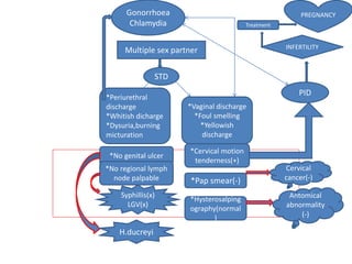
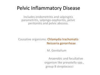
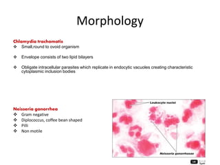
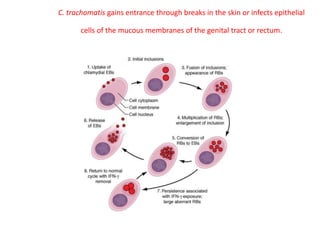
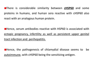
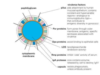
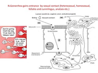
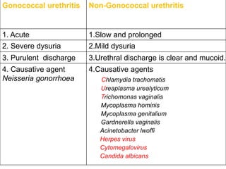
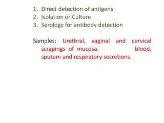
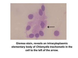
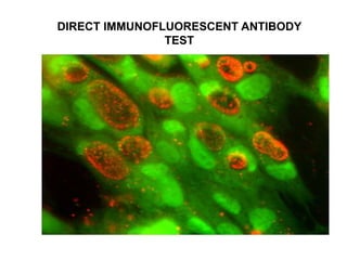
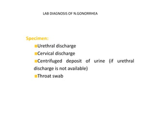
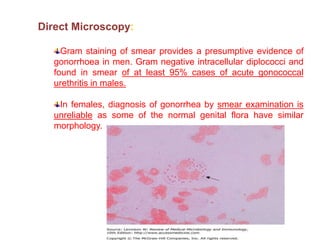
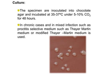
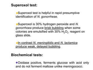
Multiple sex partner
STD
*Periurethral
discharge
*Whitish dicharge
*Dysuria,burning
micturation
*Vaginal discharge
*Foul smelling
*Yellowish
discharge
*No genital ulcer
*No regional lymph
node palpable
*Cervical motion
tenderness(+)
*Pap smear(-)
*Hysterosalping
ography(normal
)
Syphillis(x)
LGV(x)
H.ducreyi
Cervical
cancer(-)
Antomical
abnormality
(-)
PID
INFERTILITY
Gonorrhoea
Chlamydia Treatment
PREGNANCY
Pelvic Inflammatory Disease
Includes endometritis and salpingitis
parametritis, salpingo-oophoritis, pelvic
peritonitis and pelvic abscess.
Causative organisms: Chlamydia trachomatis
Neisseria gonorrheae
M. Genitalium
Anaerobic and facultative
organism like prevotella spp.,
group B streptococci
Morphology
Chlamydia trachomatis
 Small,round to ovoid organism
 Envelope consists of two lipid bilayers
 Obligate intracellular parasites which replicate in endocytic vacuoles creating characteristic
cytoplasmic inclusion bodies
Neisseria gonorrhea
 Gram negative
 Diplococcus, coffee bean shaped
 Pilli
 Non motile
C. trachomatis gains entrance through breaks in the skin or infects epithelial
cells of the mucous membranes of the genital tract or rectum.
There is considerable similarity between cHSP60 and some
proteins in humans, and human sera reactive with cHSP60 also
react with an analogous human protein.
Hence, serum antibodies reactive with HSP60 is associated with
ectopic pregnancy, infertility as well as persistent upper genital
tract infection and perihepatitis.
Hence, the pathogenesis of chlamydial disease seems to be
autoimmune, with cHSP60 being the sensitizing antigen.
N.Gonorrhea gains entrance by sexual contact (heterosexual, homosexual,
fellatio and cunnilingus, analsex etc.)
Gonococcal urethritis Non-Gonococcal urethritis
1. Acute 1.Slow and prolonged
2. Severe dysuria 2.Mild dysuria
3. Purulent discharge 3.Urethral discharge is clear and mucoid.
4. Causative agent
Neisseria gonorrhoea
4.Causative agents
Chlamydia trachomatis
Ureaplasma urealyticum
Trichomonas vaginalis
Mycoplasma hominis
Mycoplasma genitalium
Gardnerella vaginalis
Acinetobacter lwoffi
Herpes virus
Cytomegalovirus
Candida albicans
1. Direct detection of antigens
2. Isolation or Culture
3. Serology for antibody detection
Samples: Urethral, vaginal and cervical
scrapings of mucosa. In addition blood,
sputum and respiratory secretions.
Giemsa stain, reveals an intracytoplasmic
elementary body of Chlamydia trachomatis in the
cell to the left of the arrow.
DIRECT IMMUNOFLUORESCENT ANTIBODY
TEST
Iodine-stained inclusion bodies
Isolation of chlamydia is possible by yolk-sac
inoculation method and tissue culture in McCoy cells
(synovial carcinoma cell line)
Chlamydia trachomatis in McCoy cells brown colored
Serotyping is done by detecting specific
antibodies using immunofluorescene
(micro-IF) method.
Molecular methods:
PCR
Strand Displacement
Amplification
Target genes: Cryptic plasmid, the
omp 1 gene, 16srRNA gene.
LAB DIAGNOSIS OF N.GONORRHEA
Specimen:
Urethral discharge
Cervical discharge
Centrifuged deposit of urine (if urethral
discharge is not available)
Throat swab
Direct Microscopy:
Gram staining of smear provides a presumptive evidence of
gonorrhoea in men. Gram negative intracellular diplococci and
found in smear of at least 95% cases of acute gonococcal
urethritis in males.
In females, diagnosis of gonorrhea by smear examination is
unreliable as some of the normal genital flora have similar
morphology.
Culture:
The specimen are inoculated into chocolate
agar and incubated at 35-37ºC under 5-10% CO2
for 48 hours.
In chronic cases and in mixed infection such as
proctitis selective medium such as Thayer Martin
medium or modified Thayer –Martin medium is
used.
Superoxol test:
Superoxol test is helpful in rapid presumptive
identification of N. gonorrheae.
Superoxol is 30% hydrogen peroxide and N.
gonorrhoea produce brisk bubbling when some
colonies are emulsified with 30% H2O2 reagent on
glass slide.
In contrast N. meningitidis and N. lactamica
produce weak, delayed bubbling.
Biochemical tests:
Oxidase positive, ferments glucose with acid only
and do not ferment maltose unlike meningococci.
CLINICAL FEATURES
• CHLAMYDIA
TRAHOMATIS
1.Seroars D-K assosiated with
STDs and leads to
uretheritis,epididymitis,cerv
icitis,salpingitis,proctitis and
ultimately PID
2.Reiter’s
syndrome(conjunctivitis,ure
theritis,arthritis)
3.LGV by serovars L1 L2 L3
• N.gonorrhea
1.Urethritis
2.Cervicitis
3.Anoretal gonorrhea
4.Pharyngealgonorrhea
5.Oclular gonorrhea
6.Gonorrhea in pregnancy
7.Gonacoccal arthritis and
disseminated infection
QUARANTINE WITH YOUR VALENTINE!!!

pelvic inflammatory disease

  • 2.
    Multiple sex partner STD *Periurethral discharge *Whitishdicharge *Dysuria,burning micturation *Vaginal discharge *Foul smelling *Yellowish discharge *No genital ulcer *No regional lymph node palpable *Cervical motion tenderness(+) *Pap smear(-) *Hysterosalping ography(normal ) Syphillis(x) LGV(x) H.ducreyi Cervical cancer(-) Antomical abnormality (-) PID INFERTILITY Gonorrhoea Chlamydia Treatment PREGNANCY
  • 3.
    Pelvic Inflammatory Disease Includesendometritis and salpingitis parametritis, salpingo-oophoritis, pelvic peritonitis and pelvic abscess. Causative organisms: Chlamydia trachomatis Neisseria gonorrheae M. Genitalium Anaerobic and facultative organism like prevotella spp., group B streptococci
  • 4.
    Morphology Chlamydia trachomatis  Small,roundto ovoid organism  Envelope consists of two lipid bilayers  Obligate intracellular parasites which replicate in endocytic vacuoles creating characteristic cytoplasmic inclusion bodies Neisseria gonorrhea  Gram negative  Diplococcus, coffee bean shaped  Pilli  Non motile
  • 5.
    C. trachomatis gainsentrance through breaks in the skin or infects epithelial cells of the mucous membranes of the genital tract or rectum.
  • 6.
    There is considerablesimilarity between cHSP60 and some proteins in humans, and human sera reactive with cHSP60 also react with an analogous human protein. Hence, serum antibodies reactive with HSP60 is associated with ectopic pregnancy, infertility as well as persistent upper genital tract infection and perihepatitis. Hence, the pathogenesis of chlamydial disease seems to be autoimmune, with cHSP60 being the sensitizing antigen.
  • 8.
    N.Gonorrhea gains entranceby sexual contact (heterosexual, homosexual, fellatio and cunnilingus, analsex etc.)
  • 9.
    Gonococcal urethritis Non-Gonococcalurethritis 1. Acute 1.Slow and prolonged 2. Severe dysuria 2.Mild dysuria 3. Purulent discharge 3.Urethral discharge is clear and mucoid. 4. Causative agent Neisseria gonorrhoea 4.Causative agents Chlamydia trachomatis Ureaplasma urealyticum Trichomonas vaginalis Mycoplasma hominis Mycoplasma genitalium Gardnerella vaginalis Acinetobacter lwoffi Herpes virus Cytomegalovirus Candida albicans
  • 10.
    1. Direct detectionof antigens 2. Isolation or Culture 3. Serology for antibody detection Samples: Urethral, vaginal and cervical scrapings of mucosa. In addition blood, sputum and respiratory secretions.
  • 11.
    Giemsa stain, revealsan intracytoplasmic elementary body of Chlamydia trachomatis in the cell to the left of the arrow.
  • 12.
  • 13.
  • 14.
    Isolation of chlamydiais possible by yolk-sac inoculation method and tissue culture in McCoy cells (synovial carcinoma cell line) Chlamydia trachomatis in McCoy cells brown colored
  • 15.
    Serotyping is doneby detecting specific antibodies using immunofluorescene (micro-IF) method. Molecular methods: PCR Strand Displacement Amplification Target genes: Cryptic plasmid, the omp 1 gene, 16srRNA gene.
  • 16.
    LAB DIAGNOSIS OFN.GONORRHEA Specimen: Urethral discharge Cervical discharge Centrifuged deposit of urine (if urethral discharge is not available) Throat swab
  • 17.
    Direct Microscopy: Gram stainingof smear provides a presumptive evidence of gonorrhoea in men. Gram negative intracellular diplococci and found in smear of at least 95% cases of acute gonococcal urethritis in males. In females, diagnosis of gonorrhea by smear examination is unreliable as some of the normal genital flora have similar morphology.
  • 18.
    Culture: The specimen areinoculated into chocolate agar and incubated at 35-37ºC under 5-10% CO2 for 48 hours. In chronic cases and in mixed infection such as proctitis selective medium such as Thayer Martin medium or modified Thayer –Martin medium is used.
  • 19.
    Superoxol test: Superoxol testis helpful in rapid presumptive identification of N. gonorrheae. Superoxol is 30% hydrogen peroxide and N. gonorrhoea produce brisk bubbling when some colonies are emulsified with 30% H2O2 reagent on glass slide. In contrast N. meningitidis and N. lactamica produce weak, delayed bubbling. Biochemical tests: Oxidase positive, ferments glucose with acid only and do not ferment maltose unlike meningococci.
  • 20.
    CLINICAL FEATURES • CHLAMYDIA TRAHOMATIS 1.SeroarsD-K assosiated with STDs and leads to uretheritis,epididymitis,cerv icitis,salpingitis,proctitis and ultimately PID 2.Reiter’s syndrome(conjunctivitis,ure theritis,arthritis) 3.LGV by serovars L1 L2 L3 • N.gonorrhea 1.Urethritis 2.Cervicitis 3.Anoretal gonorrhea 4.Pharyngealgonorrhea 5.Oclular gonorrhea 6.Gonorrhea in pregnancy 7.Gonacoccal arthritis and disseminated infection
  • 22.