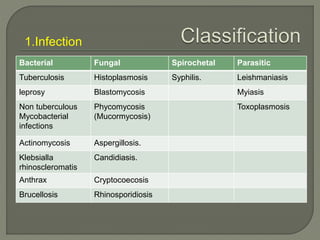
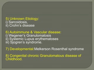
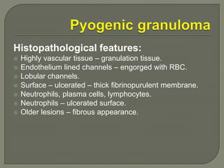
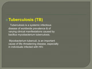
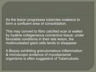
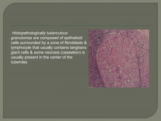
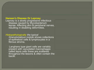
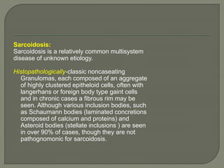
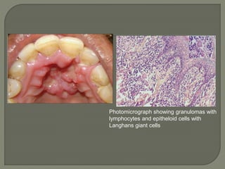
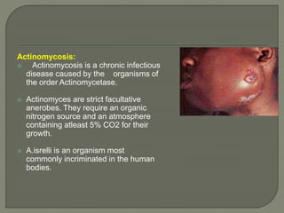
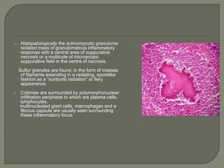
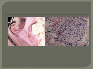
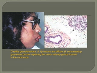
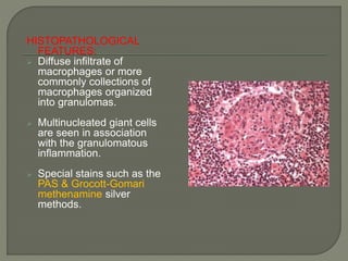
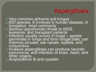
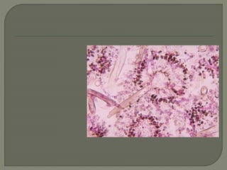
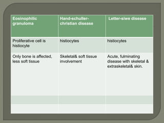
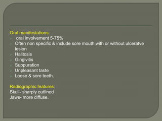
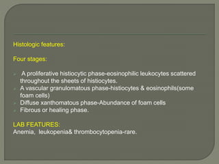
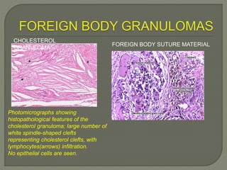
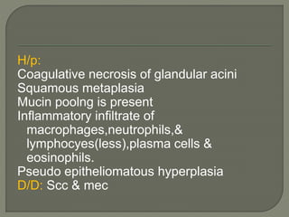
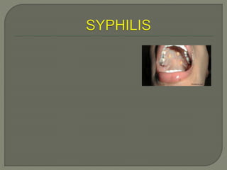
ORAL GRANULOMATOUS DISEASES
 DEFINITION 
 INTRODUCTION 
 PATHOPHYSIOLOGY OF GRANULOMA FORMATION 
 CLASSIFICATION OF GRANULOMATOUS DISEASES 
 GRANULOMATOUS LESIONS OF ORAL CAVITY 
 CONCLUSION 
 REFERENCES
DEFINITION: 
Defined as a specific variety of chronic 
inflammation in which cells of the mononuclear phagocyte 
system are predominant & take the form of macrophages, 
epithelioid cells & multinucleated cells. These cells are 
aggregated into well demarcated focal lesions called 
granulomas.
 Granulomatous diseases have plagued humans for million 
years, with evidence of tuberculosis infection in Egyptians 
mummies & description of the syphilis described by 
Hippocrates & was recognized as a venereal disease in the 
fifteenth century. 
 In seventeenth century, the minute granules (milliary) in host 
tissues were noted. Robert koch developed a method of 
staining & identified bacteria & was able to differentiate 
infectious, noninfectious granulomatous diseases. 
 Actinomycosis was isolated in vivo & associated 
with sulfur granule production in the late nineteenth century
 Granulomas are inflammatory lesions that evolve in response to irritation, 
infectious agents, and foreign bodies. 
 Localized masses of fibrovascular granulation tissue accompanied 
by a nonspecific chronic or sometimes subacute inflammatory infiltrate as seen 
microscopically are simple granulomas. 
 In the oral cavity, lesions of this nature are most frequently encountered 
intraosseously as endodontically related periapical granulomas. 
 Simple granulomas in the oral cavity are generally localized to the gingiva, yet 
may actually arise anywhere in the mouth or on the facial skin, where they 
appear as cherry-red tumefactions, termed pyogenic granulomas. 
 The peripheral giant cell granuloma of the gingiva and alveolar ridge is a reactive 
lesion without an infectious etiology. 
 specific granulomas are usually associated with mycobacteria or deep invasive 
fungi, appearing microscopically as caseating and noncaseating granulomas.
Two types: 
 Foreign body granuloma 
 Immune granuloma
Foreign body reaction: 
The foreign material is composed of particles that are usually too 
large to be phagocytosed by macrophages. Because the 
material is typically inert, it usually does not evoke an immune 
response.Instead, in an attempt to eliminate the foreign substance 
steady streams of macrophages are recruited to the site. There they 
become activated, transform into epithelioid macrophages & 
organize into granuloma.
 Distinct form of delayed-type (cell mediated) hypersensitivity. 
Immune granulomas typically develop in response to infection by 
various mycobacterial & fungal organisms. 
 initial exposure to a non-degradable antigen derived from an 
organism (usually a protein), there is an accumulation of 
predominantly, (CD4- positive) T- lymphocyte around blood vessels 
in the area where the antigens lie. Over time, the T cells 
differentiate, become activated T- helper (Th) cells, & begin 
secreting various cytokines that recruit macrophages to the site of 
the foreign antigen. 
 If the antigens persist & remain nondegradable, the recruited 
macrophages eventually transform into epitheliod cells, organize & 
form granuloma.
1.Infection 
Bacterial Fungal Spirochetal Parasitic 
Tuberculosis Histoplasmosis Syphilis. Leishmaniasis 
leprosy Blastomycosis Myiasis 
Non tuberculous 
Phycomycosis 
Mycobacterial 
(Mucormycosis) 
infections 
Toxoplasmosis 
Actinomycosis Aspergillosis. 
Klebsialla 
Candidiasis. 
rhinoscleromatis 
Anthrax Cryptocoecosis 
Brucellosis Rhinosporidiosis
2) Traumatic Etiology: 
i) Pyogenic granuloma 
ii) Reparative granuloma 
3) Foreign Body etiology: 
i) Oral foreign body reactions. 
(Suture, hair, amalgam, endodontic sealer, 
hyaluronic acid etc.) 
ii) Cholesterol granuloma 
iii) Cocaine induced midline granuloma 
iv) Gout
4) Neoplastic 
i)Histocytosis X 
a)Eosinophilic granuloma 
b) Hand schuller Christian Disease. 
c) Letterersiwe disease. 
ii) Benign Fibrous histiocytoma 
iii) Neorotizing sialometaplasia. 
iv) Polymorphic recticulosis (lethal midline 
granuloma)
5) Unknown Etiology: 
i) Sarcoidosis, 
ii) Crohn’s disease 
6) Autoimmune & Vascular disease: 
i) Wegener’s Granulomatosis 
ii) Systemic Lupus erythematoses 
iii) Sjogren’s syndrome. 
7) Developmental:Melkerson Rosenthal syndrome 
8) Congenital chronic Granulomatous disease of 
Childhood.
Histopathological features: 
 Highly vascular tissue – granulation tissue. 
 Endothelium lined channels – engorged with RBC. 
 Lobular channels. 
 Surface – ulcerated – thick fibrinopurulent membrane. 
 Neutrophils, plasma cells, lymphocytes. 
 Neutrophils – ulcerated surface. 
 Older lesions – fibrous appearance.
Clinical: 
 Peripheral giant cell granuloma. 
 Peripheral ossifying fibroma. 
 Primary or metastatic malignancy. 
Histopathological: 
 Gingival fibromas. 
 Polypoid capillary hemangioma. 
 Inflammed lobular hemangioma. 
•Conventional granulation tissue. 
•Hyperplastic gingival inflammation. 
•Kaposi’s sarcoma. 
 Granulation tissue type hemangioma. 
•Plasma cell granuloma. 
•Kaposi’s sarcoma. 
•Angiosarcoma.
Tuberculosis (TB) 
Tuberculosis is a systemic infectious 
disease of worldwide prevalence & of 
varying clinical manifestations caused by 
bacillus mycobacterium tuberculosis. 
Mycobacterium tuberculi, is an important 
cause of life–threatening disease, especially 
in individuals infected with HIV.
As the lesion progresses tubercles coalesce to 
form a confluent area of consolidation. 
This may convert to fibro calcified scar or walled 
by hyaline collageneous connective tissue, under 
favorable conditions in their late lesion, the 
multinucleated giant cells tends to disappear. 
A Biopsy exhibiting granulomatous inflammation 
& microscopic evidence of mycobacterial 
organisms is often suggestive of Tuberculosis.
Histopathologically tuberculous 
granulomas are composed of epithelioid 
cells surrounded by a zone of fibroblasts & 
lymphocyte that usually contains langhans 
giant cells & some necrosis (caseation) is 
usually present in the center of the 
tubercles
Hansen’s Disease Or Leprosy 
Leprosy is a slowly progressive infectious 
disease caused by Mycobacterium 
leprae. Affecting skin & peripheral nerves, 
resulting in disabling deformities. 
Histopathologically the typical 
Granulomatous nodule shows collections 
of epithelioid cells & lymphocytes in a 
fibrous stroma. 
Langhans type giant cells are variably 
present with vacuolated macrophages 
called lepra cells these are scattered 
throughout the lesions & often contain the 
bacilli.
Sarcoidosis: 
Sarcoidosis is a relatively common multisystem 
disease of unknown etiology. 
Histopathologically-classic noncaseating 
Granulomas, each composed of an aggregate 
of highly clustered epitheloid cells, often with 
langerhans or foreign body type gaint cells 
and in chronic cases a fibrous rim may be 
seen. Although various inclusion bodies, such 
as Schaumann bodies (laminated concretions 
composed of calcium and proteins) and 
Asteroid bodies (stellate inclusions ) are seen 
in over 90% of cases, though they are not 
pathognomonic for sarcoidosis.
Photomicrograph showing granulomas with 
lymphocytes and epitheloid cells with 
Langhans giant cells
Actinomycosis: 
 Actinomycosis is a chronic infectious 
disease caused by the organisms of 
the order Actinomycetase. 
 Actinomyces are strict facultative 
anerobes. They require an organic 
nitrogen source and an atmosphere 
containing atleast 5% CO2 for their 
growth. 
 A.isrelli is an organism most 
commonly incriminated in the human 
bodies.
 Histopatologically the actinomycotic granuloma 
isolated mass of granulomatous inflammatory 
response with a central area of suppurative 
necrosis or a multitude of microscopic 
suppurative field in the centre of necrosis. 
Sulfur granules are found, in the form of masses 
of filaments extending in a radiating, sporelike 
fashion as a “sunburst radiation” or fiery 
appearance. 
 Colonies are surrounded by polymorphonuclear 
infiltration peripheral to which are plasma cells, 
lymphocytes, 
multinucleated giant cells, macrophages and a 
fibrous capsule are usually seen surrounding 
these inflammatory focus
The filamentous mass is mineralized and cemented by host calcium 
phosphate resulting from phosphatase activity from tissue 
inflammation. 
At the end of these filaments are club shaped extensions or rossets 
formed by the adherence of neutrophils. 
They are also encased in a polysaccharide protein complex. The 
central core of the rosette stains basophilic& the peripheral clubs 
stains eosinophilic with H & E stain. 
Methanamine silver is useful because they color older filaments very 
well.
 Crohn’s Disease 
Crohn’s disease is a chronic Granulomatous disorder that may 
involve any portion of the GIT including the oral cavity. 
 The exact etiology remains unknown. 
 Crohn’s disease is a Th1 disorder, Environmental and genetic 
factors seem to play an important role in the pathogenesis. 
 Recently mutations in the CARD15 /NoD2 gene on the Chromosome 
16 also have been identified in 25% of patients with crohn’s disease. 
 Histopathology: – noncaseating Granulomatous inflammation in 
50% of cases and in the remaining cases of nonspecific perivascular 
lymphocytic infiltrate. 
 Gut biopsies characteristically show transmural inflammation.
A classic triad - nodular lip swellings, fissured 
tongue, and unilateral facial palsy 
Clinically, the lower lip is involved more 
diffusely than the upper lip, being enlarged 
with palpable nodularity throughout the 
submucosa. 
Microscopically, multiple granulomas are 
seen to replace or infiltrate the lip minor 
salivary glands. Nodular tumefactions that are 
firm to palpation may be encountered in the 
buccal mucosa, vestibule, and tongue.
Melkersson-Rosenthal syndrome. A, Fissured tongue; B, 
granulomatous cheilitis; C, facial nerve palsy.
 Granulomas located as multiple nodules throughout the 
lips constitute cheilitis granulomatosa, provided these 
lesions are the only manifestation of the condition. 
lip granulomas with identical appearance to cheilitis 
granulomatosa are encountered in the Melkersson-Rosenthal 
syndrome. 
 The lower, upper, or both lips may be involved Although the 
swelling may appear to be diffuse, palpation discloses multiple 
and often confluent nodules within the submucosa. 
 Biopsy shows multiple noncaseating granulomas replacing 
minor salivary lobules
Cheilitis granulomatosa. A, lip lesions are diffuse; B, noncaseating 
granuloma (arrow) replacing the minor salivary glands located 
in the submusoa.
 Wegener granulomatosis 
Often included as a form of midline lethal granuloma, Wegener 
granulomatosis represents an inflammatory destructive disease that 
may have widespread systemic involvement. 
 The granulomas of Wegener granulomatosis are usually localized in 
the lung and kidneys. The middle ear, gingiva, and nasal mucosa are 
also commonly affected. 
 Microscopically, vasculitis is a dominant finding, along with fibrinoid 
necrosis. The adjacent granulation tissue is often infiltrated with 
eosinophils and occasional multinucleated giant cells are seen.
 Antibodies directed to neutrophil cytoplasmic antigens (ANCA) are 
detected using indirect immunofluorescence with neutrophils as 
substrate, being positive in over 90% of cases.
HISTOPLASMOSIS: 
 Common systemic fungal 
infection in the united states. 
 Caused by histoplasma 
capsulatum, is dimorphic, 
growing as a yeast at body 
temperature in the human 
host and as a mold in its 
natural environment. 
 Air-borne spores of the 
organism are inhaled,pass 
into the terminal passages of 
the lungs &germinate. 
 TYPES : 
Acute,chronic,disseminated 
histoplasmosis.
HISTOPATHOLOGICAL 
FEATURES: 
 Diffuse infiltrate of 
macrophages or more 
commonly collections of 
macrophages organized 
into granulomas. 
 Multinucleated giant cells 
are seen in association 
with the granulomatous 
inflammation. 
 Special stains such as the 
PAS & Grocott-Gomari 
methenamine silver 
methods.
 Synonyms- Mucormycosis,Phycomycosis 
 Zygomycota are extremely abundant saprobic 
fungi found in soil, water, organic debris, and food. 
 Genera most often involved are Rhizopus, Absidia, 
and Mucor. 
 Usually harmless air contaminants invade the 
membranes of the nose, eyes, heart, and brain of 
people with diabetes and malnutrition, with severe 
consequences.
ORAL MANIFESTATIONS: 
 Rhinocerebral form is most relevant to the 
oral health care provider. 
 Zygomycosis is noted especially in insulin-dependent 
diabetics who have uncontrolled 
diabetes and are ketoacidotic. 
 Ketoacidotic inhibits the binding of iron to 
transferin, allowing serum iron levels to rise.
H/P: 
 Extensive necrosis 
with numerous 
large (6-30 micro 
m) 
 branching 
nonseptate hyphae 
at the periphery. 
 The hyphae tend to 
branch at 
90°angles.
 Coccidioides immitis - causes 
coccidioidomycosis 
 Distinctive morphology – blocklike 
arthroconidia in the free-living stage and 
spherules containing endospores in the lungs 
 Lives in alkaline soils in semiarid, hot climates 
and is endemic to southwestern U.S. 
 Arthrospores inhaled from dust, creates 
spherules and nodules in the lungs 
 Amphotericin B treatment
Blastomyces dermatitidis- causes 
blastomycosis 
Dimorphic 
Free-living species distributed in soil of a 
large section of the midwestern and 
southeastern U.S. 
Inhaled 10-100 conidia convert to yeasts 
and multiply in lungs 
Symptoms include cough and fever. 
Chronic cutaneous, bone, and nervous 
system complications 
Amphotericin B
HISTOPATHOLOGICAL FEATURES: 
 Mixture of acute inflammation and 
granulomatous inflammation surrounded 
by yeasts. 
 These organisms are 8-20 micro meter in 
diameter. 
 B.dermatitidis can be detected-special 
stains such as gromatt-Gomori 
methenamine silver&PAS.
Very common airborne soil fungus 
 600 species, 8 involved in human disease; A. 
fumigatus most commonly 
 Serious opportunistic threat to AIDS, 
leukemia, and transplant patients 
 Infection usually occurs in lungs – spores 
germinate in lungs and form fungal balls; can 
colonize sinuses, ear canals, eyelids, and 
conjunctiva 
 Invasive aspergillosis can produce necrotic 
pneumonia, and infection of brain, heart, and 
other organs. 
 Amphotericin B and nystatin
TOXOPLASMOSIS: 
 Common disease caused by the obligate intracellular 
protozoal organism T. gondii. 
 T. godii multiplies in the intestinal tract of the cat by 
means of a sexual life cycle, discharging numerous 
oocysts in the cat feces. Another animal or human can 
ingest these oocysts , resulting in the production of 
disease. 
CLINICAL FEATURES: 
 Often Asymptomatic 
 If symptomatic they are usually mild and resemble 
infectious mononucleosis. 
 Pt’s may have a low grade fever , cervical 
lymphadenopathy.
 In immunosuppressed patients , 
toxoplasmosis represent a new,primary 
infection or reactivation of previously 
encysted organism. 
 The principal groups at risk – 
AIDS 
Transplant recipients 
Cancer patients
DIAGNOSIS AND TREATMENT: 
 Identification of rising serum antibody titers 
to T.gondii within 10-14 days after 
infection. 
 Biopsy of an involved lymphnode may 
suggest the diagnosis and serologic 
studies if possible. 
 Folinic acid and sulfadiazine,if allergic to 
sulfa drugs clindamycin can be used.
HISTIOCYTIC: 
Eosinophilic granuloma(unifocal eosinophilic granuloma): 
 Lichtenstein and jaffe in 1940 
 A lesion of bone which is primarily a histiocytic proliferation with an 
abundance of eosinophilic leukocytes but no intracellular lipid 
accumulation. 
 Primarily in older children and young adults. 
 M:F- 2:1 
C/F: 
 No physical signs & symptoms 
 Incidental radiographic findings.
Common sites: skull and mandible 
Other sites: Femur,humerus,ribs & other bones. 
Radiographic: Irregular radiolucent areas usually 
involving superficial alveolar bone. 
H/P: 
Prmary cell is histocyte which grows in sheets or 
sheetlike collections. The histiocyte may coalesce 
& form multinucleated giant cells- uncommon. 
Early lesion- large no of collections of 
eosinophils,lesion matures , fibrosis occurs & less 
no of eosinophils. Then similar H/P picture of hand 
schuller christian disease.
Synonym: multifocal eosinophilic granuloma: 
Widespread skeletal & extraskeletal & a chronic clinical course. 
 It occurs primarily in early life, before the age of 5. 
 More common in boys than girls. 
C/F: 
Classic triad- single/multiple punched out bone destruction in the 
skull 
Unilateral or bilateral exophthalmos 
Diabetic insipidus 
the complete triad occurs only 25% of affected patients.
Eosinophilic 
granuloma 
Hand-schuller-christian 
disease 
Letter-siwe disease 
Proliferative cell is 
histiocyte 
histiocytes histiocytes 
Only bone is affected, 
less soft tissue 
Skeletal& soft tissue 
involvement 
Acute, fulminating 
disease with skeletal & 
extraskeletal& skin.
Oral manifestations: 
 oral involvement 5-75% 
 Often non specific & include sore mouth,with or without ulceratve 
lesion 
 Halitosis 
 Gingivitis 
 Suppuration 
 Unpleasant taste 
 Loose & sore teeth. 
Radiographic features: 
Skull- sharply outlined 
Jaws- more diffuse.
Histologic features: 
Four stages: 
 A proliferative histiocytic phase-eosinophilic leukocytes scattered 
throughout the sheets of histiocytes. 
 A vascular granulomatous phase-histiocytes & eosinophils(some 
foam cells) 
 Diffuse xanthomatous phase-Abundance of foam cells 
 Fibrous or healing phase. 
LAB FEATURES: 
Anemia, leukopenia& thrombocytopenia-rare.
CHOLESTEROL 
GRANULOMA 
Photomicrographs showing 
histopathological features of the 
cholesterol granuloma; large number of 
white spindle-shaped clefts 
representing cholesterol clefts, with 
lymphocytes(arrows) infiltration. 
No epithelial cells are seen. 
FOREIGN BODY SUTURE MATERIAL
 Triad- Keratoconjuctivitis sicca 
Xerostomia 
Rheumatoid arthritis 
Primary sjogren syndrome- 
Dry eyes & dry mouth 
Secondary sjogrens: 
SLE 
Polyarteritis nodosa 
Scleroderma 
Rheumatoid arthritis 
Etiology: 
Genetic 
Hormonal 
Infections & immunologic
c/f: 
Females are more common over 40 yrs 
Ratio- 10:1 
Radiographic : cherry blossom or branchless 
fruit laden tree effect. 
H/P: 
 intense lymphocytic infiltration 
 Lobular architecture is preserved 
 Epimyoepithelial islands are present.
The lobular architecture of the 
seromucinous glands is preserved. The 
seromucinous acini show squamous 
metaplasia with slight reactive atypia. 
 Non neoplastic inflammatory condition 
Etiology: 
Trauma 
Radiation therapy/ surgery 
Vascular ischemia 
Tobacco – risk factor 
C/F: 
Minor salivary gland-palate 
Others retromolar pad, BM,tongue,incisive 
Canal & labial mucosa 
Clinical presentation: 
Crateriform ulcer of the palate that similates 
A malignant process. 
1-3cm & unilateral
H/p: 
Coagulative necrosis of glandular acini 
Squamous metaplasia 
Mucin poolng is present 
Inflammatory infiltrate of 
macrophages,neutrophils,& 
lymphocyes(less),plasma cells & 
eosinophils. 
Pseudo epitheliomatous hyperplasia 
D/D: Scc & mec
Seminar 12

Seminar 12

  • 1.
  • 2.
     DEFINITION INTRODUCTION  PATHOPHYSIOLOGY OF GRANULOMA FORMATION  CLASSIFICATION OF GRANULOMATOUS DISEASES  GRANULOMATOUS LESIONS OF ORAL CAVITY  CONCLUSION  REFERENCES
  • 3.
    DEFINITION: Defined asa specific variety of chronic inflammation in which cells of the mononuclear phagocyte system are predominant & take the form of macrophages, epithelioid cells & multinucleated cells. These cells are aggregated into well demarcated focal lesions called granulomas.
  • 4.
     Granulomatous diseaseshave plagued humans for million years, with evidence of tuberculosis infection in Egyptians mummies & description of the syphilis described by Hippocrates & was recognized as a venereal disease in the fifteenth century.  In seventeenth century, the minute granules (milliary) in host tissues were noted. Robert koch developed a method of staining & identified bacteria & was able to differentiate infectious, noninfectious granulomatous diseases.  Actinomycosis was isolated in vivo & associated with sulfur granule production in the late nineteenth century
  • 5.
     Granulomas areinflammatory lesions that evolve in response to irritation, infectious agents, and foreign bodies.  Localized masses of fibrovascular granulation tissue accompanied by a nonspecific chronic or sometimes subacute inflammatory infiltrate as seen microscopically are simple granulomas.  In the oral cavity, lesions of this nature are most frequently encountered intraosseously as endodontically related periapical granulomas.  Simple granulomas in the oral cavity are generally localized to the gingiva, yet may actually arise anywhere in the mouth or on the facial skin, where they appear as cherry-red tumefactions, termed pyogenic granulomas.  The peripheral giant cell granuloma of the gingiva and alveolar ridge is a reactive lesion without an infectious etiology.  specific granulomas are usually associated with mycobacteria or deep invasive fungi, appearing microscopically as caseating and noncaseating granulomas.
  • 6.
    Two types: Foreign body granuloma  Immune granuloma
  • 7.
    Foreign body reaction: The foreign material is composed of particles that are usually too large to be phagocytosed by macrophages. Because the material is typically inert, it usually does not evoke an immune response.Instead, in an attempt to eliminate the foreign substance steady streams of macrophages are recruited to the site. There they become activated, transform into epithelioid macrophages & organize into granuloma.
  • 8.
     Distinct formof delayed-type (cell mediated) hypersensitivity. Immune granulomas typically develop in response to infection by various mycobacterial & fungal organisms.  initial exposure to a non-degradable antigen derived from an organism (usually a protein), there is an accumulation of predominantly, (CD4- positive) T- lymphocyte around blood vessels in the area where the antigens lie. Over time, the T cells differentiate, become activated T- helper (Th) cells, & begin secreting various cytokines that recruit macrophages to the site of the foreign antigen.  If the antigens persist & remain nondegradable, the recruited macrophages eventually transform into epitheliod cells, organize & form granuloma.
  • 9.
    1.Infection Bacterial FungalSpirochetal Parasitic Tuberculosis Histoplasmosis Syphilis. Leishmaniasis leprosy Blastomycosis Myiasis Non tuberculous Phycomycosis Mycobacterial (Mucormycosis) infections Toxoplasmosis Actinomycosis Aspergillosis. Klebsialla Candidiasis. rhinoscleromatis Anthrax Cryptocoecosis Brucellosis Rhinosporidiosis
  • 10.
    2) Traumatic Etiology: i) Pyogenic granuloma ii) Reparative granuloma 3) Foreign Body etiology: i) Oral foreign body reactions. (Suture, hair, amalgam, endodontic sealer, hyaluronic acid etc.) ii) Cholesterol granuloma iii) Cocaine induced midline granuloma iv) Gout
  • 11.
    4) Neoplastic i)HistocytosisX a)Eosinophilic granuloma b) Hand schuller Christian Disease. c) Letterersiwe disease. ii) Benign Fibrous histiocytoma iii) Neorotizing sialometaplasia. iv) Polymorphic recticulosis (lethal midline granuloma)
  • 12.
    5) Unknown Etiology: i) Sarcoidosis, ii) Crohn’s disease 6) Autoimmune & Vascular disease: i) Wegener’s Granulomatosis ii) Systemic Lupus erythematoses iii) Sjogren’s syndrome. 7) Developmental:Melkerson Rosenthal syndrome 8) Congenital chronic Granulomatous disease of Childhood.
  • 14.
    Histopathological features: Highly vascular tissue – granulation tissue.  Endothelium lined channels – engorged with RBC.  Lobular channels.  Surface – ulcerated – thick fibrinopurulent membrane.  Neutrophils, plasma cells, lymphocytes.  Neutrophils – ulcerated surface.  Older lesions – fibrous appearance.
  • 16.
    Clinical:  Peripheralgiant cell granuloma.  Peripheral ossifying fibroma.  Primary or metastatic malignancy. Histopathological:  Gingival fibromas.  Polypoid capillary hemangioma.  Inflammed lobular hemangioma. •Conventional granulation tissue. •Hyperplastic gingival inflammation. •Kaposi’s sarcoma.  Granulation tissue type hemangioma. •Plasma cell granuloma. •Kaposi’s sarcoma. •Angiosarcoma.
  • 17.
    Tuberculosis (TB) Tuberculosisis a systemic infectious disease of worldwide prevalence & of varying clinical manifestations caused by bacillus mycobacterium tuberculosis. Mycobacterium tuberculi, is an important cause of life–threatening disease, especially in individuals infected with HIV.
  • 18.
    As the lesionprogresses tubercles coalesce to form a confluent area of consolidation. This may convert to fibro calcified scar or walled by hyaline collageneous connective tissue, under favorable conditions in their late lesion, the multinucleated giant cells tends to disappear. A Biopsy exhibiting granulomatous inflammation & microscopic evidence of mycobacterial organisms is often suggestive of Tuberculosis.
  • 19.
    Histopathologically tuberculous granulomasare composed of epithelioid cells surrounded by a zone of fibroblasts & lymphocyte that usually contains langhans giant cells & some necrosis (caseation) is usually present in the center of the tubercles
  • 20.
    Hansen’s Disease OrLeprosy Leprosy is a slowly progressive infectious disease caused by Mycobacterium leprae. Affecting skin & peripheral nerves, resulting in disabling deformities. Histopathologically the typical Granulomatous nodule shows collections of epithelioid cells & lymphocytes in a fibrous stroma. Langhans type giant cells are variably present with vacuolated macrophages called lepra cells these are scattered throughout the lesions & often contain the bacilli.
  • 21.
    Sarcoidosis: Sarcoidosis isa relatively common multisystem disease of unknown etiology. Histopathologically-classic noncaseating Granulomas, each composed of an aggregate of highly clustered epitheloid cells, often with langerhans or foreign body type gaint cells and in chronic cases a fibrous rim may be seen. Although various inclusion bodies, such as Schaumann bodies (laminated concretions composed of calcium and proteins) and Asteroid bodies (stellate inclusions ) are seen in over 90% of cases, though they are not pathognomonic for sarcoidosis.
  • 22.
    Photomicrograph showing granulomaswith lymphocytes and epitheloid cells with Langhans giant cells
  • 23.
    Actinomycosis:  Actinomycosisis a chronic infectious disease caused by the organisms of the order Actinomycetase.  Actinomyces are strict facultative anerobes. They require an organic nitrogen source and an atmosphere containing atleast 5% CO2 for their growth.  A.isrelli is an organism most commonly incriminated in the human bodies.
  • 24.
     Histopatologically theactinomycotic granuloma isolated mass of granulomatous inflammatory response with a central area of suppurative necrosis or a multitude of microscopic suppurative field in the centre of necrosis. Sulfur granules are found, in the form of masses of filaments extending in a radiating, sporelike fashion as a “sunburst radiation” or fiery appearance.  Colonies are surrounded by polymorphonuclear infiltration peripheral to which are plasma cells, lymphocytes, multinucleated giant cells, macrophages and a fibrous capsule are usually seen surrounding these inflammatory focus
  • 25.
    The filamentous massis mineralized and cemented by host calcium phosphate resulting from phosphatase activity from tissue inflammation. At the end of these filaments are club shaped extensions or rossets formed by the adherence of neutrophils. They are also encased in a polysaccharide protein complex. The central core of the rosette stains basophilic& the peripheral clubs stains eosinophilic with H & E stain. Methanamine silver is useful because they color older filaments very well.
  • 26.
     Crohn’s Disease Crohn’s disease is a chronic Granulomatous disorder that may involve any portion of the GIT including the oral cavity.  The exact etiology remains unknown.  Crohn’s disease is a Th1 disorder, Environmental and genetic factors seem to play an important role in the pathogenesis.  Recently mutations in the CARD15 /NoD2 gene on the Chromosome 16 also have been identified in 25% of patients with crohn’s disease.  Histopathology: – noncaseating Granulomatous inflammation in 50% of cases and in the remaining cases of nonspecific perivascular lymphocytic infiltrate.  Gut biopsies characteristically show transmural inflammation.
  • 28.
    A classic triad- nodular lip swellings, fissured tongue, and unilateral facial palsy Clinically, the lower lip is involved more diffusely than the upper lip, being enlarged with palpable nodularity throughout the submucosa. Microscopically, multiple granulomas are seen to replace or infiltrate the lip minor salivary glands. Nodular tumefactions that are firm to palpation may be encountered in the buccal mucosa, vestibule, and tongue.
  • 29.
    Melkersson-Rosenthal syndrome. A,Fissured tongue; B, granulomatous cheilitis; C, facial nerve palsy.
  • 30.
     Granulomas locatedas multiple nodules throughout the lips constitute cheilitis granulomatosa, provided these lesions are the only manifestation of the condition. lip granulomas with identical appearance to cheilitis granulomatosa are encountered in the Melkersson-Rosenthal syndrome.  The lower, upper, or both lips may be involved Although the swelling may appear to be diffuse, palpation discloses multiple and often confluent nodules within the submucosa.  Biopsy shows multiple noncaseating granulomas replacing minor salivary lobules
  • 31.
    Cheilitis granulomatosa. A,lip lesions are diffuse; B, noncaseating granuloma (arrow) replacing the minor salivary glands located in the submusoa.
  • 32.
     Wegener granulomatosis Often included as a form of midline lethal granuloma, Wegener granulomatosis represents an inflammatory destructive disease that may have widespread systemic involvement.  The granulomas of Wegener granulomatosis are usually localized in the lung and kidneys. The middle ear, gingiva, and nasal mucosa are also commonly affected.  Microscopically, vasculitis is a dominant finding, along with fibrinoid necrosis. The adjacent granulation tissue is often infiltrated with eosinophils and occasional multinucleated giant cells are seen.
  • 33.
     Antibodies directedto neutrophil cytoplasmic antigens (ANCA) are detected using indirect immunofluorescence with neutrophils as substrate, being positive in over 90% of cases.
  • 34.
    HISTOPLASMOSIS:  Commonsystemic fungal infection in the united states.  Caused by histoplasma capsulatum, is dimorphic, growing as a yeast at body temperature in the human host and as a mold in its natural environment.  Air-borne spores of the organism are inhaled,pass into the terminal passages of the lungs &germinate.  TYPES : Acute,chronic,disseminated histoplasmosis.
  • 35.
    HISTOPATHOLOGICAL FEATURES: Diffuse infiltrate of macrophages or more commonly collections of macrophages organized into granulomas.  Multinucleated giant cells are seen in association with the granulomatous inflammation.  Special stains such as the PAS & Grocott-Gomari methenamine silver methods.
  • 36.
     Synonyms- Mucormycosis,Phycomycosis  Zygomycota are extremely abundant saprobic fungi found in soil, water, organic debris, and food.  Genera most often involved are Rhizopus, Absidia, and Mucor.  Usually harmless air contaminants invade the membranes of the nose, eyes, heart, and brain of people with diabetes and malnutrition, with severe consequences.
  • 37.
    ORAL MANIFESTATIONS: Rhinocerebral form is most relevant to the oral health care provider.  Zygomycosis is noted especially in insulin-dependent diabetics who have uncontrolled diabetes and are ketoacidotic.  Ketoacidotic inhibits the binding of iron to transferin, allowing serum iron levels to rise.
  • 38.
    H/P:  Extensivenecrosis with numerous large (6-30 micro m)  branching nonseptate hyphae at the periphery.  The hyphae tend to branch at 90°angles.
  • 39.
     Coccidioides immitis- causes coccidioidomycosis  Distinctive morphology – blocklike arthroconidia in the free-living stage and spherules containing endospores in the lungs  Lives in alkaline soils in semiarid, hot climates and is endemic to southwestern U.S.  Arthrospores inhaled from dust, creates spherules and nodules in the lungs  Amphotericin B treatment
  • 40.
    Blastomyces dermatitidis- causes blastomycosis Dimorphic Free-living species distributed in soil of a large section of the midwestern and southeastern U.S. Inhaled 10-100 conidia convert to yeasts and multiply in lungs Symptoms include cough and fever. Chronic cutaneous, bone, and nervous system complications Amphotericin B
  • 42.
    HISTOPATHOLOGICAL FEATURES: Mixture of acute inflammation and granulomatous inflammation surrounded by yeasts.  These organisms are 8-20 micro meter in diameter.  B.dermatitidis can be detected-special stains such as gromatt-Gomori methenamine silver&PAS.
  • 43.
    Very common airbornesoil fungus  600 species, 8 involved in human disease; A. fumigatus most commonly  Serious opportunistic threat to AIDS, leukemia, and transplant patients  Infection usually occurs in lungs – spores germinate in lungs and form fungal balls; can colonize sinuses, ear canals, eyelids, and conjunctiva  Invasive aspergillosis can produce necrotic pneumonia, and infection of brain, heart, and other organs.  Amphotericin B and nystatin
  • 45.
    TOXOPLASMOSIS:  Commondisease caused by the obligate intracellular protozoal organism T. gondii.  T. godii multiplies in the intestinal tract of the cat by means of a sexual life cycle, discharging numerous oocysts in the cat feces. Another animal or human can ingest these oocysts , resulting in the production of disease. CLINICAL FEATURES:  Often Asymptomatic  If symptomatic they are usually mild and resemble infectious mononucleosis.  Pt’s may have a low grade fever , cervical lymphadenopathy.
  • 46.
     In immunosuppressedpatients , toxoplasmosis represent a new,primary infection or reactivation of previously encysted organism.  The principal groups at risk – AIDS Transplant recipients Cancer patients
  • 47.
    DIAGNOSIS AND TREATMENT:  Identification of rising serum antibody titers to T.gondii within 10-14 days after infection.  Biopsy of an involved lymphnode may suggest the diagnosis and serologic studies if possible.  Folinic acid and sulfadiazine,if allergic to sulfa drugs clindamycin can be used.
  • 48.
    HISTIOCYTIC: Eosinophilic granuloma(unifocaleosinophilic granuloma):  Lichtenstein and jaffe in 1940  A lesion of bone which is primarily a histiocytic proliferation with an abundance of eosinophilic leukocytes but no intracellular lipid accumulation.  Primarily in older children and young adults.  M:F- 2:1 C/F:  No physical signs & symptoms  Incidental radiographic findings.
  • 49.
    Common sites: skulland mandible Other sites: Femur,humerus,ribs & other bones. Radiographic: Irregular radiolucent areas usually involving superficial alveolar bone. H/P: Prmary cell is histocyte which grows in sheets or sheetlike collections. The histiocyte may coalesce & form multinucleated giant cells- uncommon. Early lesion- large no of collections of eosinophils,lesion matures , fibrosis occurs & less no of eosinophils. Then similar H/P picture of hand schuller christian disease.
  • 50.
    Synonym: multifocal eosinophilicgranuloma: Widespread skeletal & extraskeletal & a chronic clinical course.  It occurs primarily in early life, before the age of 5.  More common in boys than girls. C/F: Classic triad- single/multiple punched out bone destruction in the skull Unilateral or bilateral exophthalmos Diabetic insipidus the complete triad occurs only 25% of affected patients.
  • 51.
    Eosinophilic granuloma Hand-schuller-christian disease Letter-siwe disease Proliferative cell is histiocyte histiocytes histiocytes Only bone is affected, less soft tissue Skeletal& soft tissue involvement Acute, fulminating disease with skeletal & extraskeletal& skin.
  • 52.
    Oral manifestations: oral involvement 5-75%  Often non specific & include sore mouth,with or without ulceratve lesion  Halitosis  Gingivitis  Suppuration  Unpleasant taste  Loose & sore teeth. Radiographic features: Skull- sharply outlined Jaws- more diffuse.
  • 53.
    Histologic features: Fourstages:  A proliferative histiocytic phase-eosinophilic leukocytes scattered throughout the sheets of histiocytes.  A vascular granulomatous phase-histiocytes & eosinophils(some foam cells)  Diffuse xanthomatous phase-Abundance of foam cells  Fibrous or healing phase. LAB FEATURES: Anemia, leukopenia& thrombocytopenia-rare.
  • 54.
    CHOLESTEROL GRANULOMA Photomicrographsshowing histopathological features of the cholesterol granuloma; large number of white spindle-shaped clefts representing cholesterol clefts, with lymphocytes(arrows) infiltration. No epithelial cells are seen. FOREIGN BODY SUTURE MATERIAL
  • 56.
     Triad- Keratoconjuctivitissicca Xerostomia Rheumatoid arthritis Primary sjogren syndrome- Dry eyes & dry mouth Secondary sjogrens: SLE Polyarteritis nodosa Scleroderma Rheumatoid arthritis Etiology: Genetic Hormonal Infections & immunologic
  • 57.
    c/f: Females aremore common over 40 yrs Ratio- 10:1 Radiographic : cherry blossom or branchless fruit laden tree effect. H/P:  intense lymphocytic infiltration  Lobular architecture is preserved  Epimyoepithelial islands are present.
  • 58.
    The lobular architectureof the seromucinous glands is preserved. The seromucinous acini show squamous metaplasia with slight reactive atypia.  Non neoplastic inflammatory condition Etiology: Trauma Radiation therapy/ surgery Vascular ischemia Tobacco – risk factor C/F: Minor salivary gland-palate Others retromolar pad, BM,tongue,incisive Canal & labial mucosa Clinical presentation: Crateriform ulcer of the palate that similates A malignant process. 1-3cm & unilateral
  • 59.
    H/p: Coagulative necrosisof glandular acini Squamous metaplasia Mucin poolng is present Inflammatory infiltrate of macrophages,neutrophils,& lymphocyes(less),plasma cells & eosinophils. Pseudo epitheliomatous hyperplasia D/D: Scc & mec