all about glaucoma
•
2 likes•911 views

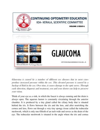
Glaucoma is caused by increased pressure in the eye due to a backup of fluid that over time damages the optic nerve. It is often asymptomatic until vision is impaired. The main types are primary open-angle glaucoma, which is the most common, as well as normal tension glaucoma, angle-closure glaucoma, and others. Treatment aims to lower pressure through medication or surgery to prevent further vision loss.

Recommended
More Related Content
What's hot
What's hot (20)
Similar to all about glaucoma
Similar to all about glaucoma (20)
More from OPTOM FASLU MUHAMMED
More from OPTOM FASLU MUHAMMED (20)
Recently uploaded
Recently uploaded (20)
all about glaucoma
- 1. Glaucoma is caused by a number of different eye diseases that in most cases produce increased pressure within the eye. This elevated pressure is caused by a backup of fluid in the eye. Over time, it causes damage to the optic nerve. Through early detection, diagnosis and treatment, you and your doctor can help to preserve your vision. Think of your eye as a sink, in which the faucet is always running and the drain is always open. The aqueous humor is constantly circulating through the anterior chamber. It is produced by a tiny gland called the ciliary body that is situated behind the iris. It flows between the iris and the lens, and after nourishing the cornea and lens, flows out through a very tiny spongy tissue called the trabecular meshwork, which is only one-fiftieth of an inch wide and serves as the drain of the eye. The trabecular meshwork is situated in the angle where the iris and cornea
- 2. meet. When this drain becomes clogged, aqueous cannot leave the eye as quickly as it is produced, causing the fluid to back up. But because the eye is a closed compartment, your `sink´ doesn´t overflow, and instead the backed up fluid causes increased pressure to build up within the eye. We call this open (wide) angle glaucoma. To understand how this increased pressure affects the eye, think of your eye as a balloon. When too much air is blown into the balloon, the pressure builds, causing it to pop. But the eye is too strong to pop. Instead, it gives at the weakest point, which is the site in the sclera at which the optic nerve leaves the eye. As we mentioned earlier, the optic nerve is the part of the eye that carries visual information to the brain. It is made up of over one million nerve cells, and while each cell is several inches long, it is extremely thin -- about one twenty-thousandth of an inch in diameter. When the pressure in the eye builds, the nerve cells become compressed, causing them to become damaged and to eventually die. The death of these cells results in permanent visual loss. Early diagnosis and treatment of glaucoma can help prevent this from happening. The Eye and How It Works The first step in understanding glaucoma is to know a few basic facts about the eye and how it works. Once you have this information, you will be better able to discuss your condition and treatment with your eye doctor. Working together, you and your doctor will be able to act as a team to protect your vision Think of Your Eye as a Camera Like a camera, the eye captures information about shape, color, and movement, and relays it in the form of impulses to the brain. The brain then processes this information into the "pictures" we see. Let us look at the various parts of our camera -- the eye.
- 3. The outer layer of the eyeball is called the sclera. The sclera is a thin, yet tough, leathery protective shell which is the "white of the eye." The front portion of the shell is called the cornea. The cornea is a clear tissue through which light rays enter the eye. The cornea is much like the lens of a camera, providing the eye with much of its light focusing power. The colored portion of the eye is called the iris. The iris not only determines whether your eyes appear blue or brown, but functions like the diaphragm of a camera. The iris contains muscles which control the size of the pupil, regulating the amount of light allowed to enter the eye. The pupil, which is the dark-colored area in the center of the iris, opens and closes depending upon how much light is present. When there is a great deal of light, like a bright, sunny day outdoors, the iris constricts the pupil, or makes it smaller. This limits the amount of light which passes through the pupil to the retina at the back of the eye. The retina may be thought of as the camera´s film. When there is little or no light, the iris dilates the pupil, widening it so that more light can enter the eye. The lens, which is behind the iris, adjusts its shape and thickness to focus the image onto the retina. The retina then delivers the image to the brain via nerve signals sent through the optic nerve to the brain, which processes these signals into a "picture," or visual image. The interior of the eye is filled with fluid. A gel-like substance called vitreous fills the center region of the eye. This region is called the vitreous cavity. The anterior chamber, or front compartment of the eye, is bounded by the cornea, iris, pupil, and lens. It is filled with a watery fluid called the aqueous humor. This fluid nourishes the cornea and the lens, providing them with oxygen and vital nutrients. The aqueous humor also provides the necessary pressure to help maintain the shape of the eye. We call this pressure the intraocular pressure (IOP). As explained on the next page, maintaining the right amount of pressure within the eye is very important to protecting your vision. Measuring the IOP is one of the ways in which your eye doctor tests for glaucoma.
- 4. TYPES OF GLAUCOMA There are a variety of different types of glaucoma. The most common forms are: Primary Open-Angle Glaucoma Normal Tension Glaucoma Angle-Closure Glaucoma Acute Glaucoma Pigmentary Glaucoma Exfoliation Syndrome Trauma-Related Glaucoma Let us look at each of these forms of glaucoma. Primary Open-Angle Glaucoma (POAG) Approximately one percent of all Americans have this form of glaucoma, making it the most common form of glaucoma in our country. It occurs mainly in the over-50 age group. There are no symptoms associated with POAG. The pressure in the eye slowly rises and the cornea adapts without swelling. If the cornea were to swell, which is usually a signal that something is wrong, symptoms would be present. But because this is not the case, this disease often goes undetected. It is painless, and the patient often does not realize that he or she is slowly losing vision until the later stages of the disease. However, by the time the vision is impaired, the damage is irreversible. In POAG, there is no visible abnormality of the trabecular meshwork. It is believed that something is wrong with the ability of the cells in the trabecular meshwork to carry out their normal function, or there may be fewer cells present as a natural result of getting older. Some believe it is due to a structural defect of the eye´s drainage system. Others believe it is caused by an enzymatic problem. These
- 5. theories, as well as others, are currently being studied and tested at numerous research centers across the country. Glaucoma is really about the problems that occur as a result of increased IOP. The average IOP in a normal population is 14-16 millimeters of mercury (mmHg). In a normal population pressures up to 20 mmHg may be within normal range. A pressure of 22 is considered to be suspicious and possibly abnormal. However, not all patients with elevated IOP develop glaucoma-related eye damage. What causes one person to develop damage while another does not is a topic of active research. As we mentioned earlier, this increased pressure can ultimately destroy the optic nerve cells. Once a sufficient number of nerve cells are destroyed, `blind spots´ begin to form in the field of vision. These blind spots usually develop first in the peripheral field of vision, the outer sides of the field of vision. In the later stages, the central vision, which we experience as `seeing,´ is affected. Once visual loss occurs, it is irreversible because once the nerve cells are dead, nothing can presently restore them. Later on, we will talk about the many ways your eye doctor can detect glaucoma in its earliest stages -- before any visual damage occurs. POAG is a chronic disease. It may be hereditary. There is no cure for it at present, but the disease can be slowed or arrested by treatment. Since there are no symptoms, many patients find it difficult to understand why lifelong treatment with expensive drugs is necessary, especially when these drugs are often bothersome to take and have a variety of side effects. Taking medications regularly, as prescribed, is crucial to preventing vision- threatening damage. That is why it is important for you to discuss these side effects with your doctor. The two of you need to act as a `team´ in the battle against glaucoma. Later on, we will discuss the medications commonly prescribed and their side effects. Normal Tension Glaucoma Normal-tension glaucoma, also known as low-tension glaucoma, is characterized by progressive optic nerve damage and visual field loss with a statistically normal intraocular pressure. This form of glaucoma, which is being increasingly
- 6. recognized, may account for as many as one-third of the cases of open-angle glaucoma in the United States. Normal-tension glaucoma is thought to be related, at least in part, to poor blood flow to the optic nerve, which leads to death of the cells that carry impulses from the retina to the brain. In addition, these eyes appear to be susceptible to pressure- related damage even in the high normal range, and therefore a pressure lower than normal is often necessary to prevent further visual loss. Research in the field of optic nerve blood flow and its role in glaucoma is a source of much excitement at the present time, and hopefully will lead to new methods of treating this disorder. Since the best therapy for normal-tension glaucoma is largely unknown, much attention is being given to a study known as the International Collaborative Low Tension Glaucoma Protocol. Angle-Closure Glaucoma Angle-closure glaucoma affects nearly half a million people in the United States. There is a tendency for this disease to be inherited, and often several members of a family will be afflicted. It is most common in people of Asian descent and people who are farsighted. In people with a tendency to angle-closure glaucoma, the anterior chamber is smaller than average. As mentioned earlier, the trabecular meshwork is situated in the angle formed where the cornea and the iris meet. In most people, this angle is about 45 degrees. The narrower the angle, the closer the iris is to the trabecular meshwork. As we age, the lens routinely grows larger. The ability of aqueous humor to pass between the iris and lens on its way to the anterior chamber becomes decreased, causing fluid pressure to build up behind the iris, further narrowing the angle. If the pressure becomes sufficiently high, the iris is forced against the trabecular meshwork, blocking drainage, similar to putting a stopper over the drain of a sink. When this space becomes completely blocked, an angleclosure glaucoma attack (acute glaucoma) results.
- 7. Acute Glaucoma Unlike POAG, where the IOP increases slowly, in acute angle-closure, it increases suddenly. This sudden rise in pressure can occur within a matter of hours and become very painful. If the pressure rises high enough, the pain may become so intense that it can cause nausea and vomiting. The eye becomes red, the cornea swells and clouds, and the patient may see haloes around lights and may experience blurred vision. An acute attack is an emergency condition. If treatment is delayed, eyesight can be permanently destroyed. Scarring of the trabecular meshwork may occur and result in chronic glaucoma, which is much more difficult to control. Cataracts may also develop. Damage to the optic nerve may occur quickly and cause permanently impaired vision. Many of these sudden `attacks´ occur in darkened rooms, such as movie theaters. If you recall, darkened environments cause the pupil to dilate, or increase in size. When this happens, there is maximum contact between the eye´s lens and the iris. This further narrows the angle and may trigger an attack. But the pupil also dilates when one is excited or anxious. Consequently, many acute glaucoma attacks occur during periods of stress. A variety of drugs can also cause dilation of the pupil and lead to an attack of glaucoma. These include anti-depressants, cold medications, antihistamines, and some medications to treat nausea. Acute glaucoma attacks are not always full blown. Sometimes a patient may have a series of minor attacks. A slight blurring of vision and haloes (rainbow-colored rings around lights) may be experienced, but without pain or redness. These attacks may end when the patient enters a well lit room or goes to sleep -- two situations which naturally cause the pupil to constrict, thereby allowing the iris to pull away from the drain.
- 8. An acute attack may be stopped with a combination of drops that constrict the pupil and drugs that help reduce the eye´s fluid production. As soon as the IOP has dropped to a safe level, your ophthalmologist will perform a laser iridotomy. A laser iridotomy is an outpatient procedure in which a laser beam is used to make a small opening in the iris. This allows the fluid to flow more freely. Drops will be used to anesthetize your eye and there is no pain involved. The entire procedure should take less than thirty minutes. Laser surgery may be performed prophylactically on the other eye, as well. Since it is common for both eyes to suffer from narrowed angles, operating on the unaffected eye is done as a preventive measure. Routine examinations using a technique called gonioscopy can predict one´s chances of having an acute attack. A special lens that contains a mirror is placed lightly on the front of the eye and the width of the angle examined visually. Patients with narrow angles can be warned of early symptoms, so that they can seek immediate treatment. In some cases, laser treatment is recommended as a preventive measure. Not all angle-closure glaucoma sufferers will experience an acute attack. Instead, some may develop what is called chronic angle-closure glaucoma. In this case, the iris gradually closes over the drain, causing no overt symptoms. When this occurs, scars slowly form between the iris and the drain and the IOP will not rise until there is a significant amount of scar tissue formed -- enough to cover the drainage area. If the patient is treated with medication, such as pilocarpine, an acute attack may be prevented, but the chronic form of the disease may still develop. Pigmentary Glaucoma Pigmentary glaucoma is a type of inherited open-angle glaucoma that develops more frequently in men than in women. It most often begins in the twenties and thirties, which makes it particularly dangerous to a lifetime of normal vision. Nearsighted patients are more typically afflicted. The anatomy of the eyes of these patients appears to play a key role in the development of this type of glaucoma. Let us examine why.
- 9. Myopic (nearsighted) eyes have a concave-shaped iris which creates an unusually wide angle. This causes the pigment layer of the eye to rub on the lens. This rubbing action causes the iris pigment to shed into the aqueous humor and onto neighboring structures, such as the trabecular meshwork. Pigment may plug the pores of the trabecular meshwork, causing it to clog, and thereby increasing the IOP. Miotic therapy is the treatment of choice, but these drugs in drop form can cause disabling visual blurring in younger patients. Fortunately, a slow-release form is available. We´ll discuss these drugs and their side effects in detail later on in this guide. Laser iridotomy is presently being investigated in the treatment of this disorder. Exfoliation Syndrome This common cause of glaucoma is found everywhere in the world, but is most common among people of European descent. In about 10% of the population over age 50, a whitish material, which upon slit-lamp examination looks somewhat like tiny flakes of dandruff, builds up on the lens of the eye. This exfoliation material is rubbed off the lens by movement of the iris and at the same time, pigment is rubbed off the iris. Both pigment and exfoliation material clogs the trabecular meshwork, leading to IOP elevation, sometimes to very high levels. Exfoliation syndrome can lead to both open-angle glaucoma and angle-closure glaucoma, often producing both kinds of glaucoma in the same individual. Not all persons with exfoliation syndrome develop glaucoma. However, if you have exfoliation syndrome, your chances of developing glaucoma are about six times as high than if you don’t have it. It often appears in one eye long before the other, for unknown reasons. If you have glaucoma in one eye only, this is the most likely cause. It can be detected before the glaucoma develops; so that you can be more carefully observed and minimize your chances of vision loss.
- 10. Trauma-Related Glaucoma A blow to the eye, chemical burn, or penetrating injury may all lead to the development of glaucoma, either acute or chronic. This can be due to a mechanical disruption or physical change within the eye´s drainage system. It is therefore crucial for anyone who has suffered eye trauma to have check-ups at regular intervals. Who is at Risk? Everyone should be concerned about glaucoma and its effects. It is important for each of us, from infants to senior citizens, to have our eyes checked regularly, because early detection and treatment of glaucoma are the only way to prevent vision impairment and blindness. There are a few conditions related to this disease which tend to put some people at greater risk: People over the age of 45. While glaucoma can develop in younger patients, it occurs more frequently as we get older. People who have a family history of glaucoma. Glaucoma appears to `run´ in families. The tendency for developing glaucoma may be inherited. However, just because someone in your family has glaucoma does not mean that you will necessarily develop the disease. People with abnormally high intraocular pressure (IOP). High IOP is the most important risk factor for glaucomatous damage. People of African descent. African-Americans have a greater tendency for developing primary open- angle glaucoma than do people of other races. People who have: Diabetes Myopia (nearsightedness) Regular, long-term Steroid/Cortisone use A previous eye injury
- 11. DIAGNOSING GLAUCOMA Your eye doctor has a variety of diagnostic tools that aid in determining whether or not you have glaucoma -- even before you have any symptoms. Let us explore these tools and what they do. The Tonometer The Tonometer measures the pressure in the eye. If your doctor were to use applanation tonometry, your eye would be anesthetized with drops. Then, you would sit at the slit lamp, and a plastic prism would lightly push against your eye in order to measure your IOP. In air tonometry a puff of air is sent onto the cornea to take the measurement. Since this instrument does not come in direct contact with your eye, no anesthetic eye drops are required. Visual Field Test Testing your visual field lets your doctor know if and how your field of vision has been affected by glaucoma. The visual field is an important measure of the extent of damage to your optic nerve from increased IOP. There are several methods of examination available to your doctor. In computerized visual field testing you will be asked to place your chin on a stand that appears before a computerized screen. Whenever you see a flash of light appear, you will be asked to press a button. At the end of this test, your doctor will receive a printout of your field of vision. Another test that is similar is the Goldmann perimeter. However, in this test, no computer is used. The examiner records your answers whenever you indicate that the light is in view. Ophthalmoscopy Using an instrument called an ophthalmoscope; your eye doctor can look directly through the pupil at the optic nerve. Its color and appearance can indicate whether or not damage from glaucoma is present and how extensive it is. TREATING GLAUCOMA Glaucoma can be treated with eye drops, pills, laser surgery, eye operations, or a combination of methods. The whole purpose of treatment is to prevent further loss
- 12. of vision. This is imperative as loss of vision due to glaucoma is irreversible. Keeping the IOP under control is the key to preventing loss of vision from glaucoma. Your doctor has several options for doing so. They include: Eye drops All eye drops may cause a burning or stinging sensation at first. This is often due to the antibacterial agent present in the drop solution and not due to the drug itself. While it can be uncomfortable, the discomfort lasts for only a few seconds. It is important that you take your medication exactly as it is prescribed if you are to control your IOP. For example, drops which are prescribed four times a day usually have a `duration of action´ of six hours. By taking these drops four times spaced through your waking hours, you ensure that you are covered by the drug´s effectiveness for a full 24- hour period. Since eye drops are absorbed into the bloodstream, it is important that you tell your doctor about all other medications you are currently taking. Some drugs can be dangerous when mixed with other medications. Ask your doctor and/or pharmacist if the medications you are taking together are safe. To minimize absorption into the bloodstream and maximize the amount of drug absorbed into the eye, close your eyes for one to two minutes after administering your drops and press lightly against the nasal corner of your eyelids to close the tear duct that drains into the nose.
- 14. * While every drug has some potential side effects, it is important to note that many patients experience NO side effects at all. Pills Sometimes, drops are not enough to control IOP. When this is the case, pills may be prescribed in addition to drops. These pills, which have more side effects than drops do, also serve to turn down the eye´s faucet and lessen the production of fluid in the eye. The medication is usually taken from two to four times daily. It is important to share this information with your other doctors. By doing so, they can best prescribe medications for you which will not cause potentially dangerous interactions. The following are some commonly prescribed carbonic anhydrase inhibitors and their more common side effects.
- 15. SURGICAL PROCEDURES When medication does not achieve the desired results, or when it has intolerable side effects, your ophthalmologist may suggest surgery. Laser Surgery Laser surgery has become increasingly popular as an intermediate step between drugs and traditional surgery. The most common type performed for open-angle glaucoma is called trabeculoplasty. This procedure takes between ten and twenty minutes, is painless, and can be performed in either a doctor´s office or in an outpatient facility. The laser beam (a high energy light beam) is focused upon the eye´s drain. Contrary to what most people think, the laser does not burn a hole through the eye. Instead, its intense heat causes some areas of the eye´s drain to shrink, resulting in adjacent areas stretching open and permitting the fluid to drain more easily. You may go home and resume normal activities following surgery. Your doctor should check your IOP one to two hours later. After this procedure, nearly eighty percent of all patients respond well enough to be able to avoid or delay surgery. While it may take a few weeks to see the full pressure-lowering effect of this procedure, during which time you may have to continue taking your medication, many patients are eventually able to discontinue some of their medications. This, however, is not true in all cases. Your doctor is the best judge of determining whether or not medication is still necessary for you. Cataracts do not occur after laser surgery and complications are minimal, which is why this has become increasingly popular. Traditional Surgery The most common of these operations is called a trabeculectomy. In this procedure, the surgeon removes a small section of the trabecular meshwork -- the eye´s drain. This allows the aqueous humor to drain more easily, reducing the pressure in the eye. This procedure is usually done under local anesthesia either as an outpatient or with a brief hospital stay. As your doctor will want to see you on the day after the operation to check both your vision and ocular pressure, some
- 16. prefer that you remain in the hospital. It is important to note that your eyes may not have their normal visual acuity for several weeks following this procedure. Although trabeculectomy is a relatively safe surgical procedure, about one-third of patients develop cataracts within five years of surgery. After trabeculectomy, most patients are able to discontinue all anti-glaucoma medications. Perhaps ten to fifteen percent of patients require additional surgery. Routine eye exams are vital for protecting the health of your eyes. If your ophthalmologist or optometrist detects glaucoma, early treatment can help prevent the loss of your vision.