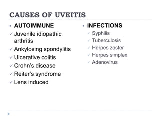
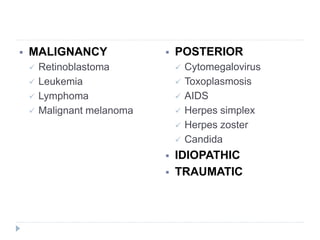
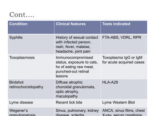
BASICS OF UVEITIS
MICAH TCHALE
INTRODUCTION
o Uveitis is one of the leading causes of blindness,
which makes it crucial for practicing optometrists to
understand how best to diagnose, differentiate and
treat it
o Uveitis was once considered as a single disease
entity; however, we now know that uveitis can be
caused by;
i. Autoimmune disorders
ii. Infections
iii. Malignancy
iv. Exposure to toxins
v. Idiopathic in nature
o Approx. 50% of uveitis are due to idiopathic
conditions, 20% due to trauma, 20% due to systemic
underlying cause eg rheumatoid arthritis or
sarcoidosis, and 10% due to localized ocular
condition eg herpes zoster or toxoplasmosis
o The sight threatening complications of uveitis;
i. Glaucoma
ii. Damage to the retina
iii. Macular edema
PATIENT EVALUATION
o The key to targeted and efficient patient evaluation is
a thorough history, physical examination and review
of systems
o When patients present with a non-resolving, painful,
red eye, our goal is to determine:
 What is the extent of the current condition?
 What ocular structures are affected?
 What is the underlying cause?
 How can the condition best be treated & managed?
o The following steps will help answer those questions:
 Acute or chronic?
• First step is determine whether the clinical course is acute
or chronic
• Acute conditions typically have an abrupt onset lasting
several weeks, while chronic conditions can wax and
wane over months or years
 What structures are affected?
• The next step is to identify the anatomical location of the
inflammation or infection
• The anatomical classification of uveitis (International
uveitis study group)
i. Anterior uveitis (primary site of inflammation is the
anterior chamber)
ii. Intermediate uveitis (primary site is ciliary body and
vitreous)
iii. Posterior uveitis (primary focus of inflammation is the
retina and choroid)
iv. Panuveitis (primary site of inflammation involving all
anterior and posterior)
• Common signs and symptoms of anterior uveitis
i. Redness
ii. Photophobia
iii. Pain
• Posterior uveitis presents with
i. Floaters
ii. Visual disturbances
 Granulomatous or nongranulomatous
• Ocular histopathology is another classification used to
differentiate the condition
• Granulomatous inflammation
 may present acutely, but more often presents as a chronic
condition with large, mutton-fat keratic precipitates (KPs) and
inflammatory cells (epitheloid cells and macrophages) in the
anterior chamber
 Often associated with systemic conditions and autoimmune
reaction eg syphilis, Lyme disease, TB or a local reactivation of
herpetic viral infection
• Nongranulomatous inflammation
 Usually involves the anterior segment
 Has an acute onset accompanied by a cellular reaction in the
AC that represents smaller cell types (lymphocytes)
 Are idiopathic or due to HLA-B27 involvement
 Occurs more frequently than does granulomatous uveitis,
especially in patients with anterior uveitis
 Unilateral or bilateral
• Laterality can help classify the type of uveitis
• Unilateral conditions are more commonly acute and can
be infectious
• Bilateral conditions are likely due to chronic, systemic
conditions
 What are the symptoms and signs?
• Patients with uveitis typically present with abrupt, dull pain
and photosensitivity
• Additional signs and symptoms
i. Pain (temple or periorbital region)
ii. Redness denser toward the limbus with no mucopurulent
discharge
iii. Blurring of vision
iv. Watery discharge or tearing (rare)
v. Mild epiphora from the pain response
o There are multiple clinical signs that are important
for clinicians to look for to help with the differential
diagnosis
• Patients may present with a decrease in vision ranging
from mild to severe
• Circumlimbal injection of the conjunctiva
• In chronic uveitis, cornea edema may be present
• Hypopyon may be present, which is highly suggestive of
diseases with HLA-B27, Behçet’s disease or infectious
uveitis (rare)
• IOP should be taken into account in the diagnosis
 Low IOP is due to decreased aqueous production in the acute
phases
 High IOP is found in viral conditions eg HSV keratouveitis
• Peripheral anterior or posterior synechiae have the
potential to lead to secondary glaucomatous conditions
• Posterior subcapsular cataracts may develop with
recurrent episodes due to chronic steroid use
 Is there a systemic cause?
• Ask the patients about their medical history and other
associated symptoms of their conditions provides
invaluable clues to possible diagnoses
• Any time a patient presents with minimal signs and
symptoms, bilateral presentation, a granulomatous
response and a posterior extension of uveitis in the eye,
be sure to consider systemic causes
STANDARDIZED GRADING SCALES FOR
UVEITIS
 standardization of uveitis nomenclature (SUN)
scheme for anterior chamber cells (using 1mm slit
beam)
GRADE CELLS IN FIELD
0 <1
0.5+ 1-5
1+ 6-15
2+ 16-25
3+ 26-50
4+ 50+
 SUN Grading scheme for anterior chamber flare
GRADE DESCRIPTION
0 None
1+ Faint
2+ Moderate (iris/lens details clear)
3+ Marked (iris/lens details hazy)
4+ Intense (fibrin/plastic aqueous)
CAUSES OF UVEITIS
 AUTOIMMUNE
 Juvenile idiopathic
arthritis
 Ankylosing spondylitis
 Ulcerative colitis
 Crohn’s disease
 Reiter’s syndrome
 Lens induced
 INFECTIONS
 Syphilis
 Tuberculosis
 Herpes zoster
 Herpes simplex
 Adenovirus
 MALIGNANCY
 Retinoblastoma
 Leukemia
 Lymphoma
 Malignant melanoma
 POSTERIOR
 Cytomegalovirus
 Toxoplasmosis
 AIDS
 Herpes simplex
 Herpes zoster
 Candida
 IDIOPATHIC
 TRAUMATIC
ANTERIOR UVEITIS
o AU is the presence of cell and flare in the anterior
chamber
o Also referred to as iritis, iridocyclitis and anterior
cyclitis
o AU can be associated with other ocular inflammatory
conditions including scleritis, episcleritis, keratitis,
posterior segment diseases, postsurgical
inflammation or lens related
o The largest number of anterior uveitis cases is
idiopathic (HLA-B27 negative)
o The next common etiology is HLA-B 27 positive
o Be sure to perform a thorough case history and a
complete review of systems before diagnosing a
patient as idiopathic
o Other associated conditions commonly seen with
anterior uveitis include;
 Juvenile idiopathic arthritis
 Ulcerative colitis
 Crohn’s disease
 Reiter’s syndrome
 Herpes simplex/zoster virus
 Syphilis
 tuberculosis
INTERMEDIATE UVEITIS
o Defined as intraocular inflammation that
predominantly involves the peripheral retina, pars
plana and vitreous
o Aka chronic cyclitis, peripheral uveitis and pars
planitis
o Proportion of patients with intermediate uveitis is
estimated to be 4% to 8% of uveitis cases
 Causes of intermediate uveitis include;
 69% of unknown etiology
 22% due to sarcoid
 1% due to multiple sclerosis and Lyme disease
o Helpful questions include asking patients about
difficulty in breathing or symptoms of eye pain,
neurological symptoms consistent with multiple
sclerosis and any history of tick bites
POSTERIOR UVEITIS
o Defined as inflammation that is limited to the
posterior segment of the eye
o Ii is associated with the highest risk of severe vision
loss
o There are many systemic causes for posterior uveitis
 Ocular toxoplasmosis is the most common cause of
posterior uveitis and accounts for 90%
o Noninfectious causes may include immunologic or
allergic origins, unknown causes and masquerade
conditions (including neoplasm)
o Ocular diseases associated with posterior uveitis
include;
 Sarcoidosis
 Toxoplasmosis
 Eales’ disease
 Frosted branch angitis
 Acute retinal necrosis
 Retinal arteritis and aneurysms
 Birdshot choroiditis
 Pars planitis
o Ancillary testing are useful for diagnosis
 Fluorescein angiography
 Optical coherence tomography
 B-scan ultrasonography
 Visual field testing
LABORATORY TESTING
o Indications for laboratory testing
 Bilateral uveitis
 Affects an atypical age group
 Recurrent, recalcitrant or hyperacute uveitis
 Patients with uveitis that worsens with tapering
 Uveitis that is causing decreased visual acuity
 Immunocompromised patients
o Minimal laboratory testing should include;
 Complete blood cell count
 Urinalysis (psoriatic arthritis, proteins)
 Lyme titers
 HLA-B27 (ankylosing spondylitis)
 Antinuclear antibody (ANA) test (juvenile idiopathic
arthritis)
 Angiotensin converting enzyme (ACE) test (sarcoidosis)
 Veneral disease research laboratory (VDRL) test
(syphilis)
 Fluorescent treponemal antibody absorption (FTA-ABS)
test (syphilis)
o Chest X-ray should also be performed when testing
LAB TESTS WHEN YOU SUSPECT
SYSTEMIC DISEASE
Condition Clinical features Tests indicated
Ankylosing spondylitis Young male, low back
pain, chest pain
HLA-B27, Sacroiliac X
ray
Reiter’s syndrome Young male, arthritis,
urethritis, conjunctivitis
HLA-B27, ESR, CRP
Juvenile idiopathic
arthritis
Slight male predilection,
sacrolitis, common
ANA, RF, knee
radiograph
Inflammatory bowel
disease
Ulcerative colitis,
diarrhea, abdominal
cramps
HLA-B27, GI referral for
endoscopy
sarcoidosis African Americans,
females, posterior
segment inflammation ie
vasculitis, vitris
ACE, chest X-ray or CT
scan
Tuberculosis Prolonged cough, fever,
chills, night sweats,
weight loss
PPD, chest X-ray
Cont….
Condition Clinical features Tests indicated
Syphilis History of sexual contact
with infected person,
rash, fever, malaise,
headache, joint pain
FTA-ABS, VDRL, RPR
Toxoplasmosis Immunocompromised
status, exposure to cats,
hx of eating raw meat,
punched-out retinal
lesions
Toxoplasma IgG or IgM
for acute acquired cases
Birdshot
retinochoroidopathy
Diffuse atrophic
choroidal granulomata,
optic atrophy,
maculopathy
HLA-A29
Lyme disease Recent tick bite Lyme Western Blot
Wegener’s Sinus, pulmonary, kidney ANCA, sinus films, chest
CURRENT TREATMENTS FOR UVEITIS
o The goal of uveitis treatment and management is to
alleviate the patient’s symptoms and control
inflammation
o Prior to initiating treatment, you must rule out any
infectious condition
o Topical steroids are the mainstay of treatment for the
majority of uveitis, but steroids injections and oral
steroids are often needed in cases of intermediate
and posterior conditions
o Steroids should always be used aggressively to treat
and suppress the inflammatory response, with initial
dose ranging from q1h to q.i.d.
o Using steroids aggressively
 In order to be effective, steroids should be pulse-dosed
frequently upon initial presentation, with a gradual taper
over several weeks depending on the response to
treatment
 Steroids should not be tapered until there is a two-grade
improvement in clinical signs (eg 4+ cells to 2+ cells), and
should not be stopped until there is a complete resolution
of all cells, flare and macular edema
o Compliance with these drops is crucial for the
resolution of uveitis, so emphasize its importance to
the patients
o Steroid treatment should be continued even after the
resolution of all cell and ocular inflammation
 Patients should a tapering regimen after resolution to
decrease the risk of any rebound effect
o Pred Forte (prednisolone acetate 1%) has been the
gold standard in the uveitis treatment and continues
to be a very effective treatment today
o Whenever you prescribe steroids, always monitor for
increased IOP and cataract formation
o Side effects of topical steroids
 Increased IOP
 Cataract formation
 Delayed wound healing or epithelialization
 Reactivate latent herpes simplex infections
 Potentiate bacterial, fungal or acanthamoebal infections
o When considering IOP lowering agents, use caution
with prostaglandin analogues because they may
exacerbate the inflammatory response
TREATMENT CONSIDERATIONS
o If you have a patient with diagnosed systemic
disease, chronic uveitis is often well controlled with
immunomodulatory or antimetabolite systemic
therapy
 Stay in contact with a rheumatologist
o Pain and photophobia are common symptoms in
uveitis that can be treated effectively with
cycloplegics
 Cycloplegics improve comfort by;
 Immobilizing the inflamed iris and ciliary body
 Stabilizing the blood aqueous barrier to prevent the leakage of
proteins
 Breaking synechiae formation
 The most popular cycloplegics used are homatropine 5%
b.i.d., scopolamine 0.25% tds, atropine 1%
o Additional treatments for uveitis include;
 Sunglasses
 Topical or oral NSAIDs (for analgesia)
 Immunosuppressants
 Periocular steroid injections
 Intravitreal steroid injections
 Long and short-acting intravitreal steroid implants
 Intravitreal VEGF inhibitor injections
 Specific surgical procedures
REFERENCE
 Whitley W, Sheppard J (2011) The Basics of Uveitis.
Review of Optometry. Available from
http://www.reviewofoptometry.com/continuing_educa
tion/tabviewtest/lessonid/107773/

BASICS OF UVEITIS

  • 1.
  • 2.
    INTRODUCTION o Uveitis isone of the leading causes of blindness, which makes it crucial for practicing optometrists to understand how best to diagnose, differentiate and treat it o Uveitis was once considered as a single disease entity; however, we now know that uveitis can be caused by;
  • 3.
    i. Autoimmune disorders ii.Infections iii. Malignancy iv. Exposure to toxins v. Idiopathic in nature
  • 4.
    o Approx. 50%of uveitis are due to idiopathic conditions, 20% due to trauma, 20% due to systemic underlying cause eg rheumatoid arthritis or sarcoidosis, and 10% due to localized ocular condition eg herpes zoster or toxoplasmosis
  • 5.
    o The sightthreatening complications of uveitis; i. Glaucoma ii. Damage to the retina iii. Macular edema
  • 6.
    PATIENT EVALUATION o Thekey to targeted and efficient patient evaluation is a thorough history, physical examination and review of systems o When patients present with a non-resolving, painful, red eye, our goal is to determine:  What is the extent of the current condition?  What ocular structures are affected?  What is the underlying cause?  How can the condition best be treated & managed?
  • 7.
    o The followingsteps will help answer those questions:  Acute or chronic? • First step is determine whether the clinical course is acute or chronic • Acute conditions typically have an abrupt onset lasting several weeks, while chronic conditions can wax and wane over months or years
  • 8.
     What structuresare affected? • The next step is to identify the anatomical location of the inflammation or infection • The anatomical classification of uveitis (International uveitis study group) i. Anterior uveitis (primary site of inflammation is the anterior chamber) ii. Intermediate uveitis (primary site is ciliary body and vitreous)
  • 9.
    iii. Posterior uveitis(primary focus of inflammation is the retina and choroid) iv. Panuveitis (primary site of inflammation involving all anterior and posterior)
  • 10.
    • Common signsand symptoms of anterior uveitis i. Redness ii. Photophobia iii. Pain • Posterior uveitis presents with i. Floaters ii. Visual disturbances
  • 11.
     Granulomatous ornongranulomatous • Ocular histopathology is another classification used to differentiate the condition • Granulomatous inflammation  may present acutely, but more often presents as a chronic condition with large, mutton-fat keratic precipitates (KPs) and inflammatory cells (epitheloid cells and macrophages) in the anterior chamber  Often associated with systemic conditions and autoimmune reaction eg syphilis, Lyme disease, TB or a local reactivation of herpetic viral infection
  • 12.
    • Nongranulomatous inflammation Usually involves the anterior segment  Has an acute onset accompanied by a cellular reaction in the AC that represents smaller cell types (lymphocytes)  Are idiopathic or due to HLA-B27 involvement  Occurs more frequently than does granulomatous uveitis, especially in patients with anterior uveitis
  • 13.
     Unilateral orbilateral • Laterality can help classify the type of uveitis • Unilateral conditions are more commonly acute and can be infectious • Bilateral conditions are likely due to chronic, systemic conditions
  • 14.
     What arethe symptoms and signs? • Patients with uveitis typically present with abrupt, dull pain and photosensitivity • Additional signs and symptoms i. Pain (temple or periorbital region) ii. Redness denser toward the limbus with no mucopurulent discharge iii. Blurring of vision iv. Watery discharge or tearing (rare) v. Mild epiphora from the pain response
  • 15.
    o There aremultiple clinical signs that are important for clinicians to look for to help with the differential diagnosis • Patients may present with a decrease in vision ranging from mild to severe • Circumlimbal injection of the conjunctiva • In chronic uveitis, cornea edema may be present • Hypopyon may be present, which is highly suggestive of diseases with HLA-B27, Behçet’s disease or infectious uveitis (rare)
  • 16.
    • IOP shouldbe taken into account in the diagnosis  Low IOP is due to decreased aqueous production in the acute phases  High IOP is found in viral conditions eg HSV keratouveitis • Peripheral anterior or posterior synechiae have the potential to lead to secondary glaucomatous conditions • Posterior subcapsular cataracts may develop with recurrent episodes due to chronic steroid use
  • 17.
     Is therea systemic cause? • Ask the patients about their medical history and other associated symptoms of their conditions provides invaluable clues to possible diagnoses • Any time a patient presents with minimal signs and symptoms, bilateral presentation, a granulomatous response and a posterior extension of uveitis in the eye, be sure to consider systemic causes
  • 18.
    STANDARDIZED GRADING SCALESFOR UVEITIS  standardization of uveitis nomenclature (SUN) scheme for anterior chamber cells (using 1mm slit beam) GRADE CELLS IN FIELD 0 <1 0.5+ 1-5 1+ 6-15 2+ 16-25 3+ 26-50 4+ 50+
  • 19.
     SUN Gradingscheme for anterior chamber flare GRADE DESCRIPTION 0 None 1+ Faint 2+ Moderate (iris/lens details clear) 3+ Marked (iris/lens details hazy) 4+ Intense (fibrin/plastic aqueous)
  • 20.
    CAUSES OF UVEITIS AUTOIMMUNE  Juvenile idiopathic arthritis  Ankylosing spondylitis  Ulcerative colitis  Crohn’s disease  Reiter’s syndrome  Lens induced  INFECTIONS  Syphilis  Tuberculosis  Herpes zoster  Herpes simplex  Adenovirus
  • 21.
     MALIGNANCY  Retinoblastoma Leukemia  Lymphoma  Malignant melanoma  POSTERIOR  Cytomegalovirus  Toxoplasmosis  AIDS  Herpes simplex  Herpes zoster  Candida  IDIOPATHIC  TRAUMATIC
  • 22.
    ANTERIOR UVEITIS o AUis the presence of cell and flare in the anterior chamber o Also referred to as iritis, iridocyclitis and anterior cyclitis o AU can be associated with other ocular inflammatory conditions including scleritis, episcleritis, keratitis, posterior segment diseases, postsurgical inflammation or lens related
  • 23.
    o The largestnumber of anterior uveitis cases is idiopathic (HLA-B27 negative) o The next common etiology is HLA-B 27 positive o Be sure to perform a thorough case history and a complete review of systems before diagnosing a patient as idiopathic
  • 24.
    o Other associatedconditions commonly seen with anterior uveitis include;  Juvenile idiopathic arthritis  Ulcerative colitis  Crohn’s disease  Reiter’s syndrome  Herpes simplex/zoster virus  Syphilis  tuberculosis
  • 25.
    INTERMEDIATE UVEITIS o Definedas intraocular inflammation that predominantly involves the peripheral retina, pars plana and vitreous o Aka chronic cyclitis, peripheral uveitis and pars planitis o Proportion of patients with intermediate uveitis is estimated to be 4% to 8% of uveitis cases
  • 26.
     Causes ofintermediate uveitis include;  69% of unknown etiology  22% due to sarcoid  1% due to multiple sclerosis and Lyme disease o Helpful questions include asking patients about difficulty in breathing or symptoms of eye pain, neurological symptoms consistent with multiple sclerosis and any history of tick bites
  • 27.
    POSTERIOR UVEITIS o Definedas inflammation that is limited to the posterior segment of the eye o Ii is associated with the highest risk of severe vision loss o There are many systemic causes for posterior uveitis  Ocular toxoplasmosis is the most common cause of posterior uveitis and accounts for 90%
  • 28.
    o Noninfectious causesmay include immunologic or allergic origins, unknown causes and masquerade conditions (including neoplasm)
  • 29.
    o Ocular diseasesassociated with posterior uveitis include;  Sarcoidosis  Toxoplasmosis  Eales’ disease  Frosted branch angitis  Acute retinal necrosis  Retinal arteritis and aneurysms  Birdshot choroiditis  Pars planitis
  • 30.
    o Ancillary testingare useful for diagnosis  Fluorescein angiography  Optical coherence tomography  B-scan ultrasonography  Visual field testing
  • 31.
    LABORATORY TESTING o Indicationsfor laboratory testing  Bilateral uveitis  Affects an atypical age group  Recurrent, recalcitrant or hyperacute uveitis  Patients with uveitis that worsens with tapering  Uveitis that is causing decreased visual acuity  Immunocompromised patients
  • 32.
    o Minimal laboratorytesting should include;  Complete blood cell count  Urinalysis (psoriatic arthritis, proteins)  Lyme titers  HLA-B27 (ankylosing spondylitis)  Antinuclear antibody (ANA) test (juvenile idiopathic arthritis)  Angiotensin converting enzyme (ACE) test (sarcoidosis)  Veneral disease research laboratory (VDRL) test (syphilis)  Fluorescent treponemal antibody absorption (FTA-ABS) test (syphilis) o Chest X-ray should also be performed when testing
  • 33.
    LAB TESTS WHENYOU SUSPECT SYSTEMIC DISEASE Condition Clinical features Tests indicated Ankylosing spondylitis Young male, low back pain, chest pain HLA-B27, Sacroiliac X ray Reiter’s syndrome Young male, arthritis, urethritis, conjunctivitis HLA-B27, ESR, CRP Juvenile idiopathic arthritis Slight male predilection, sacrolitis, common ANA, RF, knee radiograph Inflammatory bowel disease Ulcerative colitis, diarrhea, abdominal cramps HLA-B27, GI referral for endoscopy sarcoidosis African Americans, females, posterior segment inflammation ie vasculitis, vitris ACE, chest X-ray or CT scan Tuberculosis Prolonged cough, fever, chills, night sweats, weight loss PPD, chest X-ray
  • 34.
    Cont…. Condition Clinical featuresTests indicated Syphilis History of sexual contact with infected person, rash, fever, malaise, headache, joint pain FTA-ABS, VDRL, RPR Toxoplasmosis Immunocompromised status, exposure to cats, hx of eating raw meat, punched-out retinal lesions Toxoplasma IgG or IgM for acute acquired cases Birdshot retinochoroidopathy Diffuse atrophic choroidal granulomata, optic atrophy, maculopathy HLA-A29 Lyme disease Recent tick bite Lyme Western Blot Wegener’s Sinus, pulmonary, kidney ANCA, sinus films, chest
  • 35.
    CURRENT TREATMENTS FORUVEITIS o The goal of uveitis treatment and management is to alleviate the patient’s symptoms and control inflammation o Prior to initiating treatment, you must rule out any infectious condition o Topical steroids are the mainstay of treatment for the majority of uveitis, but steroids injections and oral steroids are often needed in cases of intermediate and posterior conditions
  • 36.
    o Steroids shouldalways be used aggressively to treat and suppress the inflammatory response, with initial dose ranging from q1h to q.i.d.
  • 37.
    o Using steroidsaggressively  In order to be effective, steroids should be pulse-dosed frequently upon initial presentation, with a gradual taper over several weeks depending on the response to treatment  Steroids should not be tapered until there is a two-grade improvement in clinical signs (eg 4+ cells to 2+ cells), and should not be stopped until there is a complete resolution of all cells, flare and macular edema
  • 38.
    o Compliance withthese drops is crucial for the resolution of uveitis, so emphasize its importance to the patients o Steroid treatment should be continued even after the resolution of all cell and ocular inflammation  Patients should a tapering regimen after resolution to decrease the risk of any rebound effect
  • 39.
    o Pred Forte(prednisolone acetate 1%) has been the gold standard in the uveitis treatment and continues to be a very effective treatment today o Whenever you prescribe steroids, always monitor for increased IOP and cataract formation
  • 40.
    o Side effectsof topical steroids  Increased IOP  Cataract formation  Delayed wound healing or epithelialization  Reactivate latent herpes simplex infections  Potentiate bacterial, fungal or acanthamoebal infections
  • 41.
    o When consideringIOP lowering agents, use caution with prostaglandin analogues because they may exacerbate the inflammatory response
  • 42.
    TREATMENT CONSIDERATIONS o Ifyou have a patient with diagnosed systemic disease, chronic uveitis is often well controlled with immunomodulatory or antimetabolite systemic therapy  Stay in contact with a rheumatologist
  • 43.
    o Pain andphotophobia are common symptoms in uveitis that can be treated effectively with cycloplegics  Cycloplegics improve comfort by;  Immobilizing the inflamed iris and ciliary body  Stabilizing the blood aqueous barrier to prevent the leakage of proteins  Breaking synechiae formation  The most popular cycloplegics used are homatropine 5% b.i.d., scopolamine 0.25% tds, atropine 1%
  • 44.
    o Additional treatmentsfor uveitis include;  Sunglasses  Topical or oral NSAIDs (for analgesia)  Immunosuppressants  Periocular steroid injections  Intravitreal steroid injections  Long and short-acting intravitreal steroid implants  Intravitreal VEGF inhibitor injections  Specific surgical procedures
  • 45.
    REFERENCE  Whitley W,Sheppard J (2011) The Basics of Uveitis. Review of Optometry. Available from http://www.reviewofoptometry.com/continuing_educa tion/tabviewtest/lessonid/107773/