Downloaded 143 times


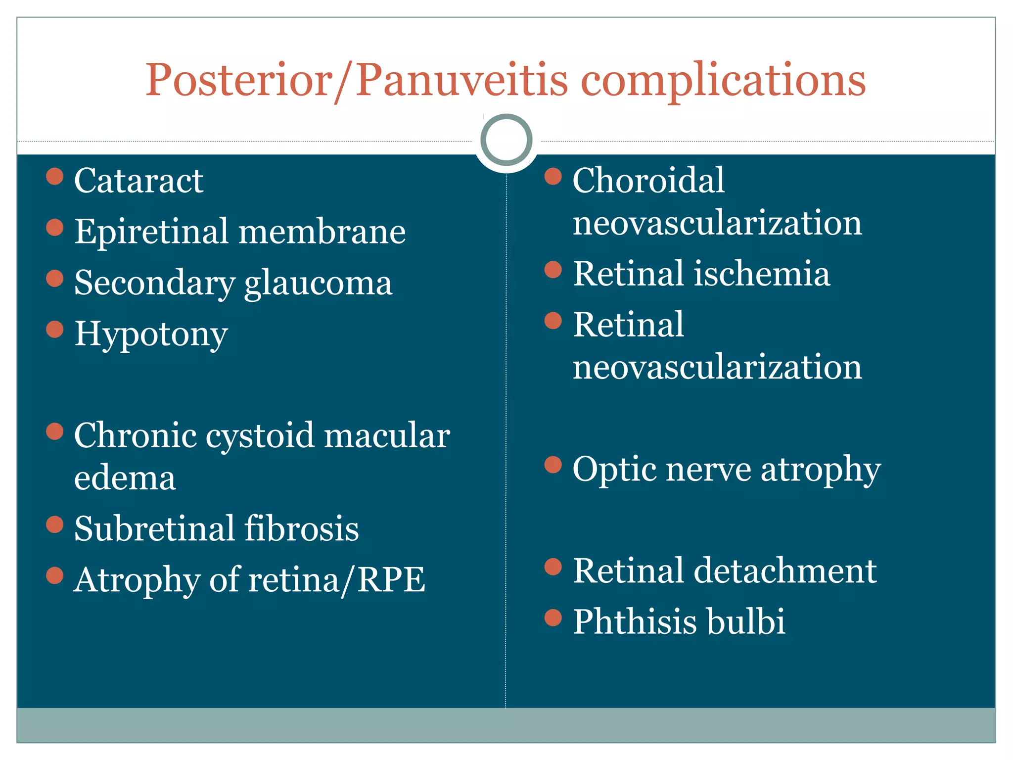


Uveitis is a general term for intraocular inflammation that does not indicate the specific site or cause of inflammation. It can be caused by autoimmune or infectious processes. Uveitis is a common cause of visual impairment and blindness worldwide. It is classified based on the site of inflammation - anterior, intermediate, posterior or panuveitis. Developing a differential diagnosis involves considering factors such as acuity of onset, laterality, associated symptoms and response to previous therapies. Common etiologies include idiopathic disease, infections like tuberculosis, and autoimmune diseases like sarcoidosis and Behcet's disease. Treatment involves corticosteroids and immunosuppressive therapies.


