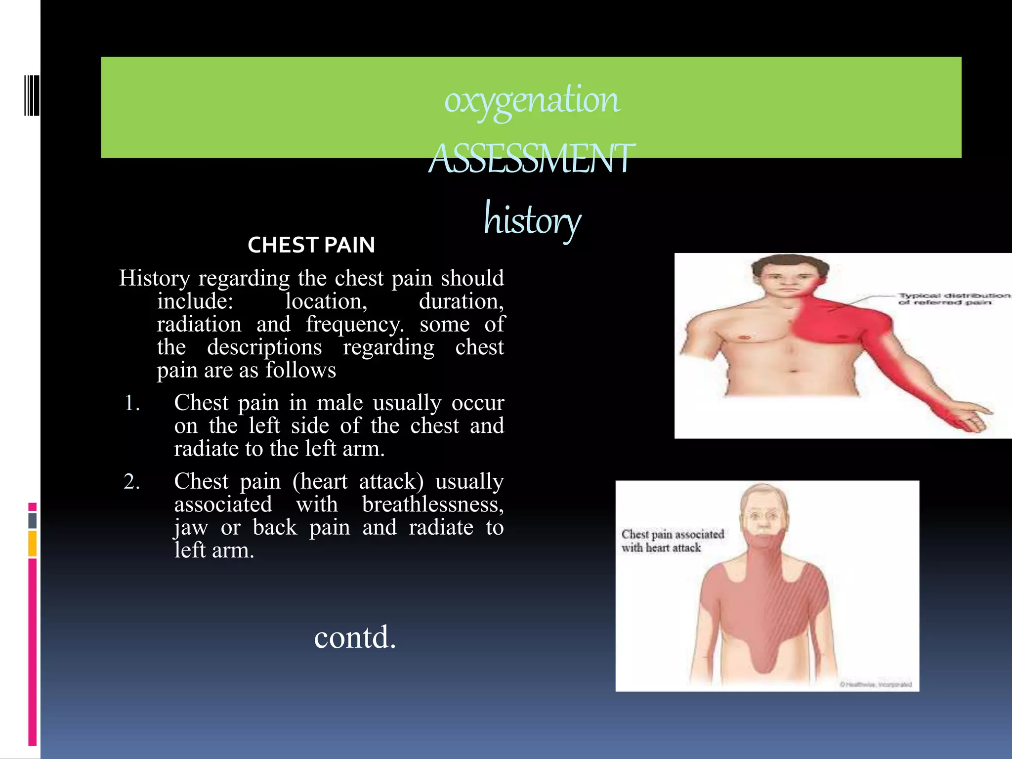
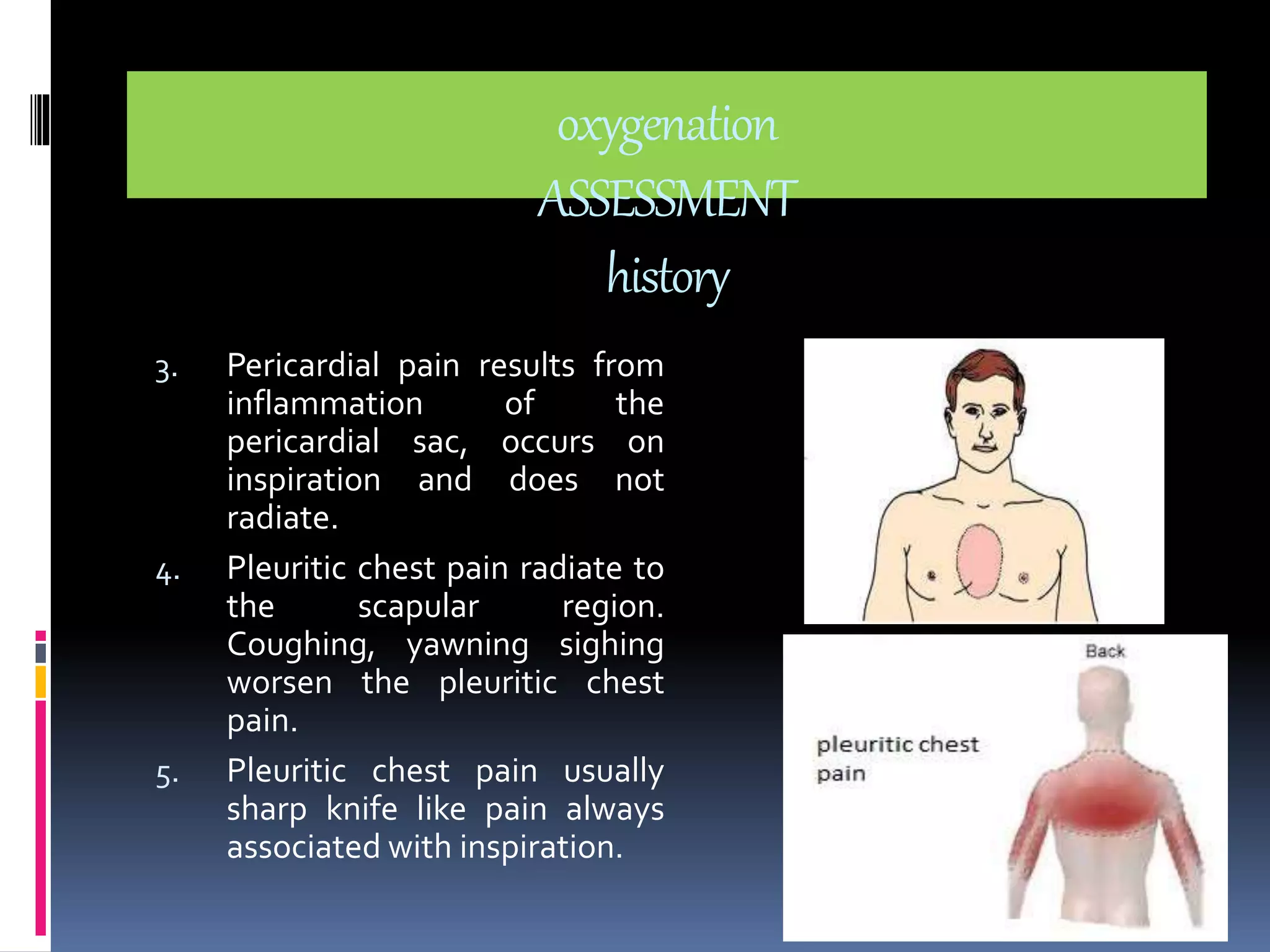
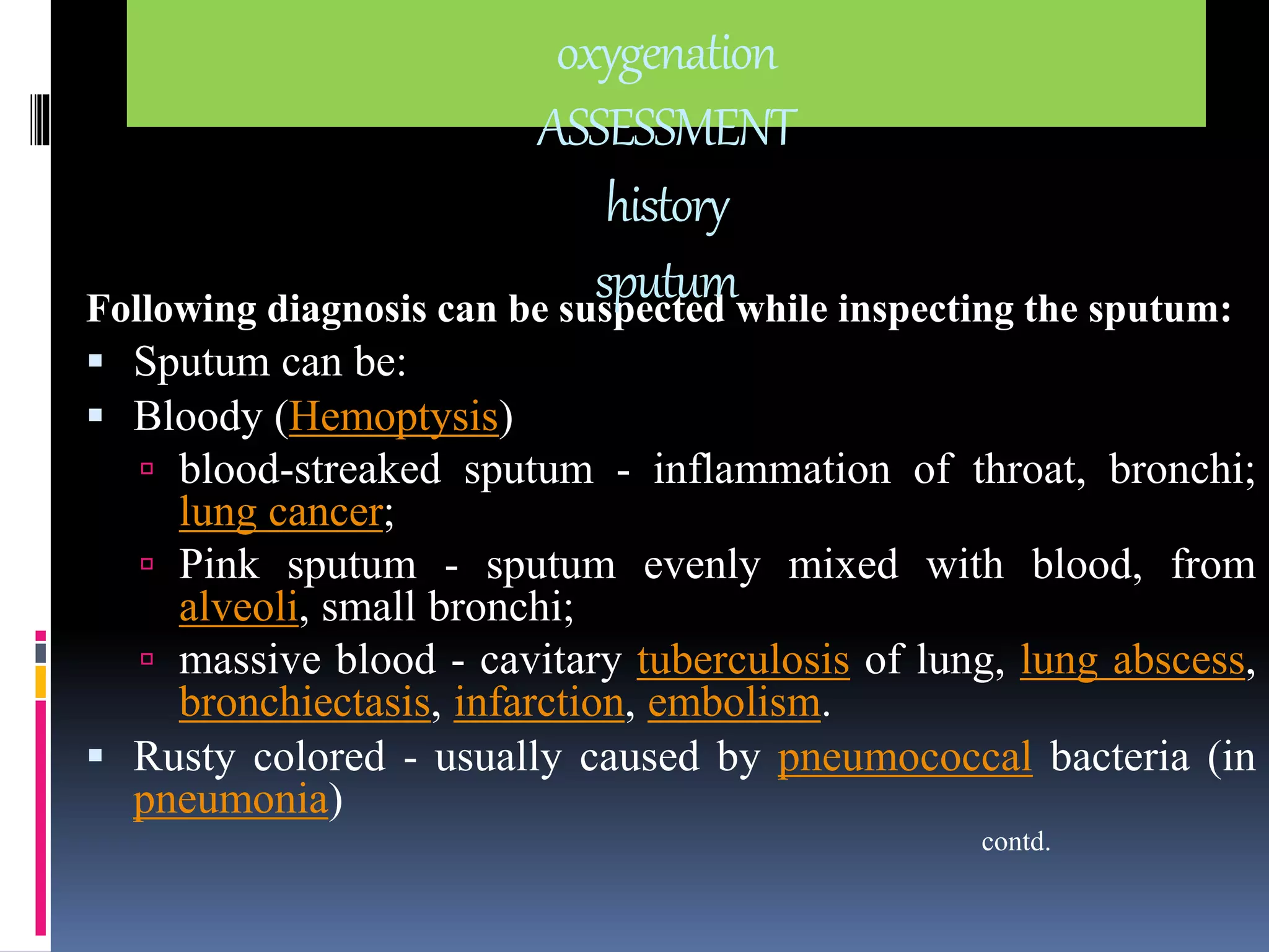
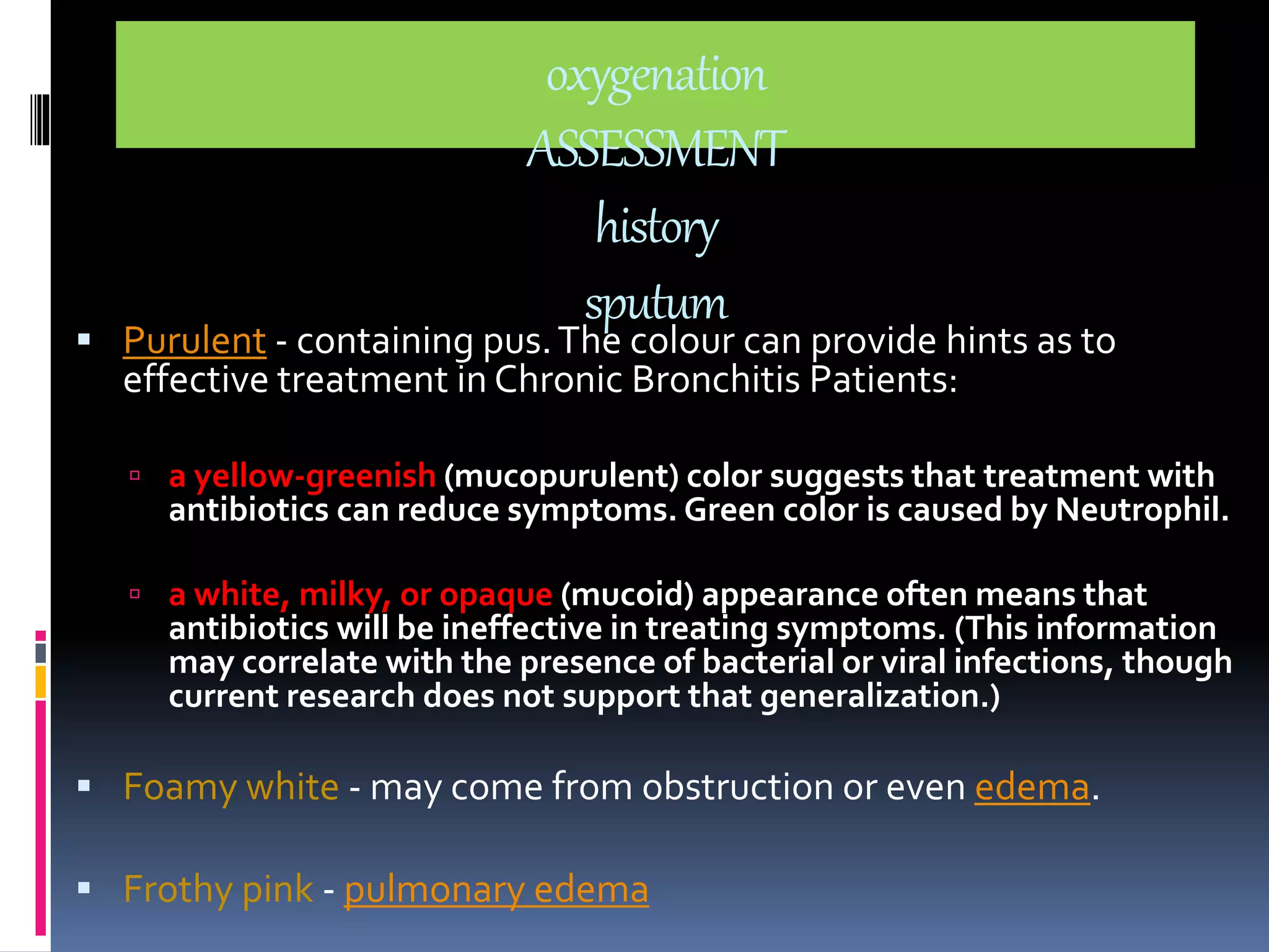
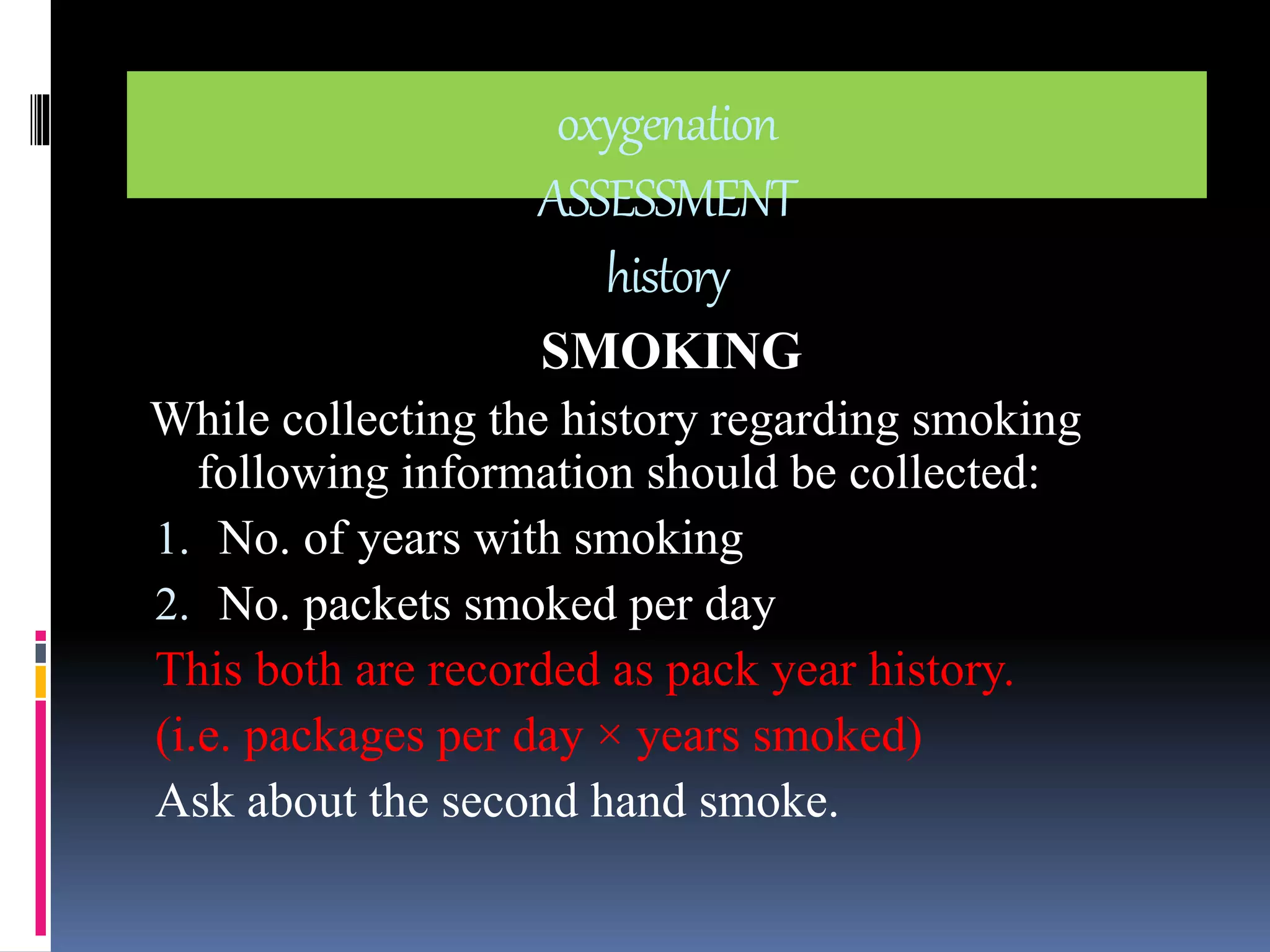
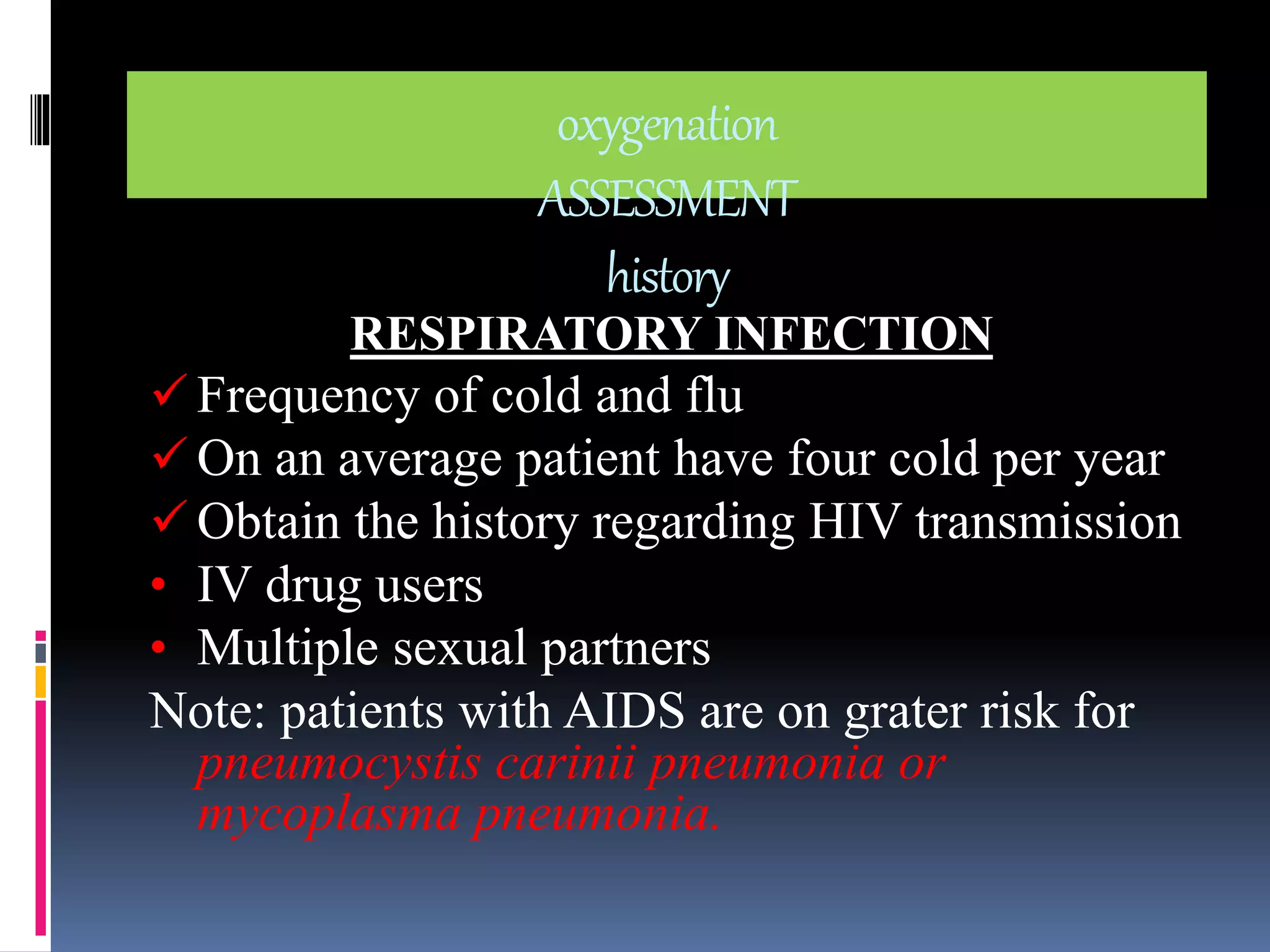
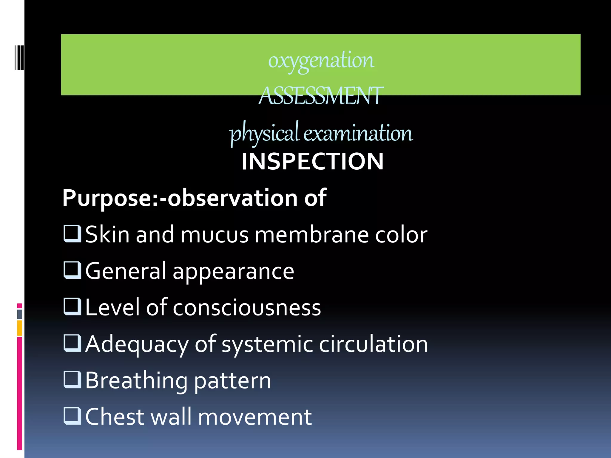
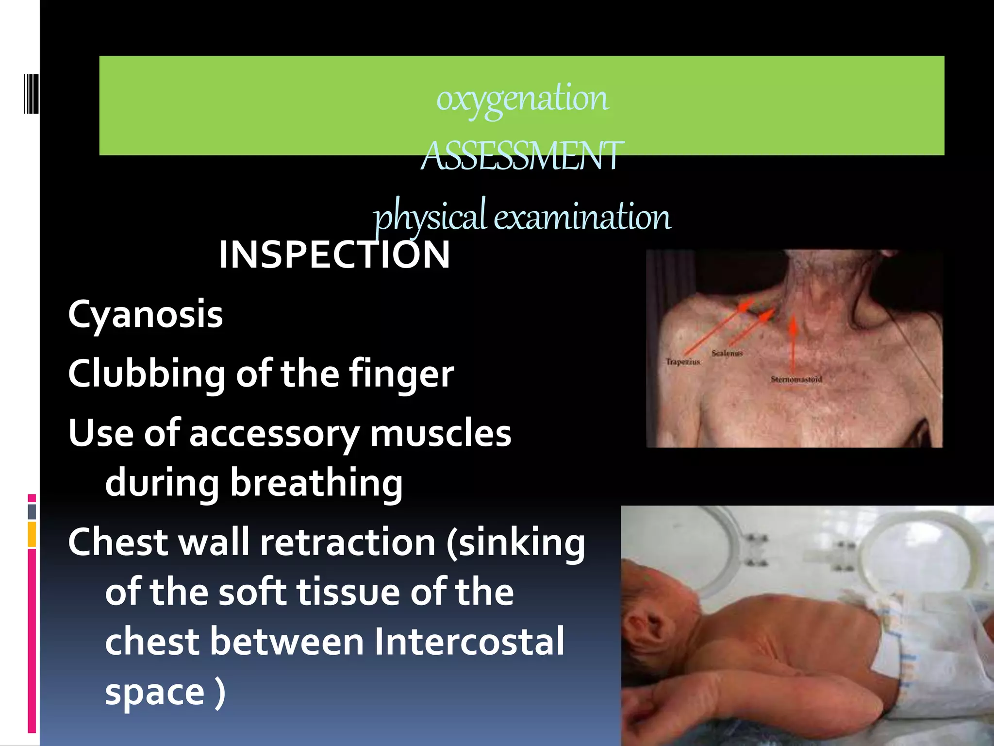
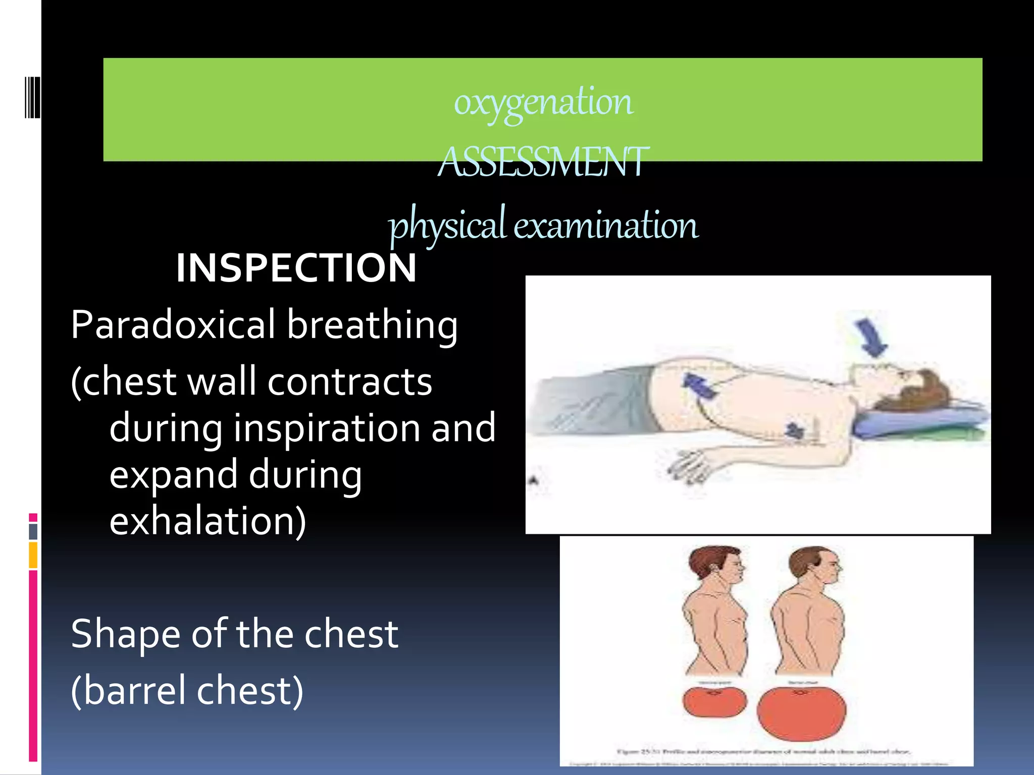
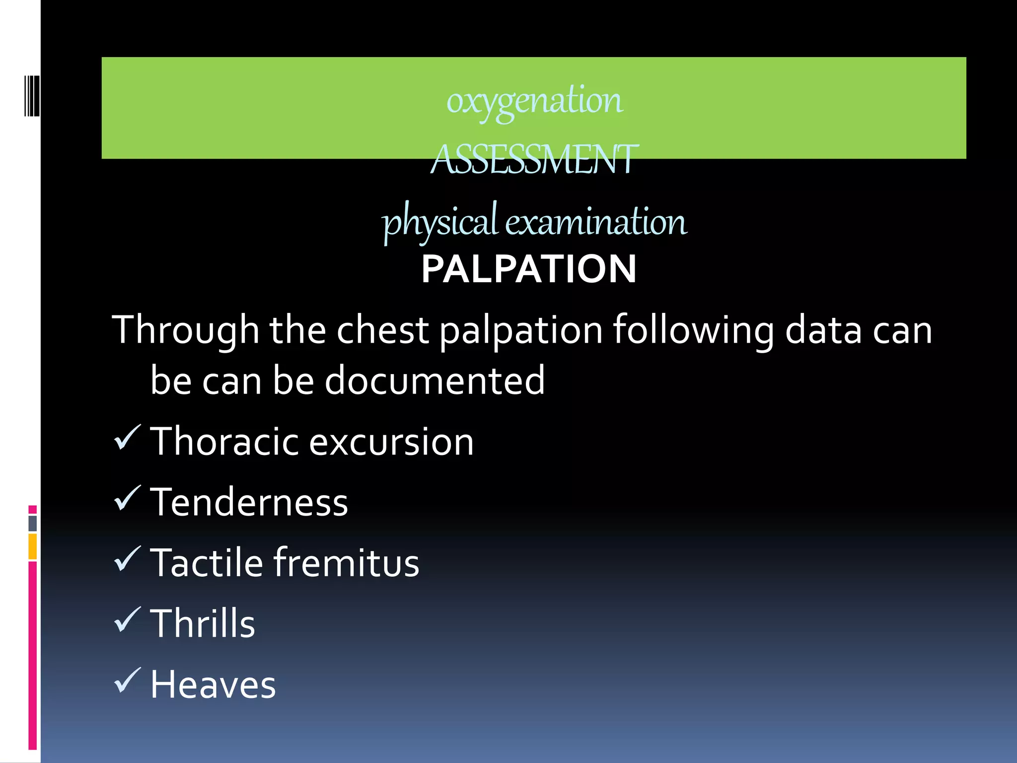
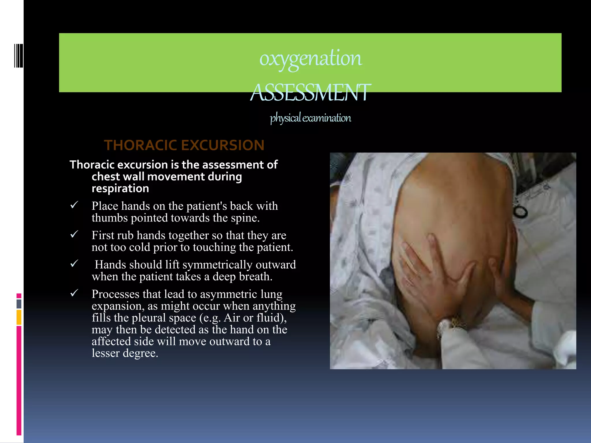
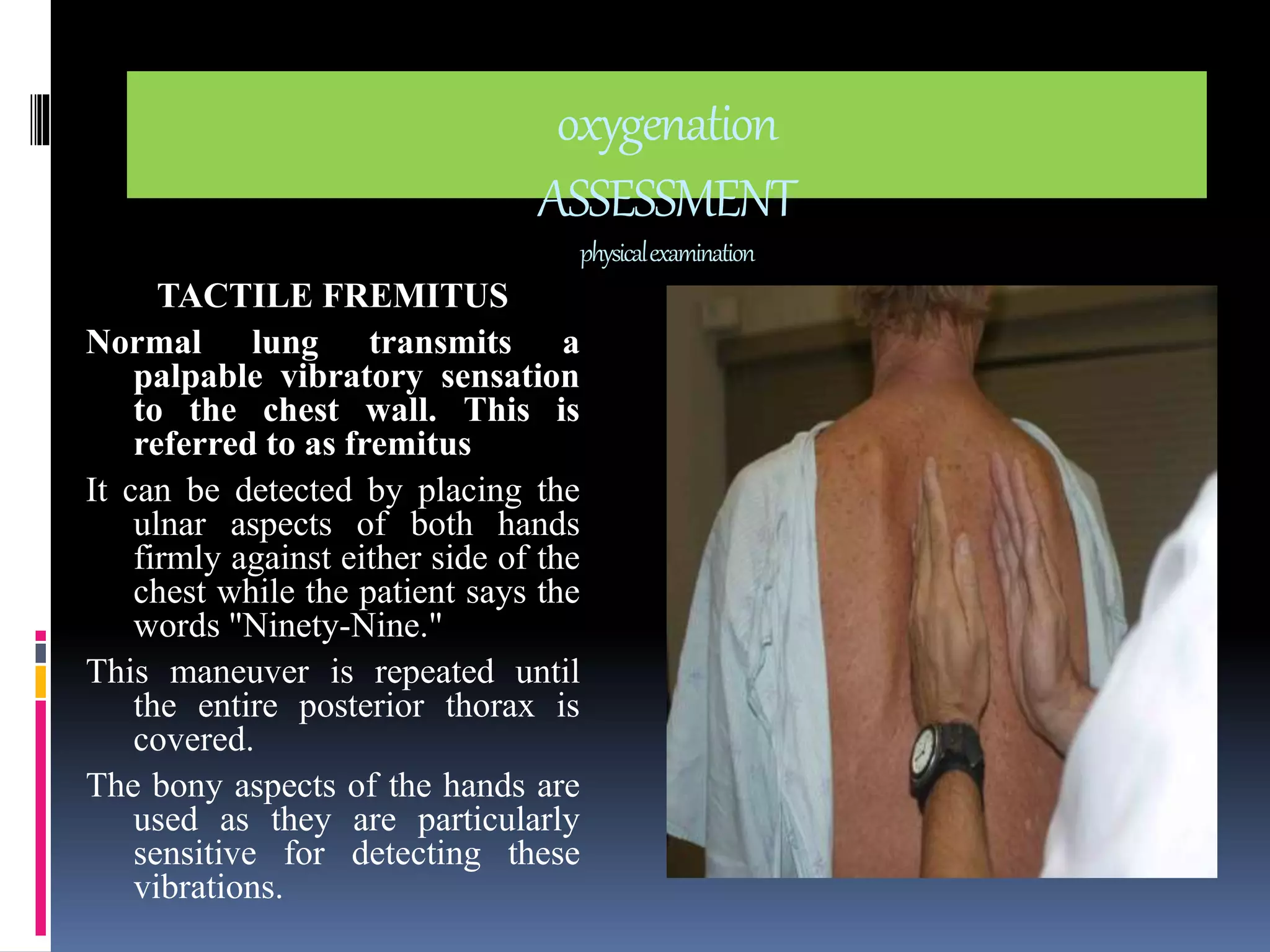
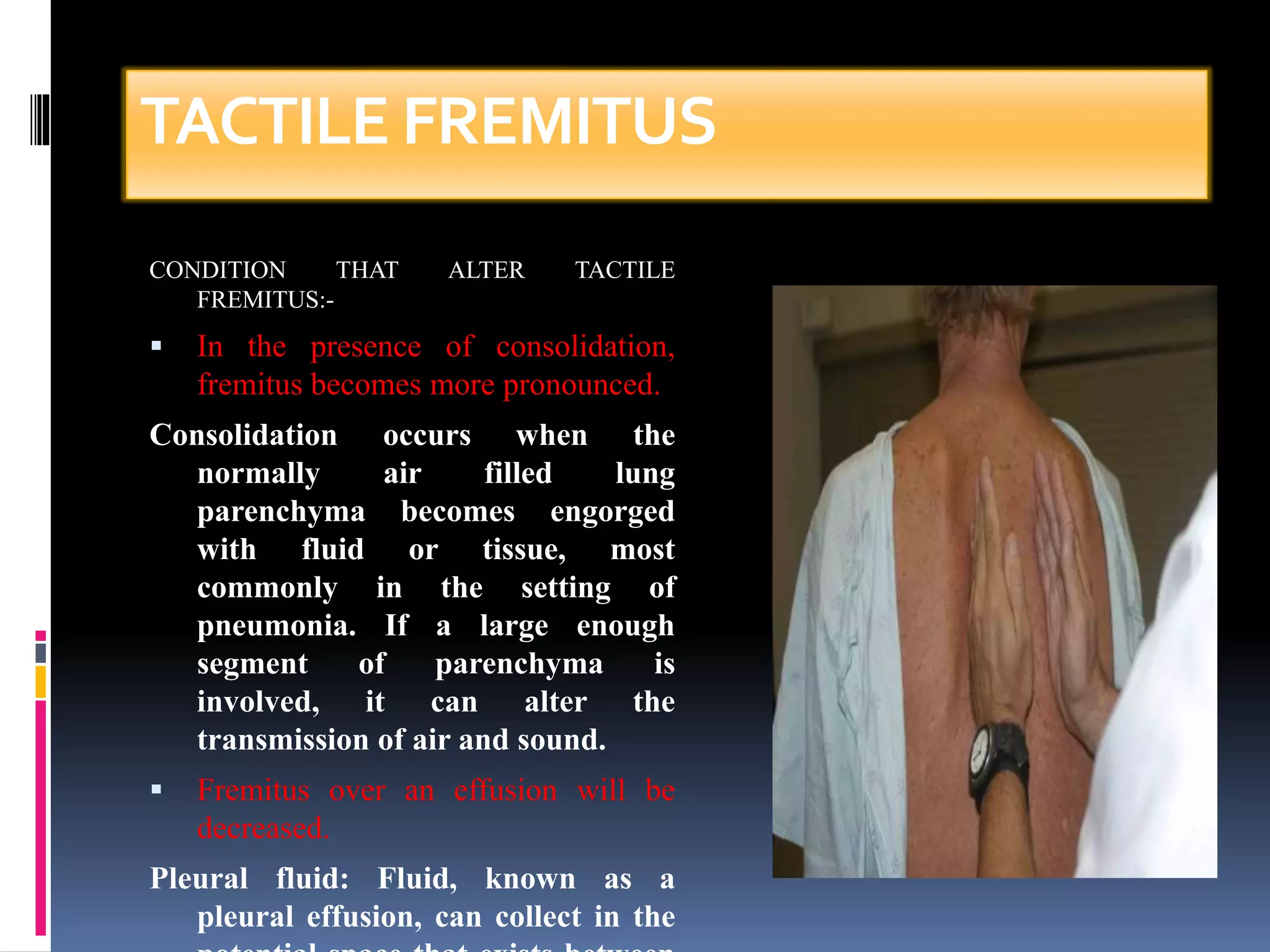
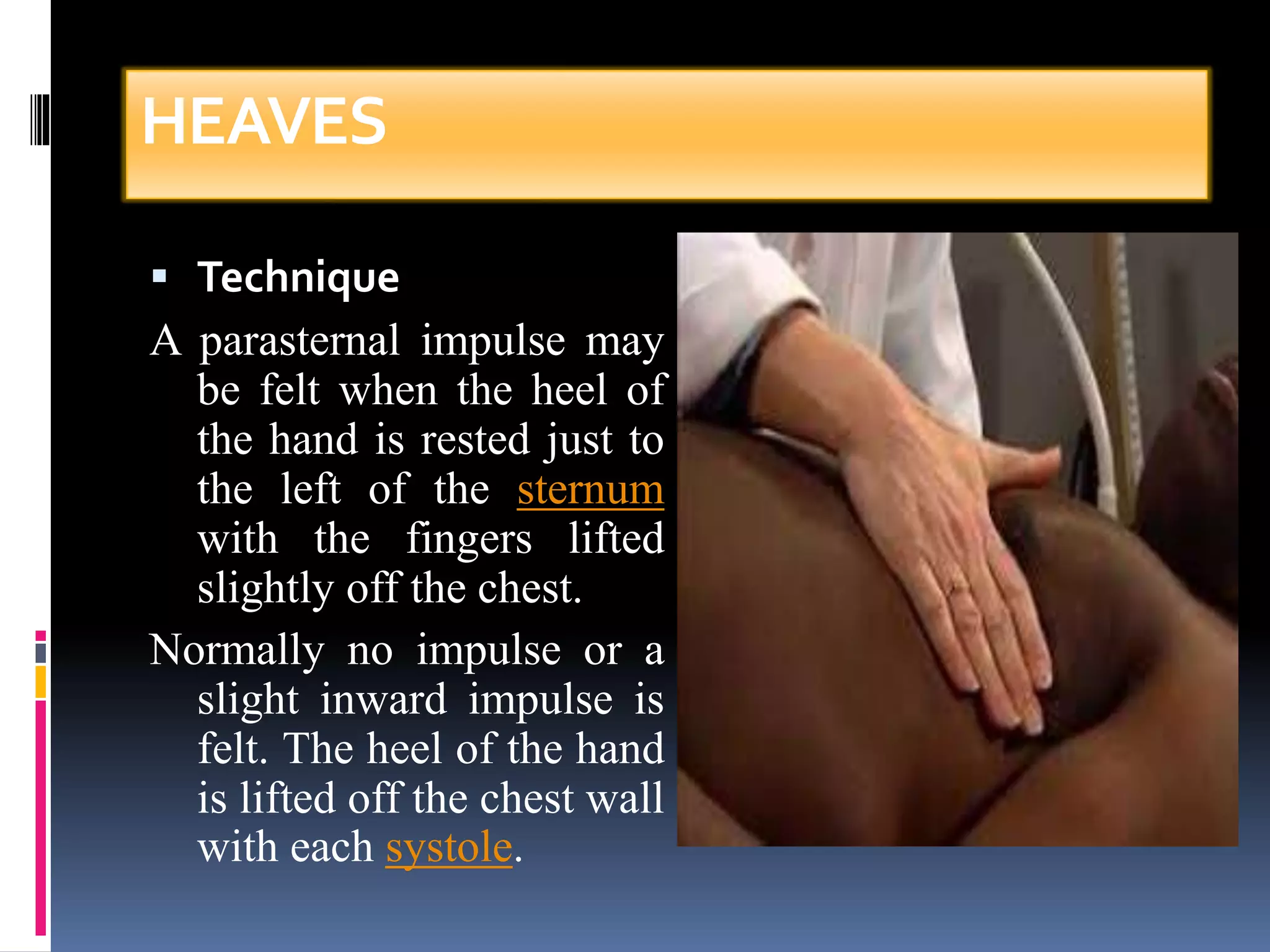
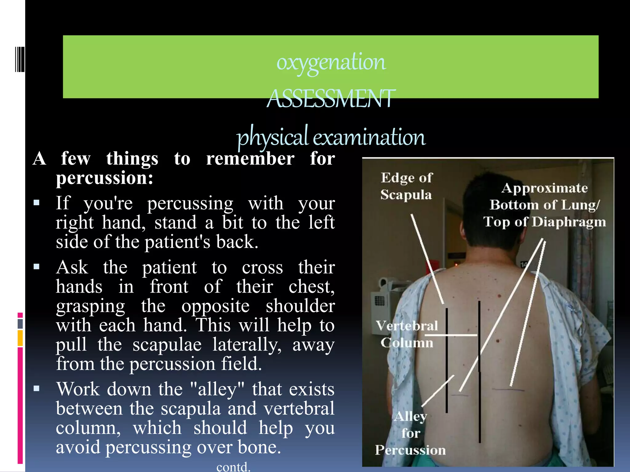
oxygenation 
Nursing process 
 ASSESSMENT 
Nursing history 
Physical examination 
Diagnostic test 
 NURSING DIAGNOSIS 
 OBJECTIVES 
 INTERVENTIONS 
 IMPLEMENTATIONS 
 EVALUATIONS
oxygenation 
ASSESSMENT 
NURSING HISTORY 
Nursing history should be focused on the patient’s 
ability to meet the oxygen needs. 
On the following points history should be collected 
from the patient to assess the respiratory status: 
1. Chest pain 
2. Fatigue 
3. Dyspnea 
4. Cough 
5. Wheezing 
6. Respiratory infection 
7. Allergies 
8. Health risks 
9. Medications 
10. Others (i.e. smoking and 
environmental and 
geographical exposure)
oxygenation 
ASSESSMENT 
history CHEST PAIN 
History regarding the chest pain should 
include: location, duration, 
radiation and frequency. some of 
the descriptions regarding chest 
pain are as follows 
1. Chest pain in male usually occur 
on the left side of the chest and 
radiate to the left arm. 
2. Chest pain (heart attack) usually 
associated with breathlessness, 
jaw or back pain and radiate to 
left arm. 
contd.
oxygenation 
ASSESSMENT 
history 
3. Pericardial pain results from 
inflammation of the 
pericardial sac, occurs on 
inspiration and does not 
radiate. 
4. Pleuritic chest pain radiate to 
the scapular region. 
Coughing, yawning sighing 
worsen the pleuritic chest 
pain. 
5. Pleuritic chest pain usually 
sharp knife like pain always 
associated with inspiration.
oxygenation 
ASSESSMENT 
history 
FATIGUE 
Fatigue is the term used for tiredness. 
It is a subjective sensation used as the early sign of 
cardiopulmonary impairment. 
It occurs in response to the decreased cellular metabolism 
due to lack or decreased oxygen supply to the tissue.
oxygenation 
ASSESSMENT 
history 
DYSPNOEA 
(difficulty in breathing) 
It is a subjective data indicates the hypoxia. 
It is associated with cardiopulmonary diseases, neuromuscular conditions 
and anemia. 
 While collecting the history about dyspnoea ask about any stressful 
event, exercises or any respiratory infections. 
 Ask the patient about sleeping pattern whether dyspnoea affects his 
ability to lie flat and feeling comfortable in semi fowler's position. 
 Dyspnoea can be made objective data if nasal flaring, use of accessory 
muscles in respiration, rate and rhythm of respiration is assessed.
oxygenation 
ASSESSMENT 
history 
WHEEZING 
Wheezing is an objective data 
Wheezing is the whistle sound produced when a high pressure of 
air moves through the narrow bronchus. 
It is usually present in the condition of the asthma, acute 
bronchitis or pneumonia. 
It may occur during inspiration, expiration or both. 
Wheezing indicates the bronchoconstricitons or bronchospasm.
oxygenation 
ASSESSMENT 
history 
COUGH 
Cough is a sudden audible expulsion of air from the lungs. 
Cough is a protective reflex to clear the trachea, bronchi and lugs of irritants 
and secretions. 
Following facts regarding the cough are helpful in assessing the respiratory 
disorder: 
1. Patient with chronic sinusitis usually cough only in morning. 
2. Patient with chronic bronchitis cough and produce the sputum all the 
day. 
3. If sputum is present inspect the sputum for color such as green or blood 
tinged, consistency is thick or thin, amount (increased or decreased), 
smell (none or foul). 
contd.
Following diagnosis can be suspected while inspecting the sputum: 
 Sputum can be: 
 Bloody (Hemoptysis) 
 blood-streaked sputum - inflammation of throat, bronchi; 
lung cancer; 
 Pink sputum - sputum evenly mixed with blood, from 
alveoli, small bronchi; 
 massive blood - cavitary tuberculosis of lung, lung abscess, 
bronchiectasis, infarction, embolism. 
 Rusty colored - usually caused by pneumococcal bacteria (in 
pneumonia) 
contd. 
oxygenation 
ASSESSMENT 
history 
sputum
oxygenation 
ASSESSMENT 
history 
sputum 
 Purulent - containing pus. The colour can provide hints as to 
effective treatment in Chronic Bronchitis Patients: 
 a yellow-greenish (mucopurulent) color suggests that treatment with 
antibiotics can reduce symptoms. Green color is caused by Neutrophil. 
 a white, milky, or opaque (mucoid) appearance often means that 
antibiotics will be ineffective in treating symptoms. (This information 
may correlate with the presence of bacterial or viral infections, though 
current research does not support that generalization.) 
 Foamy white - may come from obstruction or even edema. 
 Frothy pink - pulmonary edema
oxygenation 
ASSESSMENT 
history 
SMOKING 
While collecting the history regarding smoking 
following information should be collected: 
1. No. of years with smoking 
2. No. packets smoked per day 
This both are recorded as pack year history. 
(i.e. packages per day × years smoked) 
Ask about the second hand smoke.
oxygenation 
ASSESSMENT 
history 
RESPIRATORY INFECTION 
 Frequency of cold and flu 
 On an average patient have four cold per year 
 Obtain the history regarding HIV transmission 
• IV drug users 
• Multiple sexual partners 
Note: patients with AIDS are on grater risk for 
pneumocystis carinii pneumonia or 
mycoplasma pneumonia.
oxygenation 
ASSESSMENT 
history 
ALLERGIES 
Collect the history regarding: 
 Types of allergens 
 Response to the allergens 
 Relief measures 
Patient with allergies usually exhibits watery 
eyes, running nose, sneezing, or respiratory 
symptoms like cough or wheezing.
oxygenation 
ASSESSMENT 
history 
HEALTH RISK 
Collect the history regarding the respiratory 
disease such as emphysema, lung cancer 
If the family members have the disease ask 
about the level of health or age at the time 
death 
Obtain the data regarding any 
communicable disease in the family 
especially TB.
oxygenation 
ASSESSMENT 
history 
MEDICATIONS 
Collect the history of prescribed drug, over 
the counter drug, herbal therapies or 
alternatives therapies. 
Because such drugs may increase or decrease 
the effect of other drugs and some time may 
produce the life threatening conditions too. 
Obtain the data for any drug abuse such as 
marijuana, Opioid or cocaine.
oxygenation 
ASSESSMENT 
physical examination 
INSPECTION 
Purpose:-observation of 
Skin and mucus membrane color 
General appearance 
Level of consciousness 
Adequacy of systemic circulation 
Breathing pattern 
Chest wall movement
oxygenation 
ASSESSMENT 
physical examination 
INSPECTION 
Cyanosis 
Clubbing of the finger 
Use of accessory muscles 
during breathing 
Chest wall retraction (sinking 
of the soft tissue of the 
chest between Intercostal 
space )
oxygenation 
ASSESSMENT 
physical examination 
INSPECTION 
Paradoxical breathing 
(chest wall contracts 
during inspiration and 
expand during 
exhalation) 
Shape of the chest 
(barrel chest)
oxygenation 
ASSESSMENT 
physical examination 
PALPATION 
Through the chest palpation following data can 
be can be documented 
 Thoracic excursion 
 Tenderness 
 Tactile fremitus 
 Thrills 
 Heaves
oxygenation 
ASSESSMENT 
physical examination 
THORACIC EXCURSION 
Thoracic excursion is the assessment of 
chest wall movement during 
respiration 
 Place hands on the patient's back with 
thumbs pointed towards the spine. 
 First rub hands together so that they are 
not too cold prior to touching the patient. 
 Hands should lift symmetrically outward 
when the patient takes a deep breath. 
 Processes that lead to asymmetric lung 
expansion, as might occur when anything 
fills the pleural space (e.g. Air or fluid), 
may then be detected as the hand on the 
affected side will move outward to a 
lesser degree.
oxygenation 
ASSESSMENT 
physical examination 
TACTILE FREMITUS 
Normal lung transmits a 
palpable vibratory sensation 
to the chest wall. This is 
referred to as fremitus 
It can be detected by placing the 
ulnar aspects of both hands 
firmly against either side of the 
chest while the patient says the 
words "Ninety-Nine." 
This maneuver is repeated until 
the entire posterior thorax is 
covered. 
The bony aspects of the hands are 
used as they are particularly 
sensitive for detecting these 
vibrations.
TACTILE FREMITUS 
CONDITION THAT ALTER TACTILE 
FREMITUS:- 
 In the presence of consolidation, 
fremitus becomes more pronounced. 
Consolidation occurs when the 
normally air filled lung 
parenchyma becomes engorged 
with fluid or tissue, most 
commonly in the setting of 
pneumonia. If a large enough 
segment of parenchyma is 
involved, it can alter the 
transmission of air and sound. 
 Fremitus over an effusion will be 
decreased. 
Pleural fluid: Fluid, known as a 
pleural effusion, can collect in the 
potential space that exists between
THRILLS 
Thrills are vibratory sensations caused by the 
heart and felt on the body surface. Thrills are 
always associated with murmurs. 
Palpate for thrills as follows: 
 Place the patient in the supine position. 
 Use the proximal part of your hand (not 
fingers)and press gently over the anterior chest 
wall over the heart. 
 Note any thrills appreciated.
HEAVES 
Heave is a precordial impulse that may be felt 
(palpated) in patients with cardiac or respiratory 
disease. 
Precordial impulses are visible or palpable 
pulsations of the chest wall, which originate from 
the heart or the vena cava, pulmonary artery or 
aorta (collectively known as the great vessels). 
IT is mostly seen in right ventricular hypertrophy 
COPD, mitral stenosis, and myopathies.
HEAVES 
 Technique 
A parasternal impulse may 
be felt when the heel of 
the hand is rested just to 
the left of the sternum 
with the fingers lifted 
slightly off the chest. 
Normally no impulse or a 
slight inward impulse is 
felt. The heel of the hand 
is lifted off the chest wall 
with each systole.
oxygenation 
ASSESSMENT 
physical examination 
A few things to remember for 
percussion: 
 If you're percussing with your 
right hand, stand a bit to the left 
side of the patient's back. 
 Ask the patient to cross their 
hands in front of their chest, 
grasping the opposite shoulder 
with each hand. This will help to 
pull the scapulae laterally, away 
from the percussion field. 
 Work down the "alley" that exists 
between the scapula and vertebral 
column, which should help you 
avoid percussing over bone. 
contd.
oxygenation 
ASSESSMENT 
physical examination 
A few things to remember for percussion: 
 Strike the distal inter-phalangeal joint (i.e. the last joint) of 
your left middle finger with the tip of the right middle finger. 
 When percussing any one spot, 2 or 3 sharp taps should 
suffice, though feel free to do more if you'd like. Then move 
your hand down several inter-spaces and repeat the maneuver. 
 After you have percussed the left chest, move yours hands 
across and repeat the same procedure on the right side. 
 If you detect any abnormality on one side, it's a good idea to 
slide your hands across to the other for comparison. 
 In general, percussion is limited to the posterior lung fields. 
However, if auscultation reveals an abnormality in the anterior 
or lateral fields, percussion over these areas can help identify 
its cause.
oxygenation 
ASSESSMENT 
physical examination 
FINDINGS: 
This technique makes use of the fact that striking a surface which 
covers an air-filled structure (e.g. normal lung) will produce a 
resonant note 
Percussion over fluid or tissue filled cavity generates a 
relatively dull sound. 
If the normal, air-filled tissue has been displaced by fluid (e.g. 
pleural effusion) or infiltrated with white cells and bacteria 
(e.g. pneumonia), percussion will generate a deadened tone. 
Chronic (e.g. emphysema) or acute (e.g. pneumothorax) air 
trapping in the lung or pleural space, respectively, will 
produce hyper-resonant (i.e. more drum-like) notes on
oxygenation 
ASSESSMENT 
physical examination 
Auscultation 
Auscultation is the listening to the sounds the 
body makes to detects variations. 
Auscultation of the heart includes assessment of 
normal heart sound, murmurs, rubs and 
gallops 
Auscultation of respiratory system includes the 
normal air entry, adventitious breath sound.
oxygenation 
ASSESSMENT 
diagnostic test 
Diagnostic test confirms the findings of physical examination. 
There are certain diagnostic test which suggests the alteration in oxygenations. 
 Chest X-ray 
 Complete blood count 
 ECG 
 ABG analysis 
 Pulmonary function test 
 Thoracentasis 
 Sputum analysis 
 Serum electrolytes 
 Lung scan 
 Bronchoscopy

Oxygenation nursing process

  • 1.
    oxygenation Nursing process  ASSESSMENT Nursing history Physical examination Diagnostic test  NURSING DIAGNOSIS  OBJECTIVES  INTERVENTIONS  IMPLEMENTATIONS  EVALUATIONS
  • 2.
    oxygenation ASSESSMENT NURSINGHISTORY Nursing history should be focused on the patient’s ability to meet the oxygen needs. On the following points history should be collected from the patient to assess the respiratory status: 1. Chest pain 2. Fatigue 3. Dyspnea 4. Cough 5. Wheezing 6. Respiratory infection 7. Allergies 8. Health risks 9. Medications 10. Others (i.e. smoking and environmental and geographical exposure)
  • 3.
    oxygenation ASSESSMENT historyCHEST PAIN History regarding the chest pain should include: location, duration, radiation and frequency. some of the descriptions regarding chest pain are as follows 1. Chest pain in male usually occur on the left side of the chest and radiate to the left arm. 2. Chest pain (heart attack) usually associated with breathlessness, jaw or back pain and radiate to left arm. contd.
  • 4.
    oxygenation ASSESSMENT history 3. Pericardial pain results from inflammation of the pericardial sac, occurs on inspiration and does not radiate. 4. Pleuritic chest pain radiate to the scapular region. Coughing, yawning sighing worsen the pleuritic chest pain. 5. Pleuritic chest pain usually sharp knife like pain always associated with inspiration.
  • 5.
    oxygenation ASSESSMENT history FATIGUE Fatigue is the term used for tiredness. It is a subjective sensation used as the early sign of cardiopulmonary impairment. It occurs in response to the decreased cellular metabolism due to lack or decreased oxygen supply to the tissue.
  • 6.
    oxygenation ASSESSMENT history DYSPNOEA (difficulty in breathing) It is a subjective data indicates the hypoxia. It is associated with cardiopulmonary diseases, neuromuscular conditions and anemia.  While collecting the history about dyspnoea ask about any stressful event, exercises or any respiratory infections.  Ask the patient about sleeping pattern whether dyspnoea affects his ability to lie flat and feeling comfortable in semi fowler's position.  Dyspnoea can be made objective data if nasal flaring, use of accessory muscles in respiration, rate and rhythm of respiration is assessed.
  • 7.
    oxygenation ASSESSMENT history WHEEZING Wheezing is an objective data Wheezing is the whistle sound produced when a high pressure of air moves through the narrow bronchus. It is usually present in the condition of the asthma, acute bronchitis or pneumonia. It may occur during inspiration, expiration or both. Wheezing indicates the bronchoconstricitons or bronchospasm.
  • 8.
    oxygenation ASSESSMENT history COUGH Cough is a sudden audible expulsion of air from the lungs. Cough is a protective reflex to clear the trachea, bronchi and lugs of irritants and secretions. Following facts regarding the cough are helpful in assessing the respiratory disorder: 1. Patient with chronic sinusitis usually cough only in morning. 2. Patient with chronic bronchitis cough and produce the sputum all the day. 3. If sputum is present inspect the sputum for color such as green or blood tinged, consistency is thick or thin, amount (increased or decreased), smell (none or foul). contd.
  • 9.
    Following diagnosis canbe suspected while inspecting the sputum:  Sputum can be:  Bloody (Hemoptysis)  blood-streaked sputum - inflammation of throat, bronchi; lung cancer;  Pink sputum - sputum evenly mixed with blood, from alveoli, small bronchi;  massive blood - cavitary tuberculosis of lung, lung abscess, bronchiectasis, infarction, embolism.  Rusty colored - usually caused by pneumococcal bacteria (in pneumonia) contd. oxygenation ASSESSMENT history sputum
  • 10.
    oxygenation ASSESSMENT history sputum  Purulent - containing pus. The colour can provide hints as to effective treatment in Chronic Bronchitis Patients:  a yellow-greenish (mucopurulent) color suggests that treatment with antibiotics can reduce symptoms. Green color is caused by Neutrophil.  a white, milky, or opaque (mucoid) appearance often means that antibiotics will be ineffective in treating symptoms. (This information may correlate with the presence of bacterial or viral infections, though current research does not support that generalization.)  Foamy white - may come from obstruction or even edema.  Frothy pink - pulmonary edema
  • 11.
    oxygenation ASSESSMENT history SMOKING While collecting the history regarding smoking following information should be collected: 1. No. of years with smoking 2. No. packets smoked per day This both are recorded as pack year history. (i.e. packages per day × years smoked) Ask about the second hand smoke.
  • 12.
    oxygenation ASSESSMENT history RESPIRATORY INFECTION  Frequency of cold and flu  On an average patient have four cold per year  Obtain the history regarding HIV transmission • IV drug users • Multiple sexual partners Note: patients with AIDS are on grater risk for pneumocystis carinii pneumonia or mycoplasma pneumonia.
  • 13.
    oxygenation ASSESSMENT history ALLERGIES Collect the history regarding:  Types of allergens  Response to the allergens  Relief measures Patient with allergies usually exhibits watery eyes, running nose, sneezing, or respiratory symptoms like cough or wheezing.
  • 14.
    oxygenation ASSESSMENT history HEALTH RISK Collect the history regarding the respiratory disease such as emphysema, lung cancer If the family members have the disease ask about the level of health or age at the time death Obtain the data regarding any communicable disease in the family especially TB.
  • 15.
    oxygenation ASSESSMENT history MEDICATIONS Collect the history of prescribed drug, over the counter drug, herbal therapies or alternatives therapies. Because such drugs may increase or decrease the effect of other drugs and some time may produce the life threatening conditions too. Obtain the data for any drug abuse such as marijuana, Opioid or cocaine.
  • 16.
    oxygenation ASSESSMENT physicalexamination INSPECTION Purpose:-observation of Skin and mucus membrane color General appearance Level of consciousness Adequacy of systemic circulation Breathing pattern Chest wall movement
  • 17.
    oxygenation ASSESSMENT physicalexamination INSPECTION Cyanosis Clubbing of the finger Use of accessory muscles during breathing Chest wall retraction (sinking of the soft tissue of the chest between Intercostal space )
  • 18.
    oxygenation ASSESSMENT physicalexamination INSPECTION Paradoxical breathing (chest wall contracts during inspiration and expand during exhalation) Shape of the chest (barrel chest)
  • 19.
    oxygenation ASSESSMENT physicalexamination PALPATION Through the chest palpation following data can be can be documented  Thoracic excursion  Tenderness  Tactile fremitus  Thrills  Heaves
  • 20.
    oxygenation ASSESSMENT physicalexamination THORACIC EXCURSION Thoracic excursion is the assessment of chest wall movement during respiration  Place hands on the patient's back with thumbs pointed towards the spine.  First rub hands together so that they are not too cold prior to touching the patient.  Hands should lift symmetrically outward when the patient takes a deep breath.  Processes that lead to asymmetric lung expansion, as might occur when anything fills the pleural space (e.g. Air or fluid), may then be detected as the hand on the affected side will move outward to a lesser degree.
  • 21.
    oxygenation ASSESSMENT physicalexamination TACTILE FREMITUS Normal lung transmits a palpable vibratory sensation to the chest wall. This is referred to as fremitus It can be detected by placing the ulnar aspects of both hands firmly against either side of the chest while the patient says the words "Ninety-Nine." This maneuver is repeated until the entire posterior thorax is covered. The bony aspects of the hands are used as they are particularly sensitive for detecting these vibrations.
  • 22.
    TACTILE FREMITUS CONDITIONTHAT ALTER TACTILE FREMITUS:-  In the presence of consolidation, fremitus becomes more pronounced. Consolidation occurs when the normally air filled lung parenchyma becomes engorged with fluid or tissue, most commonly in the setting of pneumonia. If a large enough segment of parenchyma is involved, it can alter the transmission of air and sound.  Fremitus over an effusion will be decreased. Pleural fluid: Fluid, known as a pleural effusion, can collect in the potential space that exists between
  • 23.
    THRILLS Thrills arevibratory sensations caused by the heart and felt on the body surface. Thrills are always associated with murmurs. Palpate for thrills as follows:  Place the patient in the supine position.  Use the proximal part of your hand (not fingers)and press gently over the anterior chest wall over the heart.  Note any thrills appreciated.
  • 24.
    HEAVES Heave isa precordial impulse that may be felt (palpated) in patients with cardiac or respiratory disease. Precordial impulses are visible or palpable pulsations of the chest wall, which originate from the heart or the vena cava, pulmonary artery or aorta (collectively known as the great vessels). IT is mostly seen in right ventricular hypertrophy COPD, mitral stenosis, and myopathies.
  • 25.
    HEAVES  Technique A parasternal impulse may be felt when the heel of the hand is rested just to the left of the sternum with the fingers lifted slightly off the chest. Normally no impulse or a slight inward impulse is felt. The heel of the hand is lifted off the chest wall with each systole.
  • 26.
    oxygenation ASSESSMENT physicalexamination A few things to remember for percussion:  If you're percussing with your right hand, stand a bit to the left side of the patient's back.  Ask the patient to cross their hands in front of their chest, grasping the opposite shoulder with each hand. This will help to pull the scapulae laterally, away from the percussion field.  Work down the "alley" that exists between the scapula and vertebral column, which should help you avoid percussing over bone. contd.
  • 27.
    oxygenation ASSESSMENT physicalexamination A few things to remember for percussion:  Strike the distal inter-phalangeal joint (i.e. the last joint) of your left middle finger with the tip of the right middle finger.  When percussing any one spot, 2 or 3 sharp taps should suffice, though feel free to do more if you'd like. Then move your hand down several inter-spaces and repeat the maneuver.  After you have percussed the left chest, move yours hands across and repeat the same procedure on the right side.  If you detect any abnormality on one side, it's a good idea to slide your hands across to the other for comparison.  In general, percussion is limited to the posterior lung fields. However, if auscultation reveals an abnormality in the anterior or lateral fields, percussion over these areas can help identify its cause.
  • 28.
    oxygenation ASSESSMENT physicalexamination FINDINGS: This technique makes use of the fact that striking a surface which covers an air-filled structure (e.g. normal lung) will produce a resonant note Percussion over fluid or tissue filled cavity generates a relatively dull sound. If the normal, air-filled tissue has been displaced by fluid (e.g. pleural effusion) or infiltrated with white cells and bacteria (e.g. pneumonia), percussion will generate a deadened tone. Chronic (e.g. emphysema) or acute (e.g. pneumothorax) air trapping in the lung or pleural space, respectively, will produce hyper-resonant (i.e. more drum-like) notes on
  • 29.
    oxygenation ASSESSMENT physicalexamination Auscultation Auscultation is the listening to the sounds the body makes to detects variations. Auscultation of the heart includes assessment of normal heart sound, murmurs, rubs and gallops Auscultation of respiratory system includes the normal air entry, adventitious breath sound.
  • 30.
    oxygenation ASSESSMENT diagnostictest Diagnostic test confirms the findings of physical examination. There are certain diagnostic test which suggests the alteration in oxygenations.  Chest X-ray  Complete blood count  ECG  ABG analysis  Pulmonary function test  Thoracentasis  Sputum analysis  Serum electrolytes  Lung scan  Bronchoscopy