Downloaded 560 times


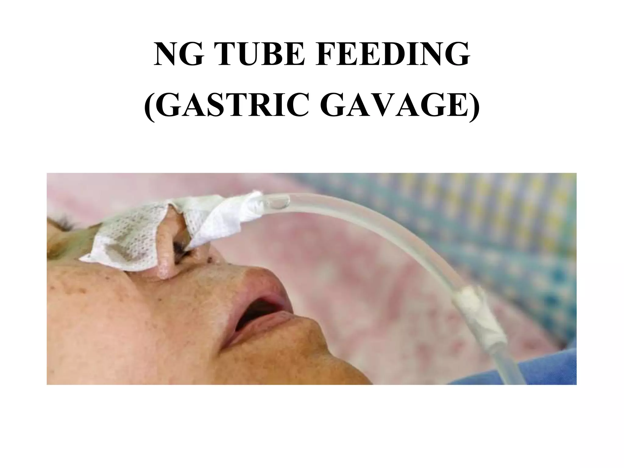


This document provides information on nasogastric tube insertion and feeding. It defines nasogastric tube insertion as the passage of a tube through the nose or mouth into the stomach. It then discusses the purposes, principles, indications, contraindications, instructions, equipment, and procedures for nasogastric tube insertion and feeding. The key steps involved in nasogastric tube feeding are confirming proper tube placement in the stomach, administering nutrients or medications through the tube slowly by gravity or pump, and providing aftercare to the patient.


