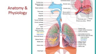
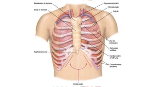
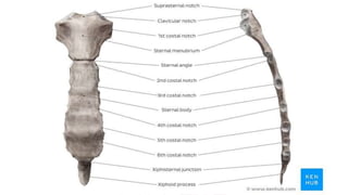
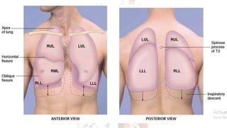
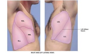
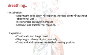
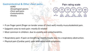
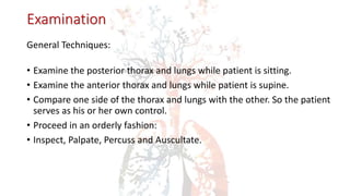
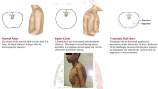
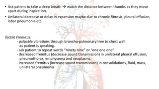
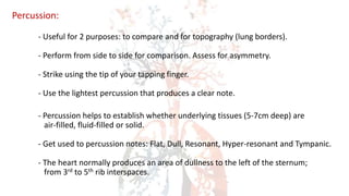
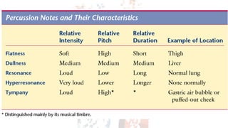
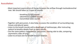
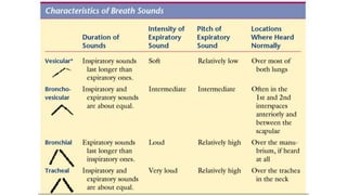
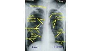
RESPIRATORY SYSTEM
DR. LAKITHA RATHNAWEERA
Anatomy &
Physiology
• Can describe local findings on chest on two dimensions.
- along the vertical axis
- around the circumference of the chest
• To make vertical locations, count the ribs and interspaces. Sternal angle is
the best guide!
• To locate findings around circumference of chest, imagine a series of vertical
lines around the chest wall.
Lungs, Fissures & Lobes
• Each lung divided (roughly) in half by an oblique (major) fissure.
• Right lung is further divided by the horizontal (minor) fissure.
• These fissures divide lungs into lobes.
- the right lung is divided into upper, middle and lower lobes.
- the left lung is divided into upper and lower lobes.
• Lung base at 6-8 ribs. T10 spinous process posteriorly.
The Trachea & Bronchi
• The Trachea and Major Bronchi:
- The trachea bifurcates into its mainstem bronchi at the levels of
sternal angle anteriorly and the T4 spinous process posteriorly.
• The Pleurae:
- The pleurae are serous membranes that cover the outer surface
of each lung (visceral pleura) and also the inner rib cage and
upper surface of the diaphragm (parietal pleura)
- Pleural fluids lubricate the surfaces.
Breathing..
• Inspiration:
- Diaphragm goes down  expands thoracic cavity  pushout
abdominal wall
- Intrathoracic pressure increases
- Scalenus and Parasternal muscles.
• Expiration:
- Chest walls and lungs recoil.
- Diaphragm relaxes  rise passively
- Chest and abdomen return to their resting position.
The Health History
• CHEST PAIN:
- Initially go for broad questions.
“do you have any discomfort or unpleasant feelings in your chest?”
- Ask the patient to point to the location of chest.
- Attempt to elicit all 7 attributes of pain.
- Aside from lung condition, pain may arise from cardiac, vascular,
gastrointestinal, musculoskeletal or skin pathology. It’s also
commonly associated with anxiety.
- Lung tissue itself has no pain fibers. Pain in lung conditions usually
arises from inflammation of the adjacent parietal pleura.
- Other surrounding structures may also irritate the parietal pleura,
causing pain.
Cardiac chest pain:
-Angina pectoris
-Myocardial Infarction
-Pericarditis
-Aortic aneurysm etc
Pulmonary chest pain:
-Tracheobronchitis
-Pleuritic chest pain
Gastrointestinal & Other chest pains:
-GORD
-Oesophageal spasms
-Chest wall pains
-Costochondritis
-Anxiety
• If can finger point (finger on tender areas of chest wall) mostly musculoskeletal pain.
• Epigastric area to neck pain: mostly GI related
• Most common in children: due to anxiety and costochondritis.
• Respiratory pain: If pain on breathing: mostly maybe due to a respiratory obstruction.
• Physical pain (Cardiac pain): pain with associated symptoms.
• Shortness of Breath (Dyspnoea/ SOB)
- Dyspnoea is a non-painful, but unpleasant, uncomfortable awareness
of breathing that is inappropriate to the level of exertion.
- Begin assessment with a broad question, such as,
“Have you had any difficulty in breathing?”
- Determine the severity of dyspnoea, based on the patients daily
activities.
• Causes may include – Left heart failure, Chronic bronchitis, COPD, BA,
Pneumonia, Spontaneous pneumothorax, Acute PE, Anxiety, Hyperventilation.
• Wheezing:
- Wheezes are musical respiratory sounds that may be audible to
patient and to others. It may sometimes be cardiovascular in origin.
• Cough:
- Cough is typically a reflex response to stimuli, that irritate receptors in larynx,
trachea or large bronchi. It may sometimes be CV in origin.
- Ask whether cough is dry or productive? (produces sputum or phlegm)
- Ask the patient to describe any volume of sputum, its colour, odor
and consistency. COUGH CAN BE A SYMPTOM OF LV FAILURE.
• Causes may include mucus, pus, blood, dust, foreign agents, extreme hot or
cold air, inflammation of respiratory mucosa, pressure or tension in airways.
ACUTE COUGH: less than 3 weeks
: viral URTI, acute bronchitis, pneumonia, LV failure,
foreign body, asthma.
SUB-ACUTE COUGH: 3 to 8 weeks
: bacterial sinusitis, asthma.
CHRONIC COUGH: more than 8 weeks
: postnasal drip, gastro-oesophageal reflux, chronic
bronchitis, bronchiectasis
• Mucoid sputum: translucent, white or gray
• Purulent sputum: yellow or greenish (in bronchiectasis, lung abscess)
• Foul smelling sputum: anaerobic lung abscess
• Tenacious sputum: Cystic fibrosis
• Haemoptysis:
- Haemoptysis is coughing up blood from lungs. It may vary from blood streaked
phlegm to frank blood. Rare in infants & children. Common in CF.
- Ask the patient to describe the blood volume produces and other sputum
attributes as well.
- Try to confirm the source of bleeding by history and examination,
before using the term “Haemoptysis”
- Blood may also originate from mouth, pharynx and GI tract.
- Stomach blood is darker than respiratory blood.
• Acute inflammations may cause hemoptysis.. Like,
- Laryngitis (dry cough)
- Tracheo-bronchitis (dry cough)
- Mycoplasma and viral pneumonias (dry cough)
- Bacterial pneumonia (pinkish red jelly – pneumococcal, klebsiella)
• Chronic inflammations may also cause hemoptysis.. Like,
- Postnasal drip (chronic cough, mucoid)
- Chronic bronchitis (chronic cough, purulent bloody)
- Bronchiectasis (chronic cough, purulent foul bloody)
- Pulmonary TB (chronic cough, purulent bloody)
- Lung abscess (chronic cough, purulent foul bloody)
- Asthma (chronic cough, mucoid)
- Gastro-oesophageal reflux (chronic cough in night and morning)
Lung cancer – cough, bloody sputum (starts with ?rusty colour)
LV failure, MS – dry cough, pink frothy sputum, pulmonary oedema, frank haemoptysis
Pulmonary emboli – dry cough, dark or bright red sputum
Tobacco Cessation:
- Smoking is a leading cause of preventable deaths in world.
- Remember the five “A” s
- Ask about smoking at each visit.
- Advice regularly to stop smoking.
- Assess patient’s readiness to quit.
- Assist patient to set stop dates and on education.
- Arrange follow-up visits to monitor and support progress.
Examination
General Techniques:
• Examine the posterior thorax and lungs while patient is sitting.
• Examine the anterior thorax and lungs while patient is supine.
• Compare one side of the thorax and lungs with the other. So the patient
serves as his or her own control.
• Proceed in an orderly fashion:
• Inspect, Palpate, Percuss and Auscultate.
Initial Survey of Respiration & Thorax:
• Healthy resting adult breaths 14-20/min
• Observe the rate, rhythm, depth and effort of breathing.
• Inspect for any signs of respiratory difficulties:
- Assess patients colour (for cyanosis)
- Listen patients breathing (audible stridor/wheeze may suggest URT obstruct)
- Inspect the patients neck (use of accessory muscles may suggest COPD)
- Observe the shape of chest (increased AP diameter may suggest COPD)
Examine the Posterior Chest:
Inspection: Note the shape of the chest and the way it moves.
Asymmetry  Pleural Effusion
Retraction  Asthma, COPD, upper airway obstruction
Unilateral impairment or lag  pleural disease from asbestosis, silicosis, phrenic nerve
damage, trauma.
Palpation:
- Identify any tender areas.
- Assess any visible abnormalities (tracheal deviation etc)
- Test chest expansion (lung excursion test) : place thumbs at the level of 10th
rib with fingers grasping loosely, and parallel to the lateral rib cage.
• Ask patient to take a deep breath  watch the distance between thumbs as they move
apart during inspiration.
• Unilateral decrease or delay in expansion maybe due to chronic fibrosis, pleural effusion,
lobar pneumonia etc.
Tactile Fremitus:
- palpable vibrations through broncho-pulmonary tree to chest wall
as patient is speaking.
- ask patient to repeat words “ninety nine” or “one one one”
- decreased fremitus (decrease sound transmission) in unilateral pleural effusion,
pneumothorax, emphysema and neoplasms.
- increased fremitus (increase sound transmission) in consolidations, fluid, mass,
unilateral pneumonia
Palpate and compare symmetric areas!
Percussion:
- Useful for 2 purposes: to compare and for topography (lung borders).
- Perform from side to side for comparison. Assess for asymmetry.
- Strike using the tip of your tapping finger.
- Use the lightest percussion that produces a clear note.
- Percussion helps to establish whether underlying tissues (5-7cm deep) are
air-filled, fluid-filled or solid.
- Get used to percussion notes: Flat, Dull, Resonant, Hyper-resonant and Tympanic.
- The heart normally produces an area of dullness to the left of the sternum;
from 3rd to 5th rib interspaces.
- Estimate the extent of diaphragmatic
excursion.
- Dullness replace resonance when, fluid or
solid tissue replaces air filled cavities!
- in lobar pn. alveoli fills with fluid and
blood
- in pleural effusion acc. of serous fluid
- in haemothorax blood
- in empyema pus etc.
- High diaphragm in Atelectasis and Phrenic
nerve paralysis.
Auscultation:
- Most important examination of thorax to assess the airflow through tracheobronchial
tree. We should lidten to 3 types of sounds.
- breathing (lungs) sounds
- adventitious (added) sounds
- transmitted voice sounds
- Together with percussion, it also helps to assess the condition of surrounding lung
tissues and pleural space.
- Listen to the breath sounds with a diaphragm of stethoscope, after instructing
patient to breathe deeply from an open mouth.
- Use the same pattern suggested for percussion, moving side to side, comparing
asymmetric sides of lungs.
- Listen to at least one full breath on each location.
Normal (lung) breath sounds:
Vesicular – soft and low pitches, usually heard over most of both lungs.
Bronchial – louder and higher in pitch, usually heard over the manubrium.
Bronchovesicular – intermediate intensity and pitch, usually heard over the 1st, 2nd ICS.
Adventitious (added) breath sounds:
Crackles – aka rales. discontinuous, intermittent non musical
- loud, low pitches, brief
- in bronchitis, pneumonia, fibrosis, left heart failure.
Wheezes - continuous, prolonged, musical
- high pitched, hissing or shrill quality
- due to narrowed airways (asthma, bronchitis, COPD)
Rhonchii - continuous, prolonged, musical
- low pitches, snoring quality
- due to secretions in large airways
Pleural Rub - aka friction rub.
- ask to cough, crackles stop when coughing. But pleural rub never stops
with cough, and pain of rub differs with position.
Mediastinal Crunch (Hammen’s Sign)
- When air is present in mediastinum.
- Scratchy sounds (Hammens sign) occurs
- Occur most frequently during systole.
- Common after cardiac post-op
Transmitted Voice Sounds:
- Only if you hear abnormally located bronchial or bronchovesicular sound,
ask pt to say “ee”
- increased transmission of voice sound (louder voice) in airless lungs and
suggests consolidation (known as bronchophony)
- if voice become nasal quality or when “ee” sounds like “aa”
scenario knowns as egophony.
Examination of the Anterior Chest:
- As examination of the posterior chest, proceed in an orderly fashion:
inspection, palpation, percussion and auscultation.
- With percussion of anterior chest, heart normally produces an area
of dullness to the left of the sternum; from 3rd to 5th rib interspaces.
- Supraclavicular retraction is normally present.
CHEST X RAY (CXR) Check for..
A – airways
B – basal lungs & pleura
C – cardio mediastinum
D – disabilities (visible fractures etc)
E – everything else
• R > L diaphragm gap (not more than 3cm)
• Cardiothoracic ratio not more than <55%
• Trachea diameter < 25mm in males/ < 21mm in females.
• Aortic notch should be visible.
• Any gastric abnormalities (bubbles)
THANK YOU

5- RESPIRATORY SYSTEM (1).pptx

  • 1.
  • 2.
  • 6.
    • Can describelocal findings on chest on two dimensions. - along the vertical axis - around the circumference of the chest • To make vertical locations, count the ribs and interspaces. Sternal angle is the best guide! • To locate findings around circumference of chest, imagine a series of vertical lines around the chest wall.
  • 9.
    Lungs, Fissures &Lobes • Each lung divided (roughly) in half by an oblique (major) fissure. • Right lung is further divided by the horizontal (minor) fissure. • These fissures divide lungs into lobes. - the right lung is divided into upper, middle and lower lobes. - the left lung is divided into upper and lower lobes. • Lung base at 6-8 ribs. T10 spinous process posteriorly.
  • 12.
    The Trachea &Bronchi • The Trachea and Major Bronchi: - The trachea bifurcates into its mainstem bronchi at the levels of sternal angle anteriorly and the T4 spinous process posteriorly. • The Pleurae: - The pleurae are serous membranes that cover the outer surface of each lung (visceral pleura) and also the inner rib cage and upper surface of the diaphragm (parietal pleura) - Pleural fluids lubricate the surfaces.
  • 16.
    Breathing.. • Inspiration: - Diaphragmgoes down  expands thoracic cavity  pushout abdominal wall - Intrathoracic pressure increases - Scalenus and Parasternal muscles. • Expiration: - Chest walls and lungs recoil. - Diaphragm relaxes  rise passively - Chest and abdomen return to their resting position.
  • 17.
    The Health History •CHEST PAIN: - Initially go for broad questions. “do you have any discomfort or unpleasant feelings in your chest?” - Ask the patient to point to the location of chest. - Attempt to elicit all 7 attributes of pain. - Aside from lung condition, pain may arise from cardiac, vascular, gastrointestinal, musculoskeletal or skin pathology. It’s also commonly associated with anxiety.
  • 19.
    - Lung tissueitself has no pain fibers. Pain in lung conditions usually arises from inflammation of the adjacent parietal pleura. - Other surrounding structures may also irritate the parietal pleura, causing pain. Cardiac chest pain: -Angina pectoris -Myocardial Infarction -Pericarditis -Aortic aneurysm etc Pulmonary chest pain: -Tracheobronchitis -Pleuritic chest pain
  • 20.
    Gastrointestinal & Otherchest pains: -GORD -Oesophageal spasms -Chest wall pains -Costochondritis -Anxiety • If can finger point (finger on tender areas of chest wall) mostly musculoskeletal pain. • Epigastric area to neck pain: mostly GI related • Most common in children: due to anxiety and costochondritis. • Respiratory pain: If pain on breathing: mostly maybe due to a respiratory obstruction. • Physical pain (Cardiac pain): pain with associated symptoms.
  • 22.
    • Shortness ofBreath (Dyspnoea/ SOB) - Dyspnoea is a non-painful, but unpleasant, uncomfortable awareness of breathing that is inappropriate to the level of exertion. - Begin assessment with a broad question, such as, “Have you had any difficulty in breathing?” - Determine the severity of dyspnoea, based on the patients daily activities. • Causes may include – Left heart failure, Chronic bronchitis, COPD, BA, Pneumonia, Spontaneous pneumothorax, Acute PE, Anxiety, Hyperventilation.
  • 23.
    • Wheezing: - Wheezesare musical respiratory sounds that may be audible to patient and to others. It may sometimes be cardiovascular in origin. • Cough: - Cough is typically a reflex response to stimuli, that irritate receptors in larynx, trachea or large bronchi. It may sometimes be CV in origin. - Ask whether cough is dry or productive? (produces sputum or phlegm) - Ask the patient to describe any volume of sputum, its colour, odor and consistency. COUGH CAN BE A SYMPTOM OF LV FAILURE.
  • 24.
    • Causes mayinclude mucus, pus, blood, dust, foreign agents, extreme hot or cold air, inflammation of respiratory mucosa, pressure or tension in airways. ACUTE COUGH: less than 3 weeks : viral URTI, acute bronchitis, pneumonia, LV failure, foreign body, asthma. SUB-ACUTE COUGH: 3 to 8 weeks : bacterial sinusitis, asthma. CHRONIC COUGH: more than 8 weeks : postnasal drip, gastro-oesophageal reflux, chronic bronchitis, bronchiectasis
  • 25.
    • Mucoid sputum:translucent, white or gray • Purulent sputum: yellow or greenish (in bronchiectasis, lung abscess) • Foul smelling sputum: anaerobic lung abscess • Tenacious sputum: Cystic fibrosis • Haemoptysis: - Haemoptysis is coughing up blood from lungs. It may vary from blood streaked phlegm to frank blood. Rare in infants & children. Common in CF. - Ask the patient to describe the blood volume produces and other sputum attributes as well. - Try to confirm the source of bleeding by history and examination, before using the term “Haemoptysis”
  • 26.
    - Blood mayalso originate from mouth, pharynx and GI tract. - Stomach blood is darker than respiratory blood. • Acute inflammations may cause hemoptysis.. Like, - Laryngitis (dry cough) - Tracheo-bronchitis (dry cough) - Mycoplasma and viral pneumonias (dry cough) - Bacterial pneumonia (pinkish red jelly – pneumococcal, klebsiella) • Chronic inflammations may also cause hemoptysis.. Like, - Postnasal drip (chronic cough, mucoid) - Chronic bronchitis (chronic cough, purulent bloody) - Bronchiectasis (chronic cough, purulent foul bloody) - Pulmonary TB (chronic cough, purulent bloody) - Lung abscess (chronic cough, purulent foul bloody) - Asthma (chronic cough, mucoid) - Gastro-oesophageal reflux (chronic cough in night and morning)
  • 27.
    Lung cancer –cough, bloody sputum (starts with ?rusty colour) LV failure, MS – dry cough, pink frothy sputum, pulmonary oedema, frank haemoptysis Pulmonary emboli – dry cough, dark or bright red sputum Tobacco Cessation: - Smoking is a leading cause of preventable deaths in world. - Remember the five “A” s - Ask about smoking at each visit. - Advice regularly to stop smoking. - Assess patient’s readiness to quit. - Assist patient to set stop dates and on education. - Arrange follow-up visits to monitor and support progress.
  • 30.
    Examination General Techniques: • Examinethe posterior thorax and lungs while patient is sitting. • Examine the anterior thorax and lungs while patient is supine. • Compare one side of the thorax and lungs with the other. So the patient serves as his or her own control. • Proceed in an orderly fashion: • Inspect, Palpate, Percuss and Auscultate.
  • 31.
    Initial Survey ofRespiration & Thorax: • Healthy resting adult breaths 14-20/min • Observe the rate, rhythm, depth and effort of breathing. • Inspect for any signs of respiratory difficulties: - Assess patients colour (for cyanosis) - Listen patients breathing (audible stridor/wheeze may suggest URT obstruct) - Inspect the patients neck (use of accessory muscles may suggest COPD) - Observe the shape of chest (increased AP diameter may suggest COPD)
  • 32.
    Examine the PosteriorChest: Inspection: Note the shape of the chest and the way it moves. Asymmetry  Pleural Effusion Retraction  Asthma, COPD, upper airway obstruction Unilateral impairment or lag  pleural disease from asbestosis, silicosis, phrenic nerve damage, trauma. Palpation: - Identify any tender areas. - Assess any visible abnormalities (tracheal deviation etc) - Test chest expansion (lung excursion test) : place thumbs at the level of 10th rib with fingers grasping loosely, and parallel to the lateral rib cage.
  • 36.
    • Ask patientto take a deep breath  watch the distance between thumbs as they move apart during inspiration. • Unilateral decrease or delay in expansion maybe due to chronic fibrosis, pleural effusion, lobar pneumonia etc. Tactile Fremitus: - palpable vibrations through broncho-pulmonary tree to chest wall as patient is speaking. - ask patient to repeat words “ninety nine” or “one one one” - decreased fremitus (decrease sound transmission) in unilateral pleural effusion, pneumothorax, emphysema and neoplasms. - increased fremitus (increase sound transmission) in consolidations, fluid, mass, unilateral pneumonia
  • 37.
    Palpate and comparesymmetric areas!
  • 38.
    Percussion: - Useful for2 purposes: to compare and for topography (lung borders). - Perform from side to side for comparison. Assess for asymmetry. - Strike using the tip of your tapping finger. - Use the lightest percussion that produces a clear note. - Percussion helps to establish whether underlying tissues (5-7cm deep) are air-filled, fluid-filled or solid. - Get used to percussion notes: Flat, Dull, Resonant, Hyper-resonant and Tympanic. - The heart normally produces an area of dullness to the left of the sternum; from 3rd to 5th rib interspaces.
  • 39.
    - Estimate theextent of diaphragmatic excursion. - Dullness replace resonance when, fluid or solid tissue replaces air filled cavities! - in lobar pn. alveoli fills with fluid and blood - in pleural effusion acc. of serous fluid - in haemothorax blood - in empyema pus etc. - High diaphragm in Atelectasis and Phrenic nerve paralysis.
  • 43.
    Auscultation: - Most importantexamination of thorax to assess the airflow through tracheobronchial tree. We should lidten to 3 types of sounds. - breathing (lungs) sounds - adventitious (added) sounds - transmitted voice sounds - Together with percussion, it also helps to assess the condition of surrounding lung tissues and pleural space. - Listen to the breath sounds with a diaphragm of stethoscope, after instructing patient to breathe deeply from an open mouth. - Use the same pattern suggested for percussion, moving side to side, comparing asymmetric sides of lungs. - Listen to at least one full breath on each location.
  • 44.
    Normal (lung) breathsounds: Vesicular – soft and low pitches, usually heard over most of both lungs. Bronchial – louder and higher in pitch, usually heard over the manubrium. Bronchovesicular – intermediate intensity and pitch, usually heard over the 1st, 2nd ICS. Adventitious (added) breath sounds: Crackles – aka rales. discontinuous, intermittent non musical - loud, low pitches, brief - in bronchitis, pneumonia, fibrosis, left heart failure.
  • 46.
    Wheezes - continuous,prolonged, musical - high pitched, hissing or shrill quality - due to narrowed airways (asthma, bronchitis, COPD) Rhonchii - continuous, prolonged, musical - low pitches, snoring quality - due to secretions in large airways Pleural Rub - aka friction rub. - ask to cough, crackles stop when coughing. But pleural rub never stops with cough, and pain of rub differs with position. Mediastinal Crunch (Hammen’s Sign) - When air is present in mediastinum. - Scratchy sounds (Hammens sign) occurs - Occur most frequently during systole. - Common after cardiac post-op
  • 48.
    Transmitted Voice Sounds: -Only if you hear abnormally located bronchial or bronchovesicular sound, ask pt to say “ee” - increased transmission of voice sound (louder voice) in airless lungs and suggests consolidation (known as bronchophony) - if voice become nasal quality or when “ee” sounds like “aa” scenario knowns as egophony. Examination of the Anterior Chest: - As examination of the posterior chest, proceed in an orderly fashion: inspection, palpation, percussion and auscultation. - With percussion of anterior chest, heart normally produces an area of dullness to the left of the sternum; from 3rd to 5th rib interspaces. - Supraclavicular retraction is normally present.
  • 54.
    CHEST X RAY(CXR) Check for.. A – airways B – basal lungs & pleura C – cardio mediastinum D – disabilities (visible fractures etc) E – everything else • R > L diaphragm gap (not more than 3cm) • Cardiothoracic ratio not more than <55% • Trachea diameter < 25mm in males/ < 21mm in females. • Aortic notch should be visible. • Any gastric abnormalities (bubbles)
  • 71.