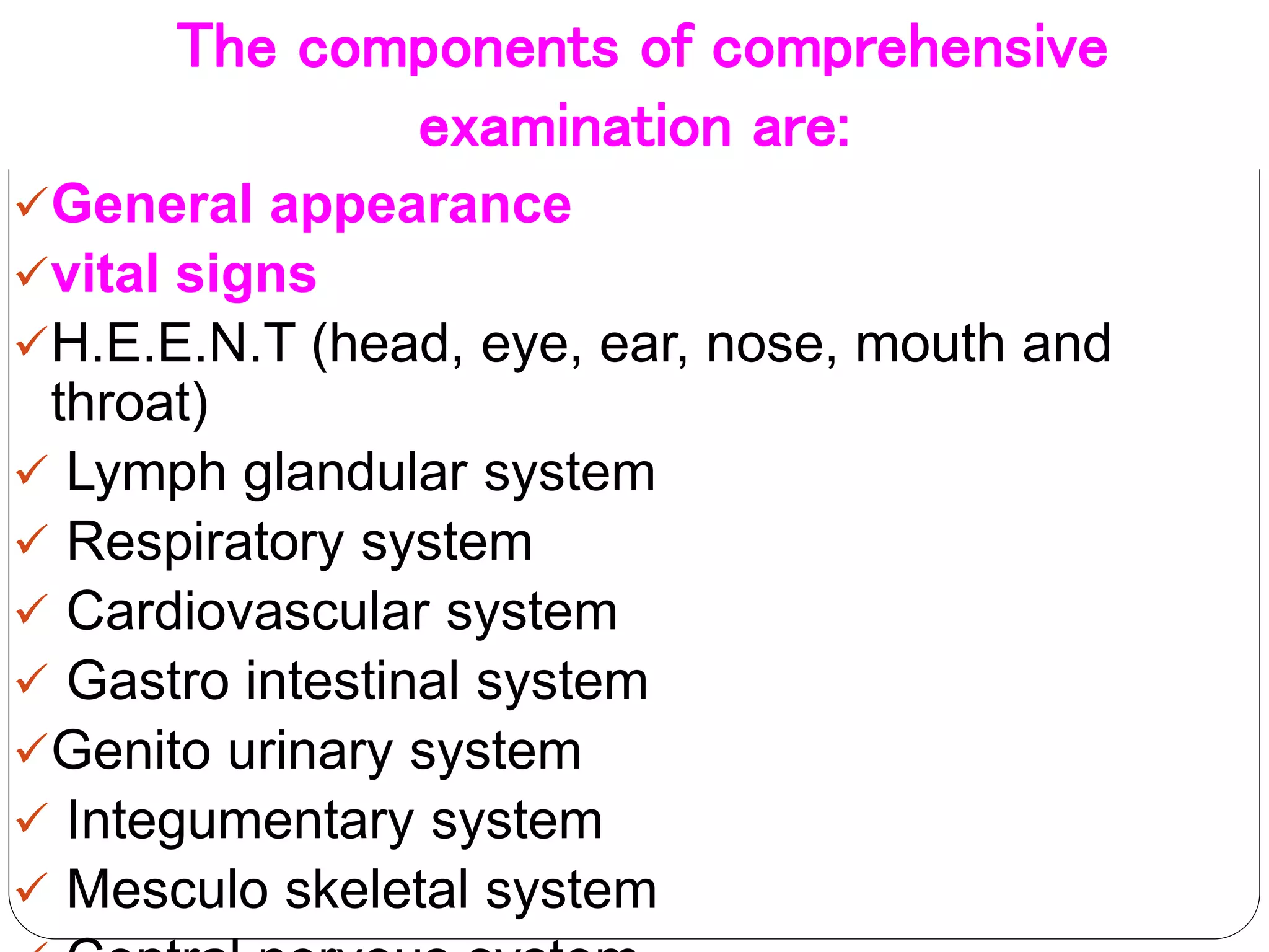
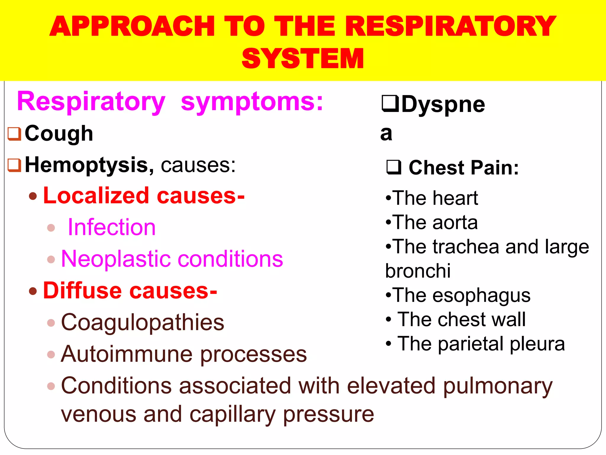
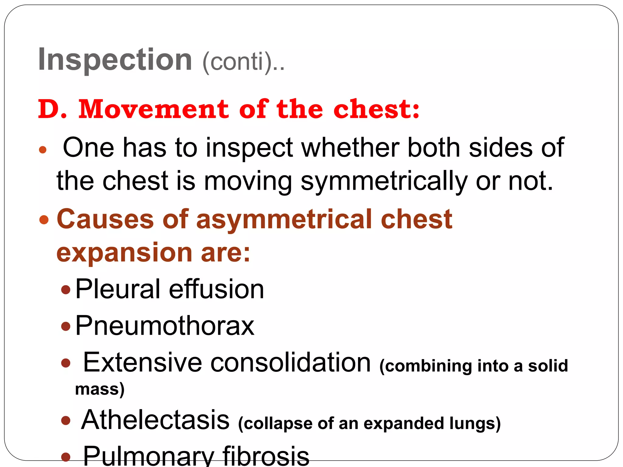
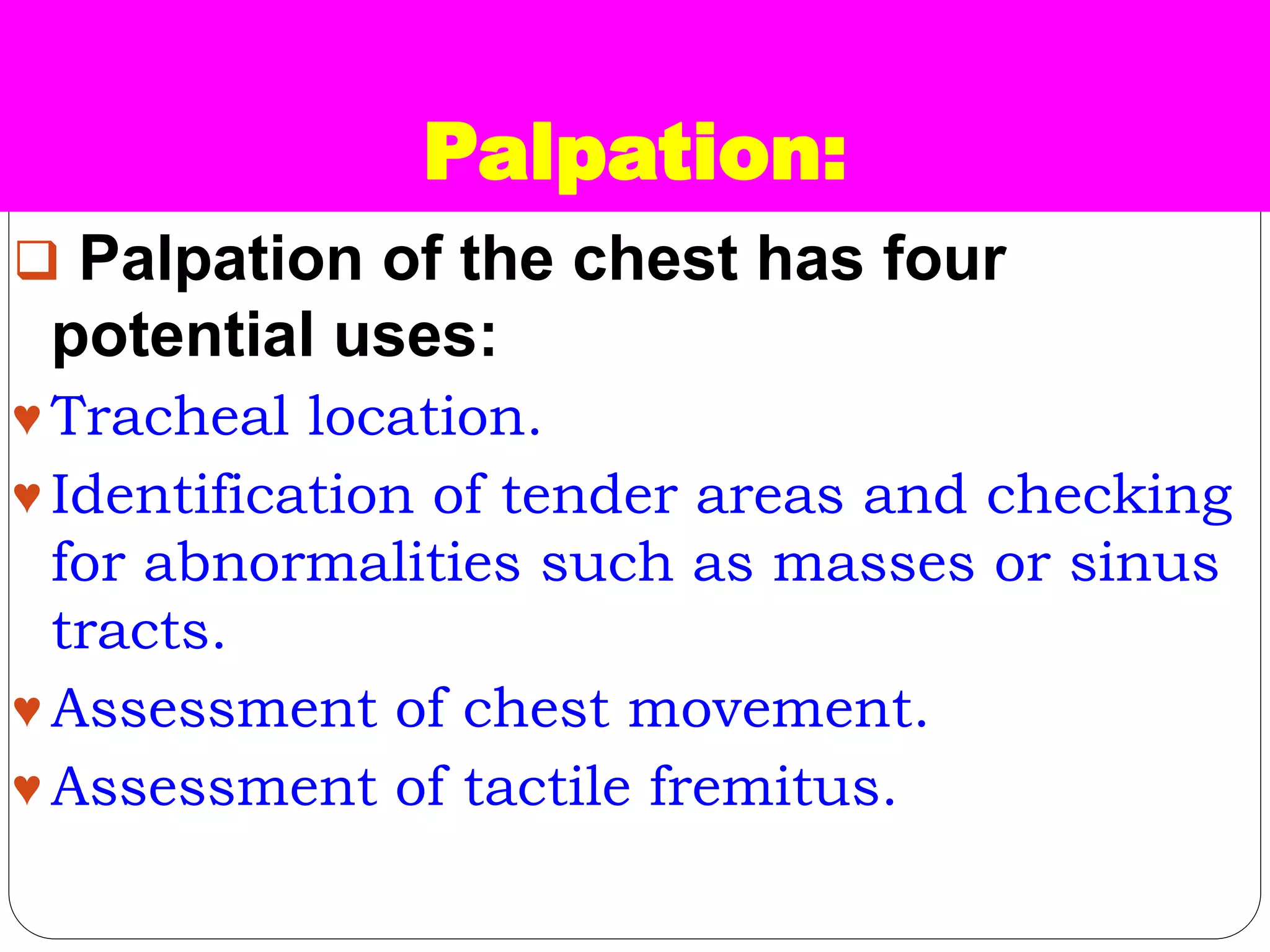
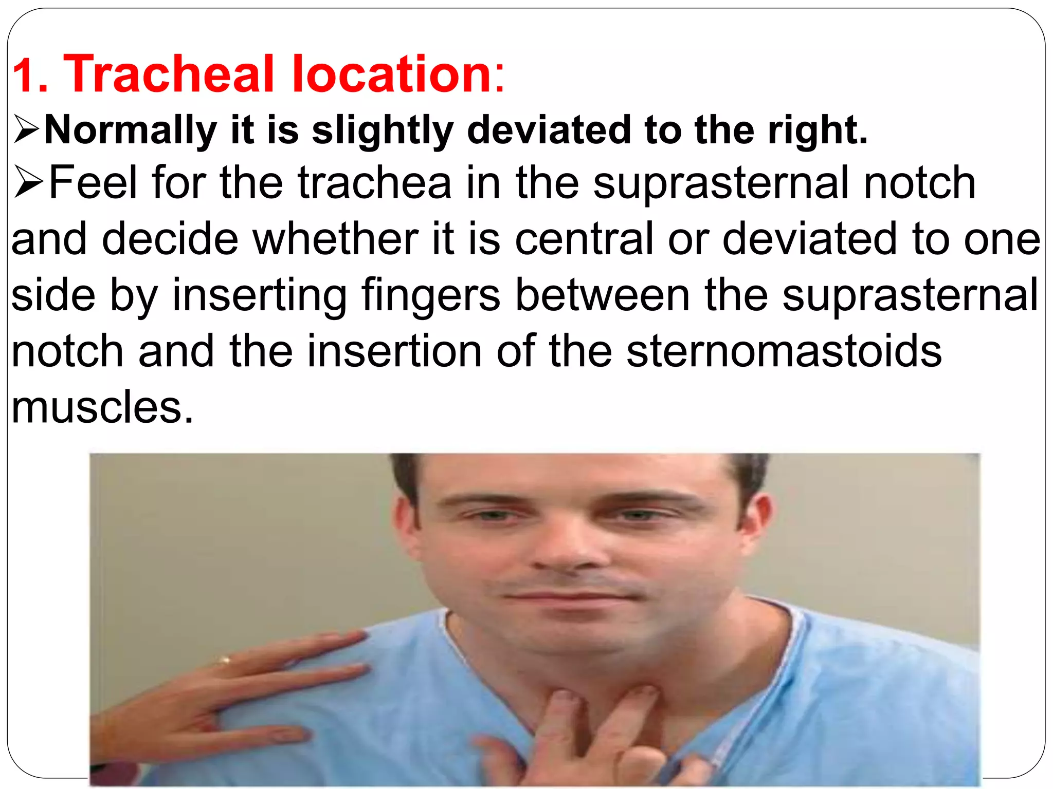
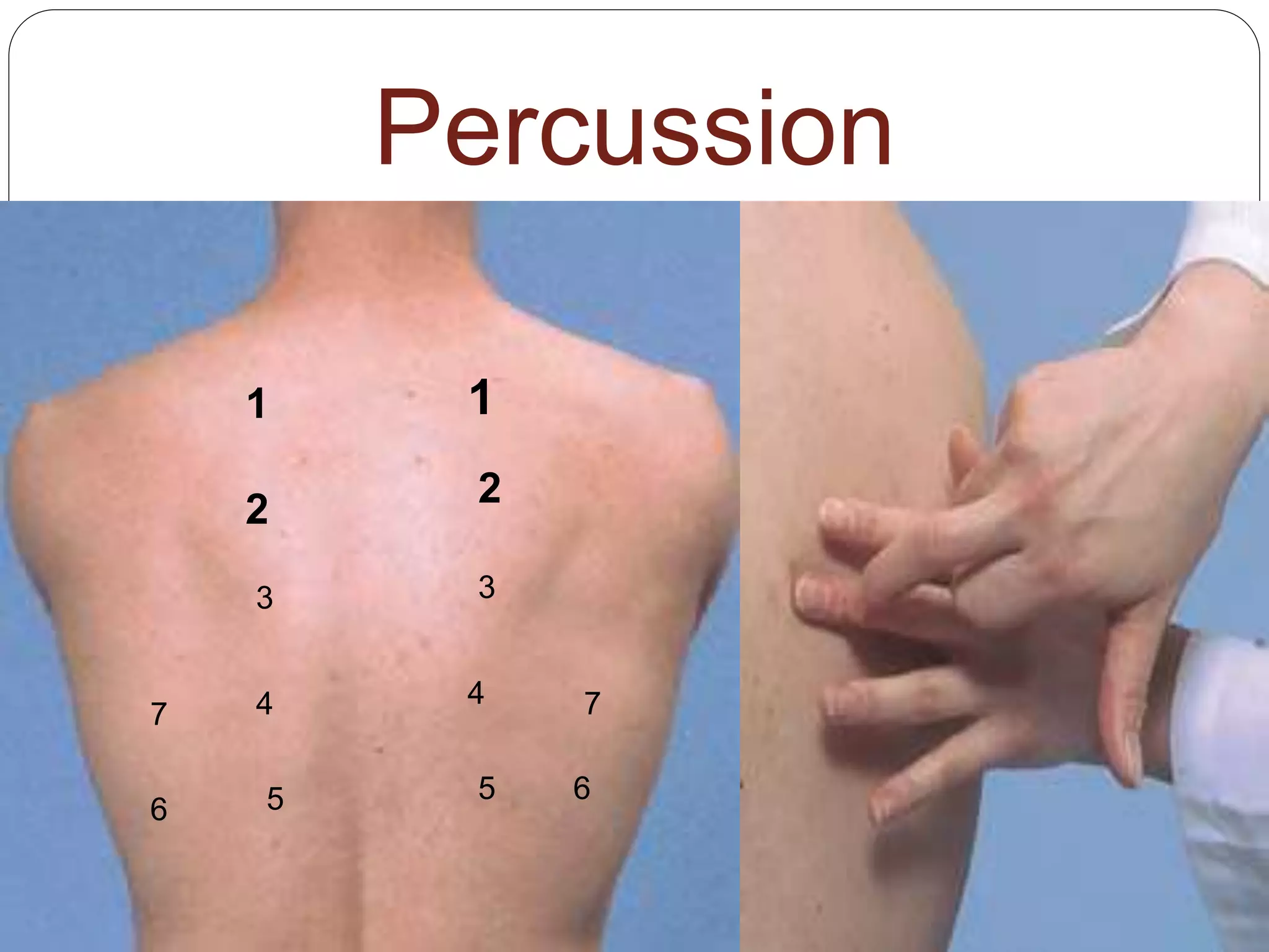
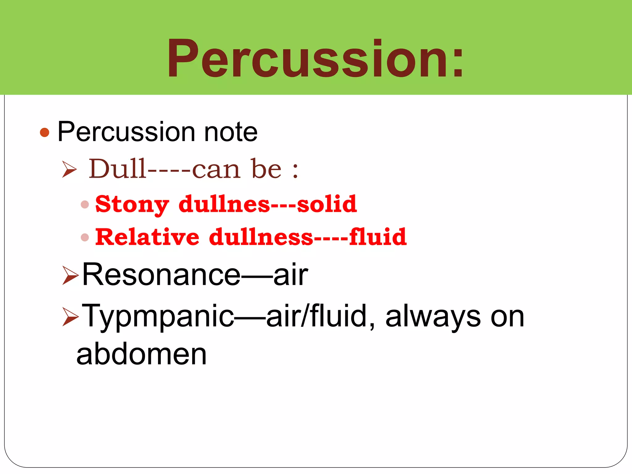
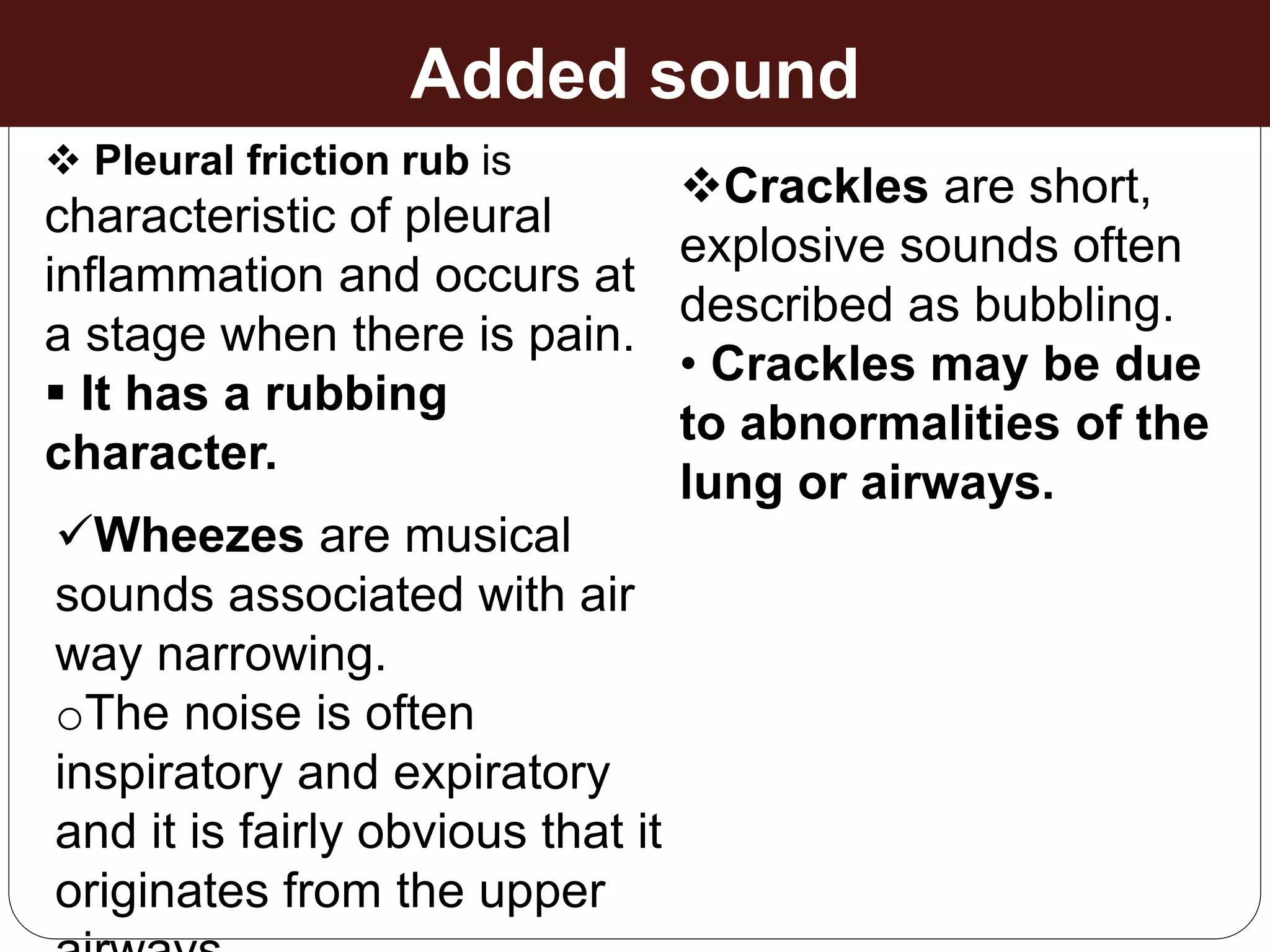
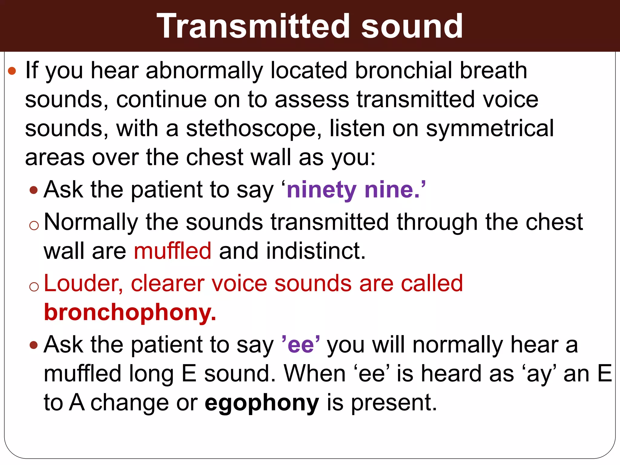
Dr. Adanwali Hassan Ahmed
Rn-Mw, Medical doctor (MD), Health Officer (HO)
Gyn/Obest
Physical Diagnosis
Physical examination
Overview of Physical
examination
 It is always advisable to follow the points
below while examining the patient:
Examination should take place with good
lighting and in a quite environment.
 It is advisable to examine a supine patient
from the patient’s right side
By words or gestures, be as clear as possible
in your instructions.
Cont---
If possible try to demonstrate the
patient what to do rather than giving
verbal instructions alone.
 Keep the patient informed as you
proceed with your examination.
While examining the patient, it is
helpful to move “from head to toe.”
The basic steps of P/E are:
 Inspection
 Palpation
 Percussion
 Auscultation
The components of comprehensive
examination are:
General appearance
vital signs
H.E.E.N.T (head, eye, ear, nose, mouth and
throat)
 Lymph glandular system
 Respiratory system
 Cardiovascular system
 Gastro intestinal system
Genito urinary system
 Integumentary system
 Mesculo skeletal system
cont---
1. General appearance:
Is the patient acutely sick,
Chronically sick looking or not sick looking at
all?
 Is the patient in cardio respiratory distress
or not?
2. Vital signs:
Pulse (rate, volume, character, radio femoral
delay)
Blood pressure (specify arm and the position it
was taken)
Respiratory rate
Temperature (Specify the location it was taken)
APPROACH TO THE RESPIRATORY
SYSTEM
Cough
Hemoptysis, causes:
 Localized causes-
 Infection
 Neoplastic conditions
 Diffuse causes-
 Coagulopathies
 Autoimmune processes
 Conditions associated with elevated pulmonary
venous and capillary pressure
Respiratory symptoms: Dyspne
a
 Chest Pain:
•The heart
•The aorta
•The trachea and large
bronchi
•The esophagus
• The chest wall
• The parietal pleura
General approach
 Arrange the patient’s gown so that you can see
the chest fully, but cover a woman’s anterior
chest while you are examining the back.
 Ideally the patient should be comfortably resting
on a bed sitting at angle of 450 and supported
by pillows.
• Proceed in an orderly fashion: inspection,
palpation, percussion, and auscultation.
 Compare one side of the chest with the other.
With this side-to side comparison, a patient
serves as his or her own control.
Conti..
 Examine the posterior thorax and lungs while
the patient is still in the sitting position.
 The patient’s arms should be folded across the
chest with hands resting, if possible, on the
opposite shoulders. This position moves the
scapulae partly out of the way and increases
your access to the lung fields.
• Then ask the patient to lie down so that you can
examine the anterior thorax and lungs.
SURVEY OF THORAX
General Assessment
• Observation of the patient’s physique and
form.
• Listening to the voice of the patient for any
hoarseness and change of voice.
• Observation of the hands for pallor,
cyanosis and clubbing.
• Inspecting the lips and tongue for central
cyanosis.
• Listening to the patient’s breathing if there
are additional sounds like wheezes or
strider.
Inspection
This part of physical examination includes:
Looking for signs of respiratory distress.
The respiratory rate and rhythm.
Observing for the shape of the chest.
Observing for the movement of the chest.
A. Signs of respiratory distress:
• Flaring of ala-nasae
• Intercostal retraction
• Subcostal retraction
• The use of accessory muscles in
the neck
B. Shape of the chest:
 The normal chest is bilaterally
symmetrical and elliptical in cross-
section.
The most common chest deformities
are:
• Kyphosis: posterior curvature of the spine.
• Lordosis: anterior curvature of the spine.
• Scoliosis: lateral curvature of the spine
Cont--
. Gibbus: a posterior angular or wedge
shaped deformity of the spine. It is caused
by fracture or spinal tuberculosis (Pott’s
disease).
• Flattening of the chest: Example:
Pulmonary fibrosis
• Barrel-shaped chest:
• Pigeon chest: This is seen in bronchial
asthma and chronic obstructive lung
diseases.
C. Respiratory rate and rhythm:
 The normal rate of respiration in a relaxed
adult is about 14-16 breaths per minute.
Tachypnea is an increased respiratory rate
observed by the clinician.
Bradypnea is decreases respiratory rate
Chyne-Stokes breathing is a deep and fast
breathing followed by diminishing respiratory
effort and rate, sometimes associated with a
short apnea.
Inspection (conti)..
D. Movement of the chest:
 One has to inspect whether both sides of
the chest is moving symmetrically or not.
 Causes of asymmetrical chest
expansion are:
Pleural effusion
Pneumothorax
 Extensive consolidation (combining into a solid
mass)
 Athelectasis (collapse of an expanded lungs)
 Pulmonary fibrosis
Palpation:
 Palpation of the chest has four
potential uses:
♥ Tracheal location.
♥ Identification of tender areas and checking
for abnormalities such as masses or sinus
tracts.
♥ Assessment of chest movement.
♥ Assessment of tactile fremitus.
1. Tracheal location:
Normally it is slightly deviated to the right.
Feel for the trachea in the suprasternal notch
and decide whether it is central or deviated to one
side by inserting fingers between the suprasternal
notch and the insertion of the sternomastoids
muscles.
2. Assessment of chest
expansion:
Manually: by placing the
finger tips of both hands on
either side of the lower rib
cage, so that the tip of the
thumbs meet in the midline
in front of but not touching
the chest, then a deep
breath by the patient will
increase the distance
between the thumbs and
indicates the degree of
expansion.
If one thumb remains closer
3. Assessment of tactile
fremitus:
Ask the patient to repeat the word
’ninety–nine’ compare both sides
of the chest symmetrically.
o If fremitus is faint, ask the patient
to speak more loudly or in a deep
voice.
 Fremitus is decreased /
absent:
 When the voice is soft
 When transmission of
vibrations from the larynx to the
surface of the chest is impeded.
•The cause can be an obstructed
bronchus, pleural effusion,
pulmonary fibrosis,
Fremitus is increased when
the transmission of sound
is increased, as through
the consolidated lung of
lobar pneumonia.
Percussion
1
1
2
2
3
3
4
4
5
5 6
6
7
7
Percussion:
 Percussion note
 Dull----can be :
 Stony dullnes---solid
 Relative dullness----fluid
Resonance—air
Typmpanic—air/fluid, always on
abdomen
Percussion
 Percussion is a useful technique and without an
X-ray can be used to distinguish reliably between
the presence of pneumothorax and pleural
effusion.
 It helps to determine whether the underlying
tissues are air filled, fluid filled or solid.
 One should systematically compare the
percussion note on two equivalent sides of the
chest.
Cont---
 It is usually sufficient to percuss three
or four areas anteriorly, three or four
areas on the back, and two in the
axillae.
 The findings on both sides of the chest
should be compared symmetrically.
 In a healthy individual, a resonant noise
is produced upon percussion of the
chest.
 Resonant sound is of low pitch and
clear in character.
 Dullness is an abnormal on chest
percussion.
 It can be stony dullness or relative dullness.
Causes of dullness to
percussion are:
 Tumors
 Lobar pneumonia
 hydrothorax
 Hemothorax
 Empyema
 Fibrosis
 Athelectasis
 Thick chest wall
Causes of hyper resonance
are:
Bilateral causes:
􀂃 Emphysema
􀂃 Bronchial asthma
Unilateral causes:
􀂃 Pneumothorax
􀂃 Large air filled bulla in the
lungs.
Auscultation:
 Auscultation involves:
Listening to the sounds generated by
breathing.
 Listening for any adventitious (added)
sounds.
 Listening to the patient’s spoken or
whispered voice as they are transmitted
through the chest wall.
Breath sounds
Breath sounds have both intensity and
quality.
 The intensity (loudness) of the breath sound may
be categorized as normal, reduced or increased
 The quality of breath sounds have been classified
in to three categories:
a) Vesicular
b) Bronchial
c) Bronchovesicular
Cont
 Vesicular:
 These are soft and low pitched sounds.
 They are heard through out inspiration, continue
without pause in to expiration, and then fade
away about one third of the way through
expiration.
 Vesicular breath sounds are the normal
breath sounds.
 Bronchial:
 These are louder and higher in pitch.
 There is a short silent period between
the inspiratory and expiratory sounds,
and the expiratory sound lasts longer
than the inspiratory one.
 Classically they are heard over an area
of consolidated lung in cases of
pneumonia.
 Bronchovesicular:
 These are intermediate.
 Inspiratory and expiratory sounds are
about equal in length, and a silent period
between them may or may not be
present.
 These are often heard in the first and
second interspaces anteriorly and
between the scapulae.
Added sound
 Pleural friction rub is
characteristic of pleural
inflammation and occurs at
a stage when there is pain.
 It has a rubbing
character.
Wheezes are musical
sounds associated with air
way narrowing.
oThe noise is often
inspiratory and expiratory
and it is fairly obvious that it
originates from the upper
Crackles are short,
explosive sounds often
described as bubbling.
• Crackles may be due
to abnormalities of the
lung or airways.
Transmitted sound
 If you hear abnormally located bronchial breath
sounds, continue on to assess transmitted voice
sounds, with a stethoscope, listen on symmetrical
areas over the chest wall as you:
 Ask the patient to say ‘ninety nine.’
o Normally the sounds transmitted through the chest
wall are muffled and indistinct.
o Louder, clearer voice sounds are called
bronchophony.
 Ask the patient to say ’ee’ you will normally hear a
muffled long E sound. When ‘ee’ is heard as ‘ay’ an E
to A change or egophony is present.
oThis can be appreciated over the area of
consolidation or above the level of pleural
effusion.
 Ask the patient to whisper ‘ninety nine’ or
‘one-two- three’ the whispered voice is
normally heard faintly and indistinctly, if at all.
oLouder, clearer whispered sounds are called
whispered pectoriloquy.
oThis can be appreciated over an area of
consolidation.
END

RESPIRATORY EXAMINATION.pptx

  • 1.
    Dr. Adanwali HassanAhmed Rn-Mw, Medical doctor (MD), Health Officer (HO) Gyn/Obest Physical Diagnosis Physical examination
  • 2.
    Overview of Physical examination It is always advisable to follow the points below while examining the patient: Examination should take place with good lighting and in a quite environment.  It is advisable to examine a supine patient from the patient’s right side By words or gestures, be as clear as possible in your instructions.
  • 3.
    Cont--- If possible tryto demonstrate the patient what to do rather than giving verbal instructions alone.  Keep the patient informed as you proceed with your examination. While examining the patient, it is helpful to move “from head to toe.”
  • 4.
    The basic stepsof P/E are:  Inspection  Palpation  Percussion  Auscultation
  • 5.
    The components ofcomprehensive examination are: General appearance vital signs H.E.E.N.T (head, eye, ear, nose, mouth and throat)  Lymph glandular system  Respiratory system  Cardiovascular system  Gastro intestinal system Genito urinary system  Integumentary system  Mesculo skeletal system
  • 6.
    cont--- 1. General appearance: Isthe patient acutely sick, Chronically sick looking or not sick looking at all?  Is the patient in cardio respiratory distress or not? 2. Vital signs: Pulse (rate, volume, character, radio femoral delay) Blood pressure (specify arm and the position it was taken) Respiratory rate Temperature (Specify the location it was taken)
  • 7.
    APPROACH TO THERESPIRATORY SYSTEM Cough Hemoptysis, causes:  Localized causes-  Infection  Neoplastic conditions  Diffuse causes-  Coagulopathies  Autoimmune processes  Conditions associated with elevated pulmonary venous and capillary pressure Respiratory symptoms: Dyspne a  Chest Pain: •The heart •The aorta •The trachea and large bronchi •The esophagus • The chest wall • The parietal pleura
  • 8.
    General approach  Arrangethe patient’s gown so that you can see the chest fully, but cover a woman’s anterior chest while you are examining the back.  Ideally the patient should be comfortably resting on a bed sitting at angle of 450 and supported by pillows. • Proceed in an orderly fashion: inspection, palpation, percussion, and auscultation.  Compare one side of the chest with the other. With this side-to side comparison, a patient serves as his or her own control.
  • 9.
    Conti..  Examine theposterior thorax and lungs while the patient is still in the sitting position.  The patient’s arms should be folded across the chest with hands resting, if possible, on the opposite shoulders. This position moves the scapulae partly out of the way and increases your access to the lung fields. • Then ask the patient to lie down so that you can examine the anterior thorax and lungs.
  • 10.
  • 12.
    General Assessment • Observationof the patient’s physique and form. • Listening to the voice of the patient for any hoarseness and change of voice. • Observation of the hands for pallor, cyanosis and clubbing. • Inspecting the lips and tongue for central cyanosis. • Listening to the patient’s breathing if there are additional sounds like wheezes or strider.
  • 14.
    Inspection This part ofphysical examination includes: Looking for signs of respiratory distress. The respiratory rate and rhythm. Observing for the shape of the chest. Observing for the movement of the chest. A. Signs of respiratory distress: • Flaring of ala-nasae • Intercostal retraction • Subcostal retraction • The use of accessory muscles in the neck
  • 15.
    B. Shape ofthe chest:  The normal chest is bilaterally symmetrical and elliptical in cross- section. The most common chest deformities are: • Kyphosis: posterior curvature of the spine. • Lordosis: anterior curvature of the spine. • Scoliosis: lateral curvature of the spine
  • 16.
    Cont-- . Gibbus: aposterior angular or wedge shaped deformity of the spine. It is caused by fracture or spinal tuberculosis (Pott’s disease). • Flattening of the chest: Example: Pulmonary fibrosis • Barrel-shaped chest: • Pigeon chest: This is seen in bronchial asthma and chronic obstructive lung diseases.
  • 17.
    C. Respiratory rateand rhythm:  The normal rate of respiration in a relaxed adult is about 14-16 breaths per minute. Tachypnea is an increased respiratory rate observed by the clinician. Bradypnea is decreases respiratory rate Chyne-Stokes breathing is a deep and fast breathing followed by diminishing respiratory effort and rate, sometimes associated with a short apnea.
  • 20.
    Inspection (conti).. D. Movementof the chest:  One has to inspect whether both sides of the chest is moving symmetrically or not.  Causes of asymmetrical chest expansion are: Pleural effusion Pneumothorax  Extensive consolidation (combining into a solid mass)  Athelectasis (collapse of an expanded lungs)  Pulmonary fibrosis
  • 21.
    Palpation:  Palpation ofthe chest has four potential uses: ♥ Tracheal location. ♥ Identification of tender areas and checking for abnormalities such as masses or sinus tracts. ♥ Assessment of chest movement. ♥ Assessment of tactile fremitus.
  • 22.
    1. Tracheal location: Normallyit is slightly deviated to the right. Feel for the trachea in the suprasternal notch and decide whether it is central or deviated to one side by inserting fingers between the suprasternal notch and the insertion of the sternomastoids muscles.
  • 23.
    2. Assessment ofchest expansion: Manually: by placing the finger tips of both hands on either side of the lower rib cage, so that the tip of the thumbs meet in the midline in front of but not touching the chest, then a deep breath by the patient will increase the distance between the thumbs and indicates the degree of expansion. If one thumb remains closer
  • 24.
    3. Assessment oftactile fremitus: Ask the patient to repeat the word ’ninety–nine’ compare both sides of the chest symmetrically. o If fremitus is faint, ask the patient to speak more loudly or in a deep voice.  Fremitus is decreased / absent:  When the voice is soft  When transmission of vibrations from the larynx to the surface of the chest is impeded. •The cause can be an obstructed bronchus, pleural effusion, pulmonary fibrosis, Fremitus is increased when the transmission of sound is increased, as through the consolidated lung of lobar pneumonia.
  • 25.
  • 26.
    Percussion:  Percussion note Dull----can be :  Stony dullnes---solid  Relative dullness----fluid Resonance—air Typmpanic—air/fluid, always on abdomen
  • 27.
    Percussion  Percussion isa useful technique and without an X-ray can be used to distinguish reliably between the presence of pneumothorax and pleural effusion.  It helps to determine whether the underlying tissues are air filled, fluid filled or solid.  One should systematically compare the percussion note on two equivalent sides of the chest.
  • 28.
    Cont---  It isusually sufficient to percuss three or four areas anteriorly, three or four areas on the back, and two in the axillae.  The findings on both sides of the chest should be compared symmetrically.  In a healthy individual, a resonant noise is produced upon percussion of the chest.  Resonant sound is of low pitch and clear in character.
  • 29.
     Dullness isan abnormal on chest percussion.  It can be stony dullness or relative dullness. Causes of dullness to percussion are:  Tumors  Lobar pneumonia  hydrothorax  Hemothorax  Empyema  Fibrosis  Athelectasis  Thick chest wall Causes of hyper resonance are: Bilateral causes: 􀂃 Emphysema 􀂃 Bronchial asthma Unilateral causes: 􀂃 Pneumothorax 􀂃 Large air filled bulla in the lungs.
  • 30.
    Auscultation:  Auscultation involves: Listeningto the sounds generated by breathing.  Listening for any adventitious (added) sounds.  Listening to the patient’s spoken or whispered voice as they are transmitted through the chest wall.
  • 31.
    Breath sounds Breath soundshave both intensity and quality.  The intensity (loudness) of the breath sound may be categorized as normal, reduced or increased  The quality of breath sounds have been classified in to three categories: a) Vesicular b) Bronchial c) Bronchovesicular
  • 32.
    Cont  Vesicular:  Theseare soft and low pitched sounds.  They are heard through out inspiration, continue without pause in to expiration, and then fade away about one third of the way through expiration.  Vesicular breath sounds are the normal breath sounds.
  • 33.
     Bronchial:  Theseare louder and higher in pitch.  There is a short silent period between the inspiratory and expiratory sounds, and the expiratory sound lasts longer than the inspiratory one.  Classically they are heard over an area of consolidated lung in cases of pneumonia.
  • 34.
     Bronchovesicular:  Theseare intermediate.  Inspiratory and expiratory sounds are about equal in length, and a silent period between them may or may not be present.  These are often heard in the first and second interspaces anteriorly and between the scapulae.
  • 35.
    Added sound  Pleuralfriction rub is characteristic of pleural inflammation and occurs at a stage when there is pain.  It has a rubbing character. Wheezes are musical sounds associated with air way narrowing. oThe noise is often inspiratory and expiratory and it is fairly obvious that it originates from the upper Crackles are short, explosive sounds often described as bubbling. • Crackles may be due to abnormalities of the lung or airways.
  • 36.
    Transmitted sound  Ifyou hear abnormally located bronchial breath sounds, continue on to assess transmitted voice sounds, with a stethoscope, listen on symmetrical areas over the chest wall as you:  Ask the patient to say ‘ninety nine.’ o Normally the sounds transmitted through the chest wall are muffled and indistinct. o Louder, clearer voice sounds are called bronchophony.  Ask the patient to say ’ee’ you will normally hear a muffled long E sound. When ‘ee’ is heard as ‘ay’ an E to A change or egophony is present.
  • 37.
    oThis can beappreciated over the area of consolidation or above the level of pleural effusion.  Ask the patient to whisper ‘ninety nine’ or ‘one-two- three’ the whispered voice is normally heard faintly and indistinctly, if at all. oLouder, clearer whispered sounds are called whispered pectoriloquy. oThis can be appreciated over an area of consolidation.
  • 38.