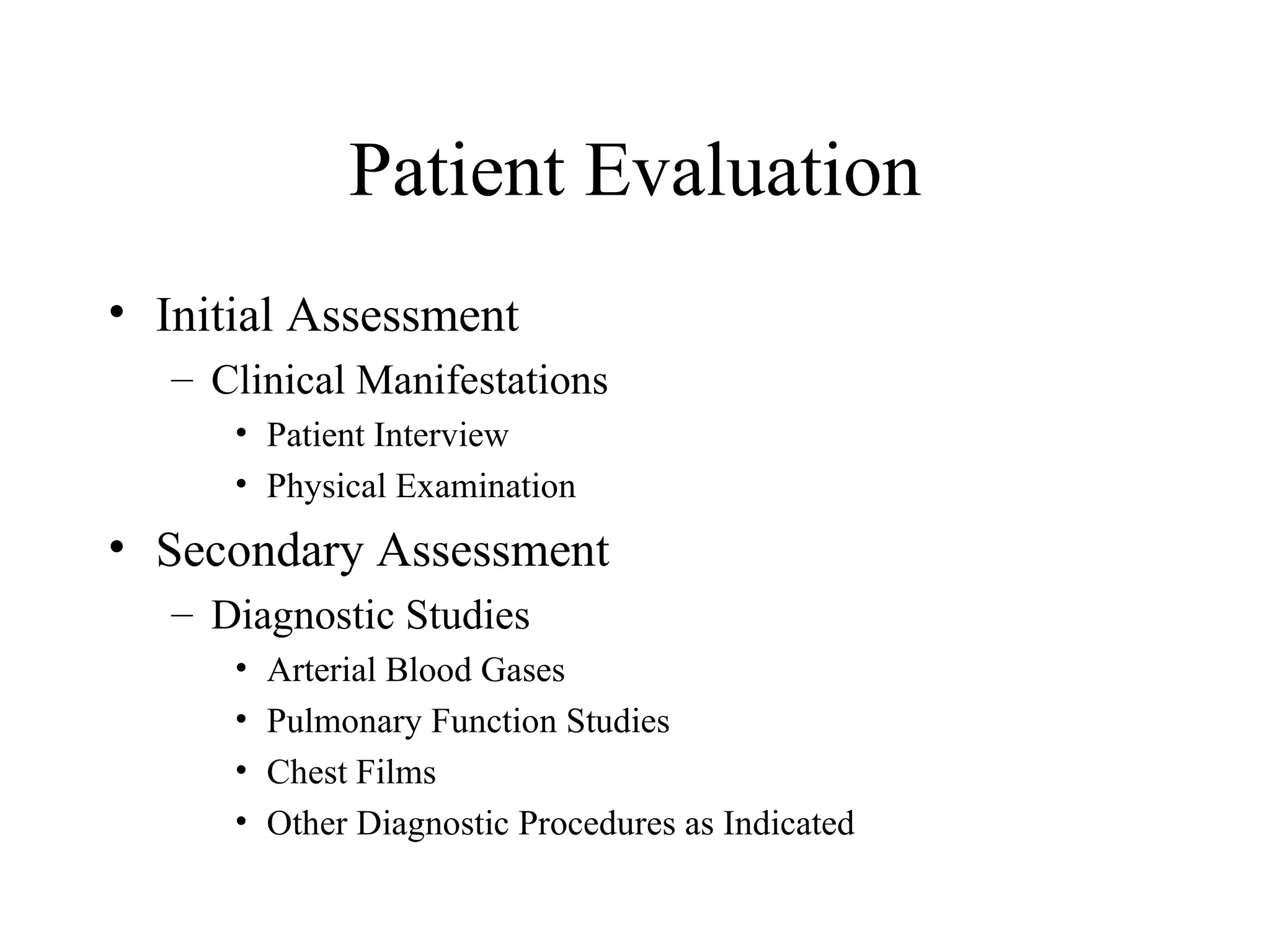
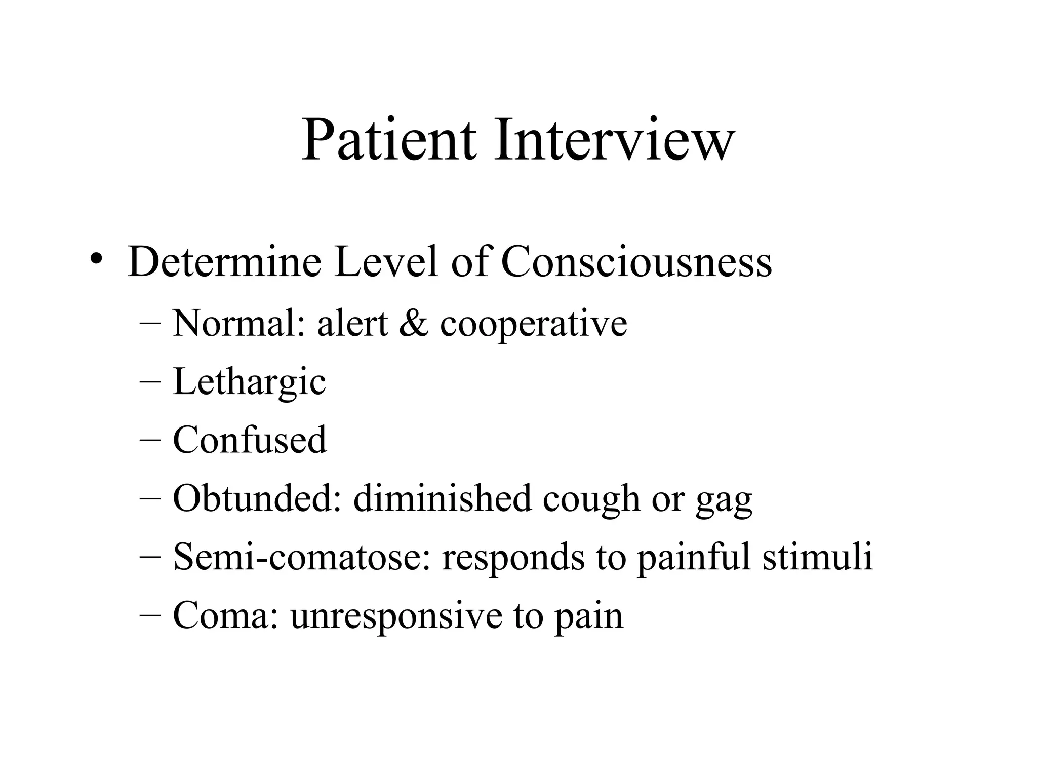
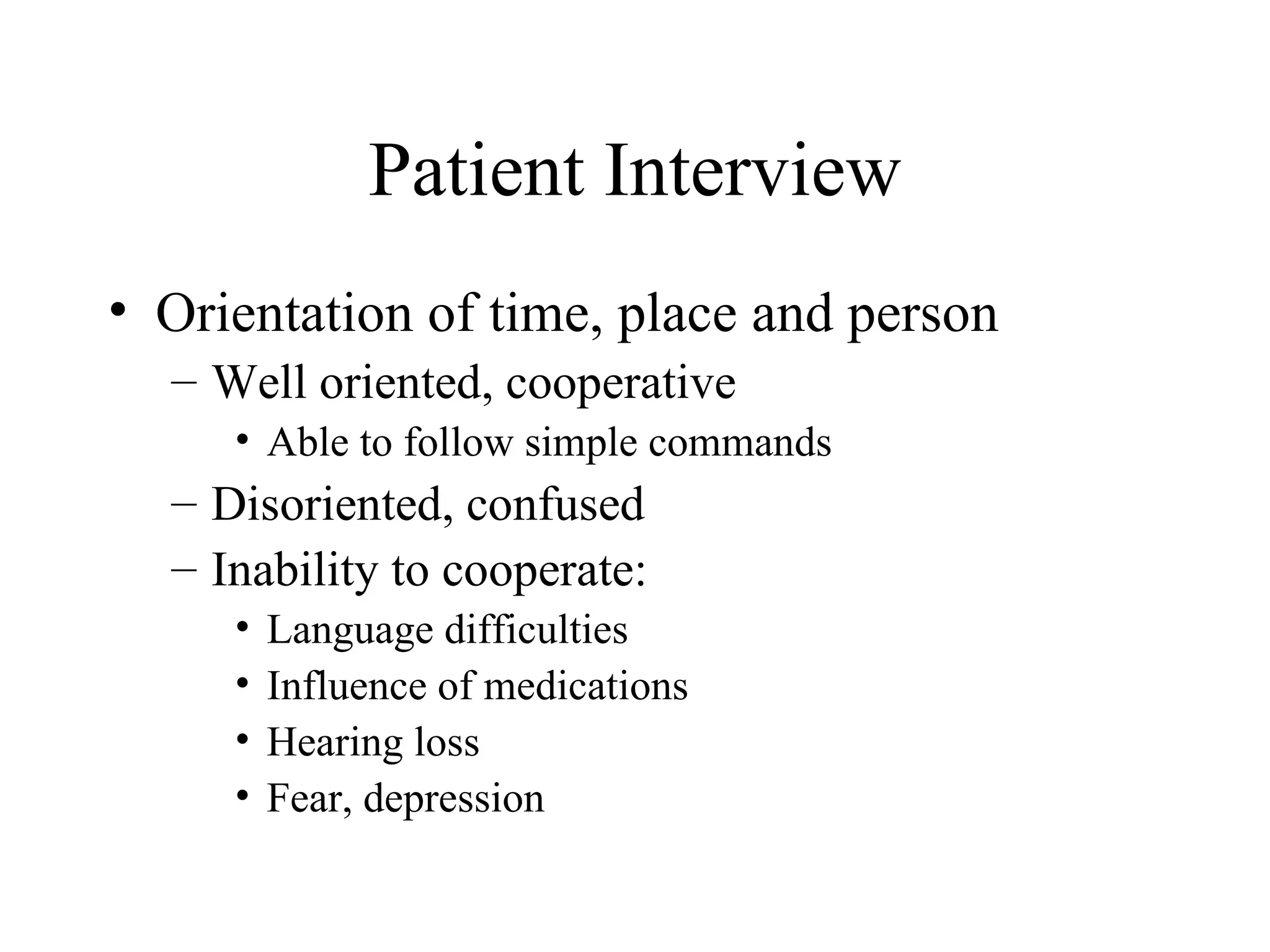
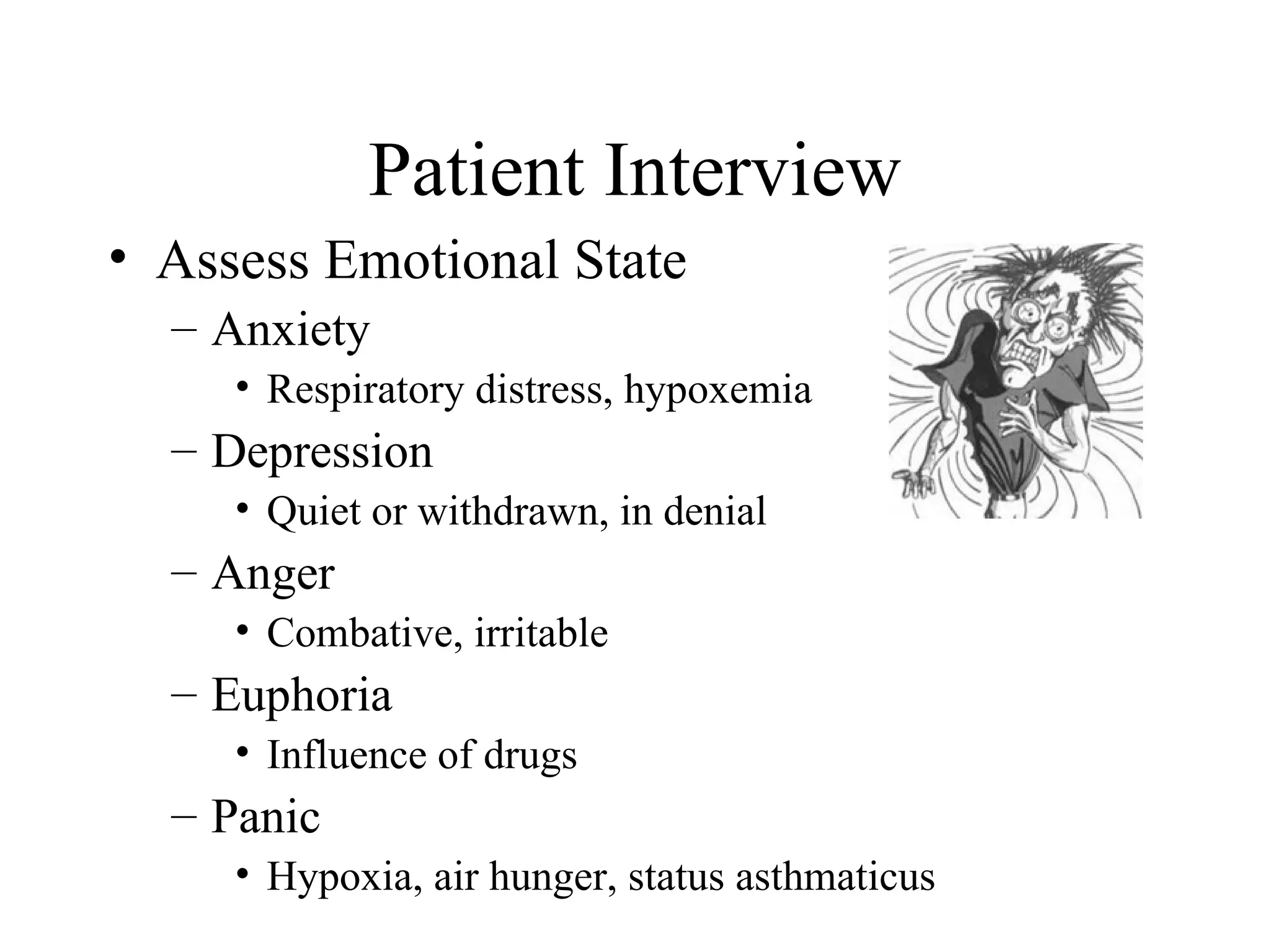
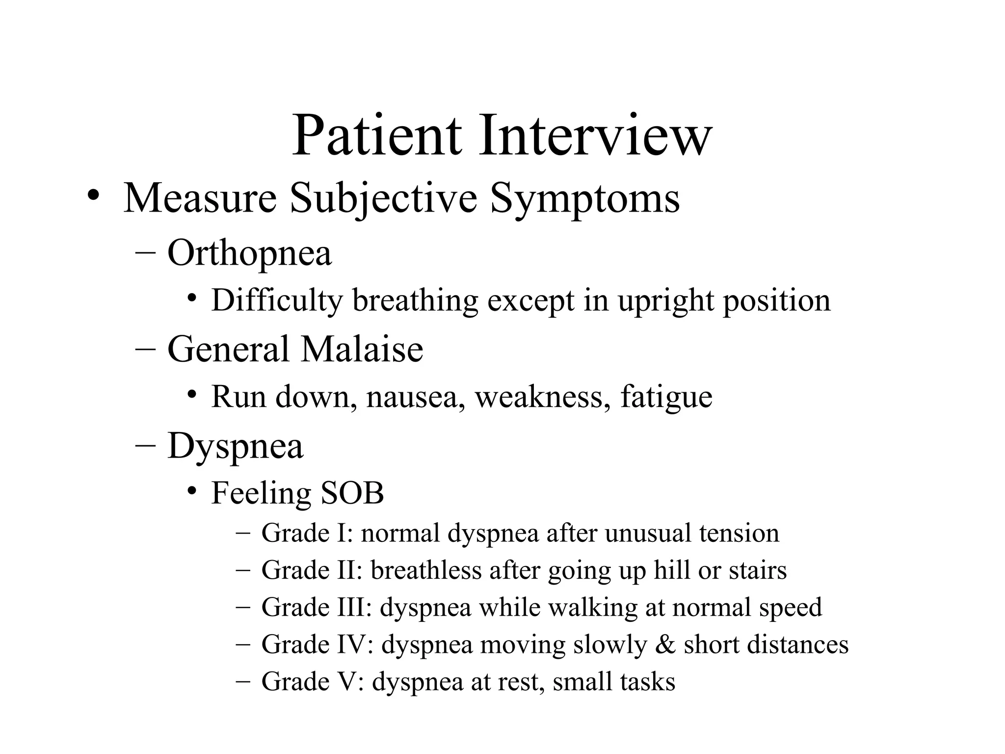
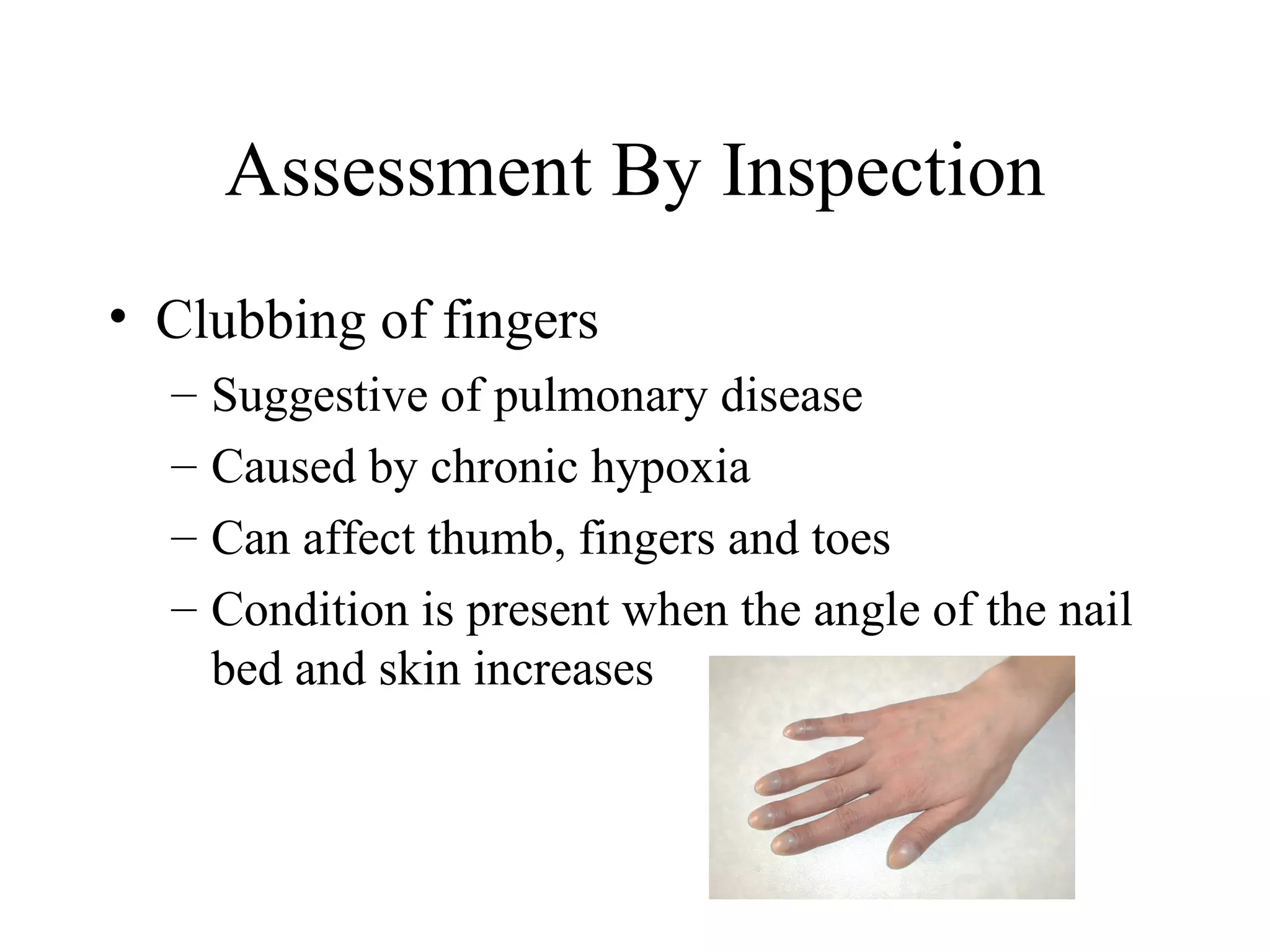
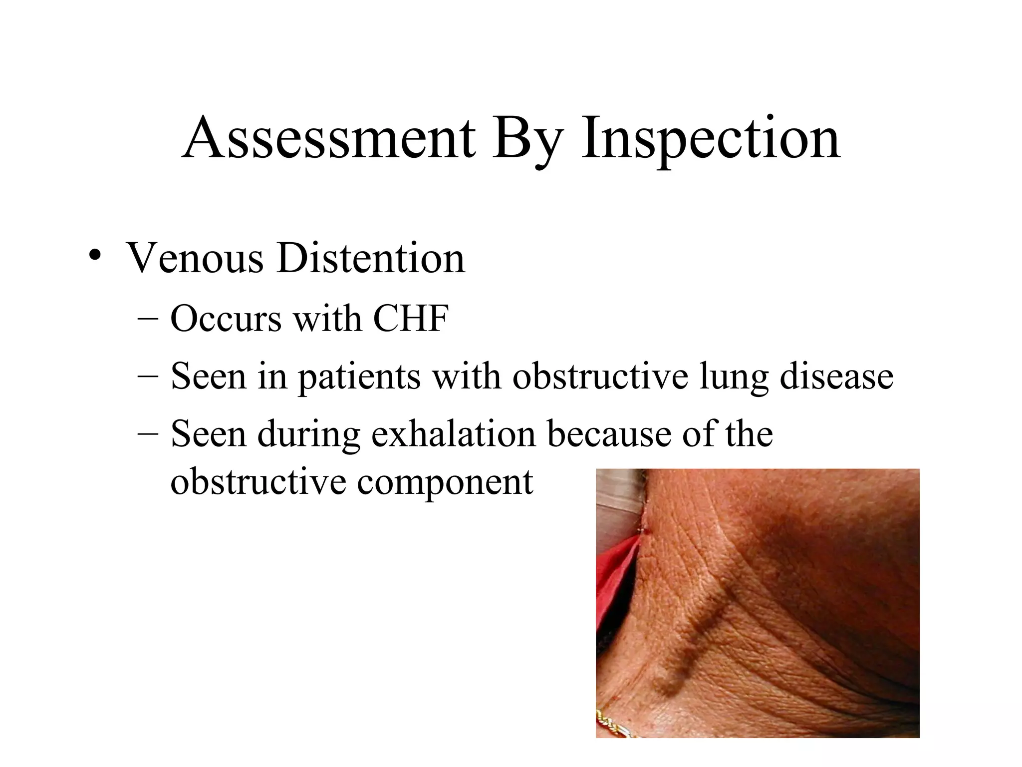
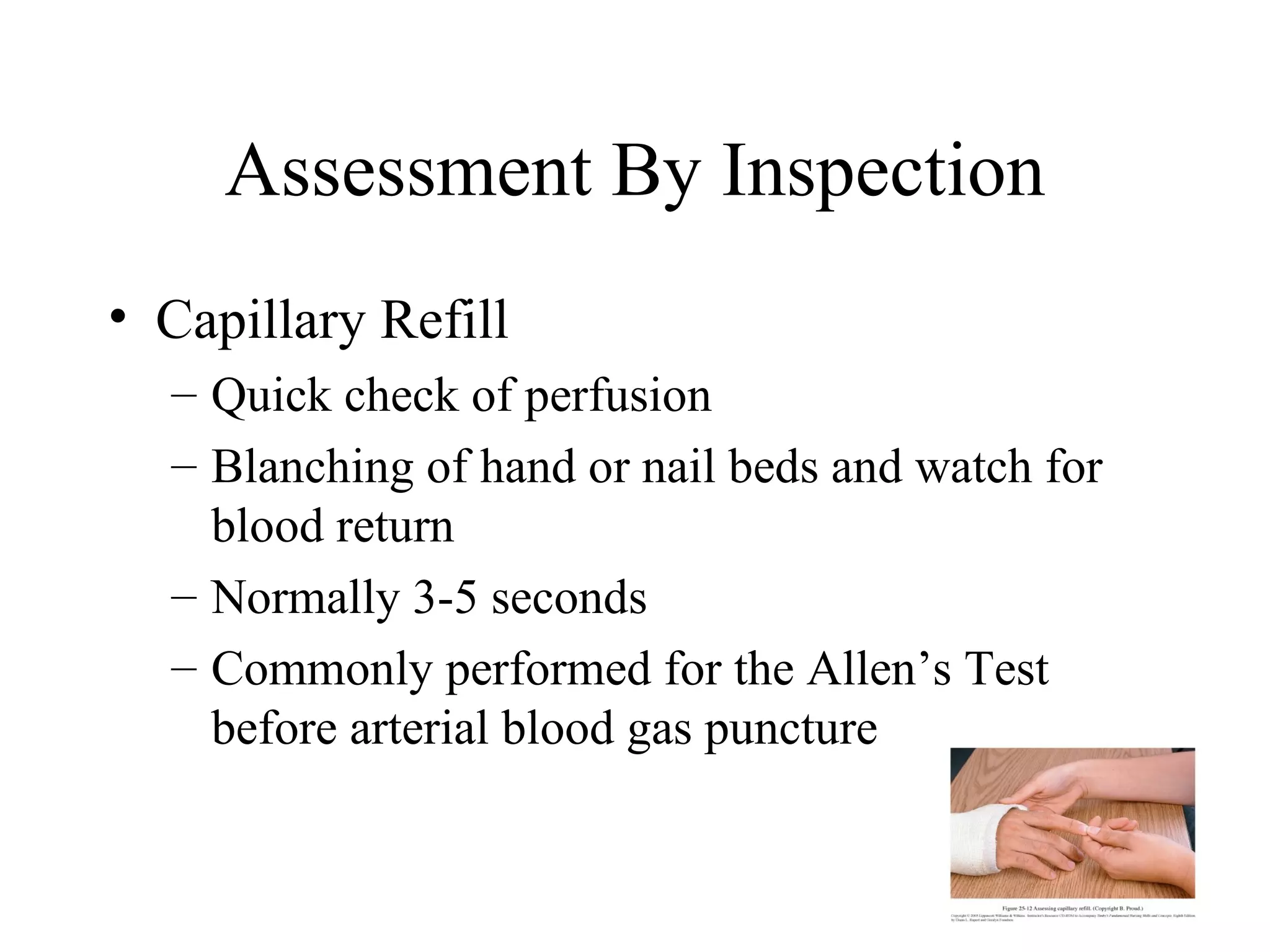
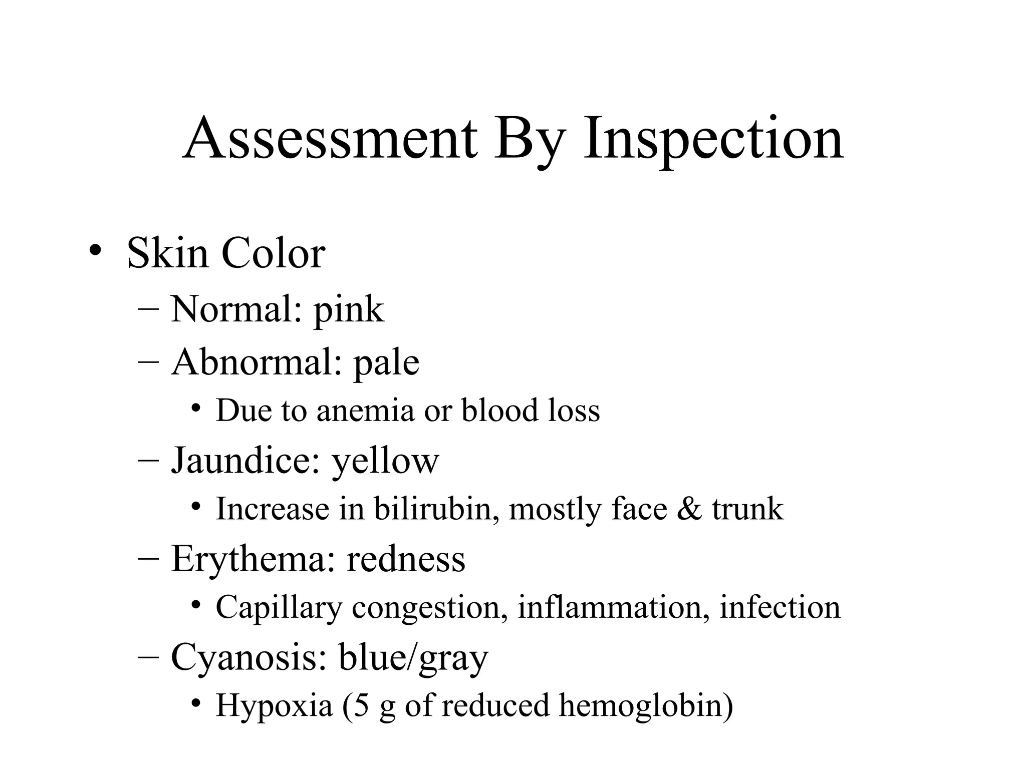
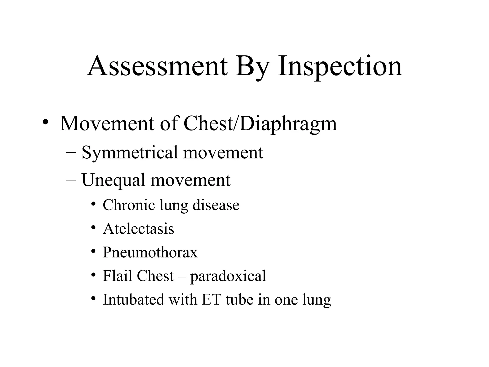
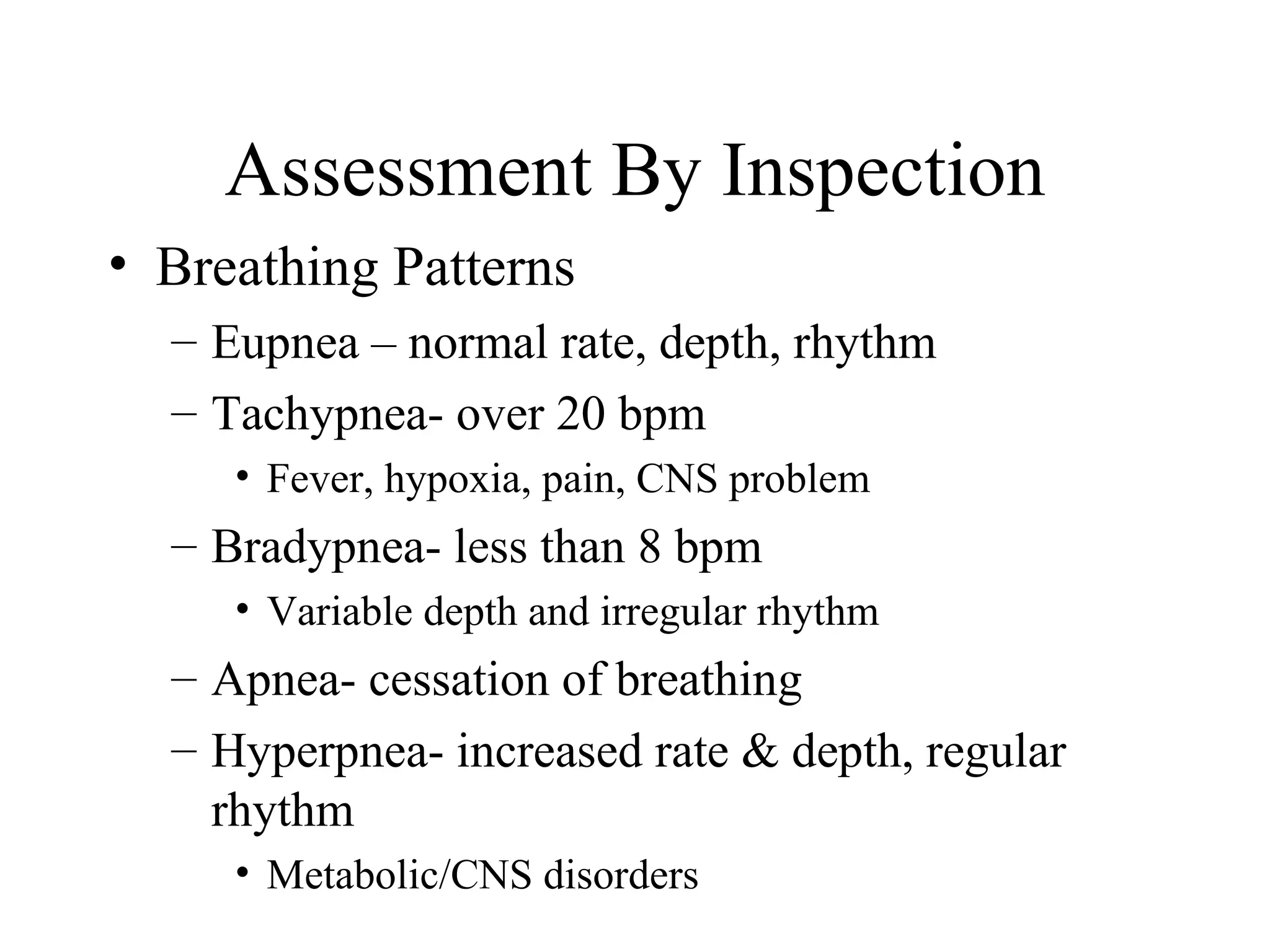
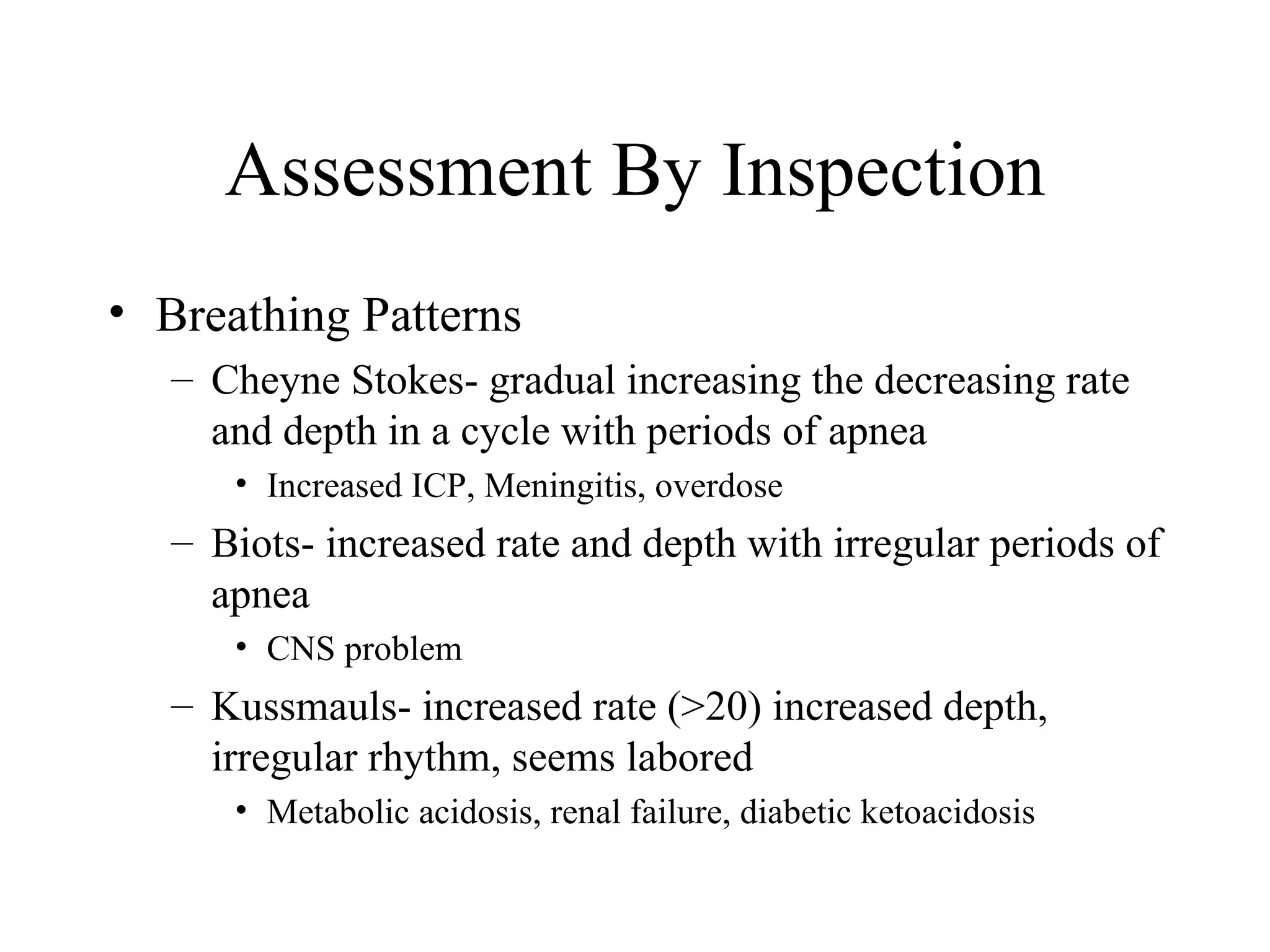
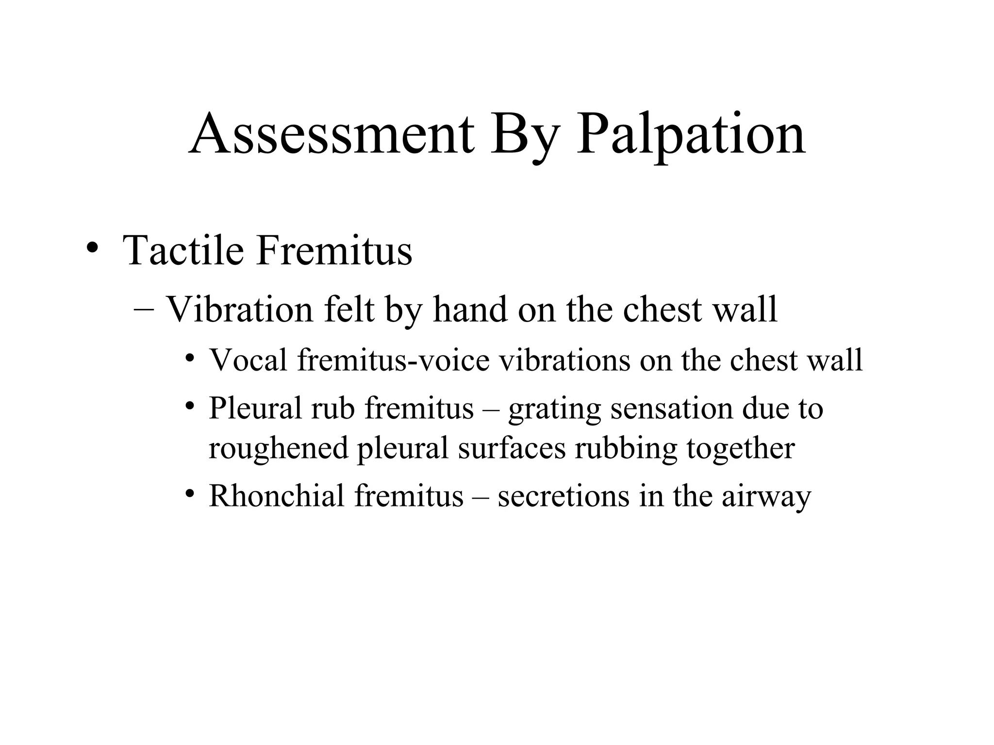
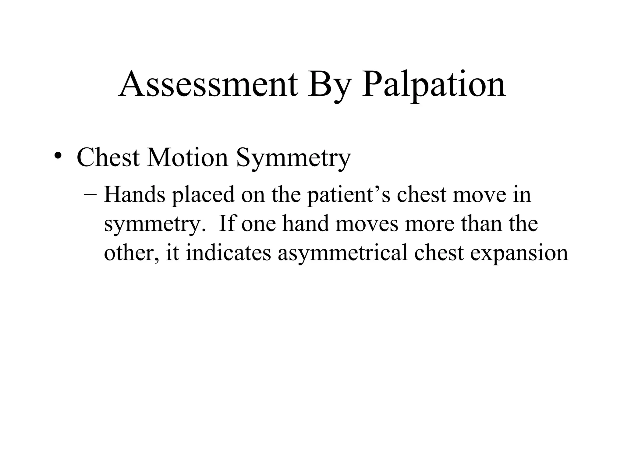
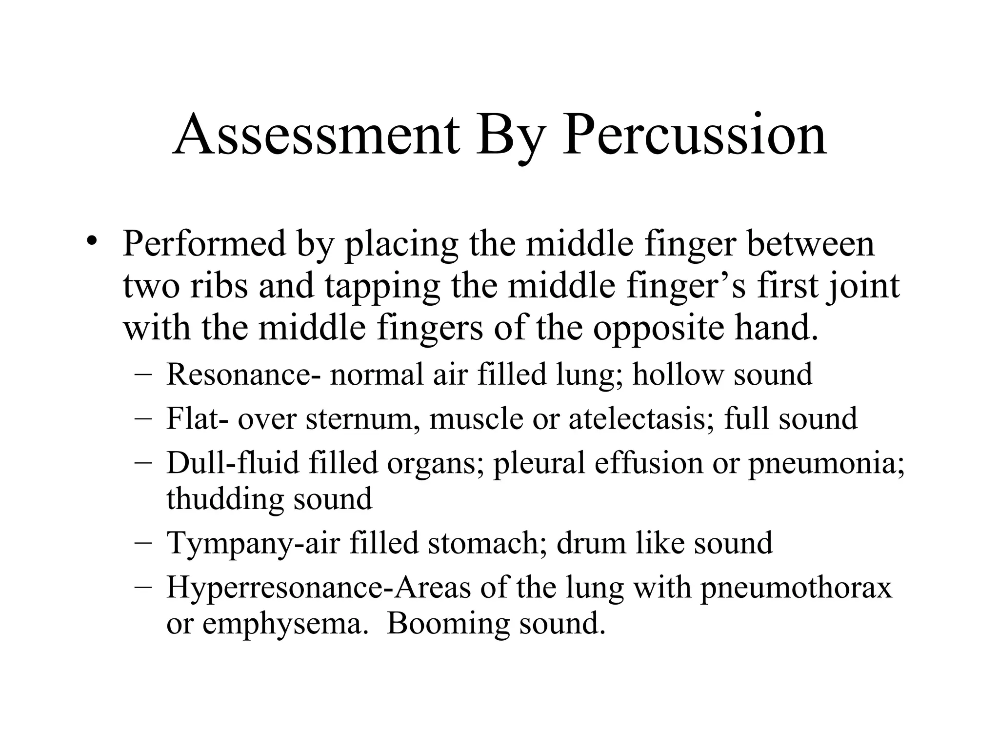
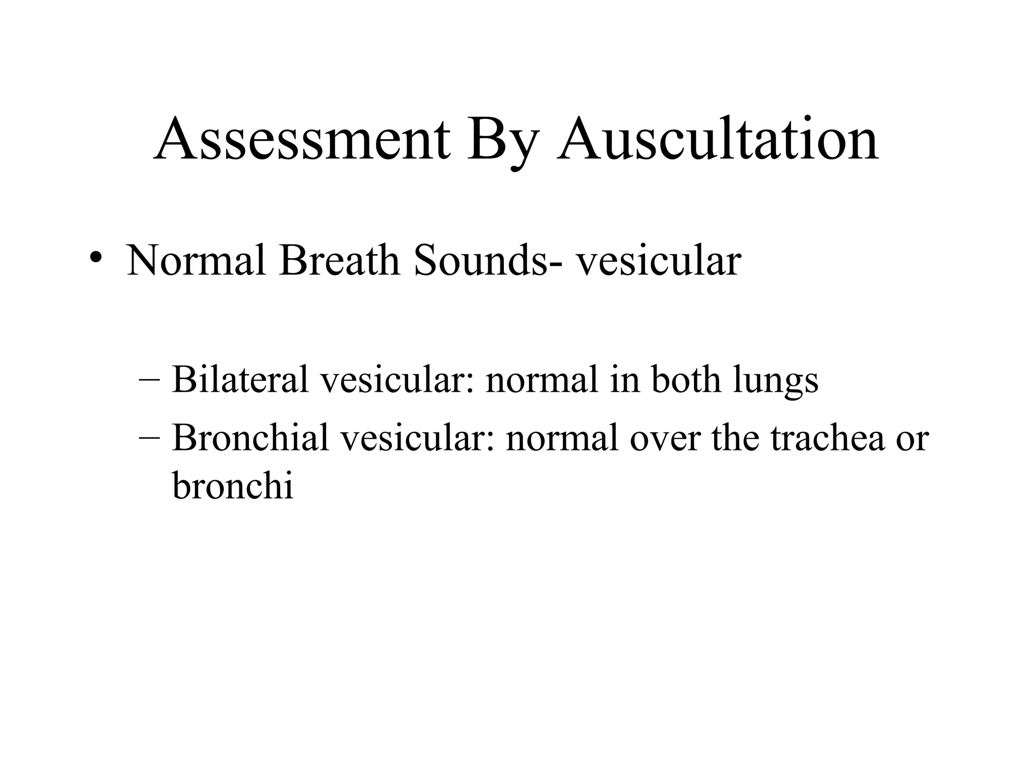
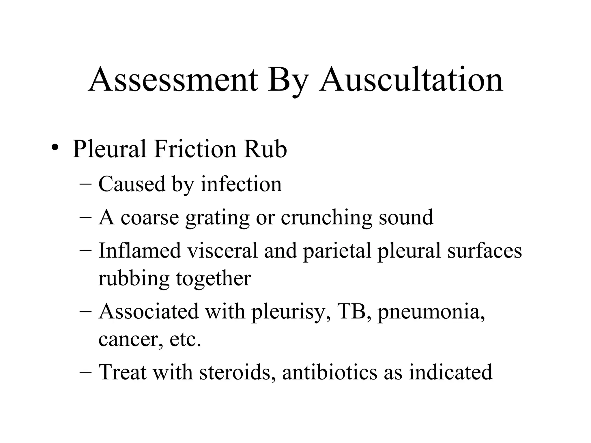
RTH 112

Assessment

  Unit 2
Text Reference
• Egan 9th Edition
  – Chapter 15 “Bedside Assessment of the
                 Patient”
Objectives
• Write a brief summary explaining the importance
  of conducting a patient interview in 500 words or
  less
• Without the use references, list the major
  components of a health history
• Using the notes, review the techniques to conduct
  a patient interview
• Without the use of references, summarize the
  importance of conducting a physical examination
• Verbally describe the four major examination
  techniques
Patient Evaluation
• Initial Assessment
   – Clinical Manifestations
      • Patient Interview
      • Physical Examination
• Secondary Assessment
   – Diagnostic Studies
      •   Arterial Blood Gases
      •   Pulmonary Function Studies
      •   Chest Films
      •   Other Diagnostic Procedures as Indicated
Patient Interview
• Determine Level of Consciousness
  –   Normal: alert & cooperative
  –   Lethargic
  –   Confused
  –   Obtunded: diminished cough or gag
  –   Semi-comatose: responds to painful stimuli
  –   Coma: unresponsive to pain
Patient Interview
• Orientation of time, place and person
  – Well oriented, cooperative
     • Able to follow simple commands
  – Disoriented, confused
  – Inability to cooperate:
     •   Language difficulties
     •   Influence of medications
     •   Hearing loss
     •   Fear, depression
Patient Interview
• Assess Emotional State
  – Anxiety
     • Respiratory distress, hypoxemia
  – Depression
     • Quiet or withdrawn, in denial
  – Anger
     • Combative, irritable
  – Euphoria
     • Influence of drugs
  – Panic
     • Hypoxia, air hunger, status asthmaticus
Patient Interview
• Measure Subjective Symptoms
  – Orthopnea
    • Difficulty breathing except in upright position
  – General Malaise
    • Run down, nausea, weakness, fatigue
  – Dyspnea
    • Feeling SOB
       –   Grade I: normal dyspnea after unusual tension
       –   Grade II: breathless after going up hill or stairs
       –   Grade III: dyspnea while walking at normal speed
       –   Grade IV: dyspnea moving slowly & short distances
       –   Grade V: dyspnea at rest, small tasks
Patient Interview
• Pain
  –   Location
  –   Quality (what kind is it)
  –   Severity
  –   Aggravating factors
  –   Relieving factors
  –   History (when did it start and how did it progress)
  –   Context (circumstances of onset)
  –   Accompanying Symptoms
Patient Interview
• Symptoms of Nose and Throat
  – Nasal secretions
     • Amount
     • Irritants, allergies
  – Itching or burning sensation of nose and throat
  – Dysphagia
     • Difficulty swallowing
     • Hoarseness
Patient Interview
• History of present illness
  – Current medical/physical problems
  – Current meds, including herbs, etc.
• Past medical history
  – Previous medical problems, hospitalizations,
    surgeries, drug allergies, etc
• Family history
  – Heart disease, diabetes, COPD, etc.
Interview Techniques
• Ask open ended questions
  – No yes or no questions

• Communicate using simple language
  – KISS Method
  – Use pictures, diagrams
  – Interpreter for those with language barriers
Physical Examination
• Begin with Assessment by Inspection
  – What Can You See?
Assessment By Inspection
• General Appearance
  –   Age
  –   Height
  –   Weight
  –   Sex
  –   Nourishment
Assessment By Inspection
• Peripheral Edema
  –   Presence of excessive fluid in the tissue
  –   Pitting Edema
  –   Occurs primarily in arms and ankles
  –   Caused by CHF, Renal insufficiency/failure
  –   Rated +1, +2, +3
       • The higher the number, the greater the swelling
Peripheral Edema
Abnormal buildup of fluid in the ankles, feet, and
legs is called peripheral edema.
             Foot with Edmea      Normal Foot
Assessment By Inspection
• Clubbing of fingers
  –   Suggestive of pulmonary disease
  –   Caused by chronic hypoxia
  –   Can affect thumb, fingers and toes
  –   Condition is present when the angle of the nail
      bed and skin increases
Assessment By Inspection
• Venous Distention
  – Occurs with CHF
  – Seen in patients with obstructive lung disease
  – Seen during exhalation because of the
    obstructive component
Assessment By Inspection
• Capillary Refill
  – Quick check of perfusion
  – Blanching of hand or nail beds and watch for
    blood return
  – Normally 3-5 seconds
  – Commonly performed for the Allen’s Test
    before arterial blood gas puncture
Assessment By Inspection
• Diaphoresis
  –   State of perfuse/heavy sweating
  –   Heart failure
  –   Fever, infection
  –   Anxiety, nervousness
  –   Tuberculosis (night sweats)
Assessment By Inspection
• Skin Color
  – Normal: pink
  – Abnormal: pale
     • Due to anemia or blood loss
  – Jaundice: yellow
     • Increase in bilirubin, mostly face & trunk
  – Erythema: redness
     • Capillary congestion, inflammation, infection
  – Cyanosis: blue/gray
     • Hypoxia (5 g of reduced hemoglobin)
Assessment By Inspection
• Chest Configuration
  – Normal: A-P diameter
     • Straight spine, no alterations in chest size
  – Pectus Carinatum
     • Anterior protrusion of the sternum
  – Pectus Excavatum
     • Depression of the sternum
  – Kyphosis
     • Hunchback or convex spine curve
  – Scoliosis
     • Lateral curve of the spine
  – Kyphoscoliosis: combination of both
  – Barrel Chest
     • Increased A-P diameter resulting form air trapping
Assessment By Inspection
• Movement of Chest/Diaphragm
  – Symmetrical movement
  – Unequal movement
    •   Chronic lung disease
    •   Atelectasis
    •   Pneumothorax
    •   Flail Chest – paradoxical
    •   Intubated with ET tube in one lung
Assessment By Inspection
• Breathing Patterns
  – Eupnea – normal rate, depth, rhythm
  – Tachypnea- over 20 bpm
     • Fever, hypoxia, pain, CNS problem
  – Bradypnea- less than 8 bpm
     • Variable depth and irregular rhythm
  – Apnea- cessation of breathing
  – Hyperpnea- increased rate & depth, regular
    rhythm
     • Metabolic/CNS disorders
Assessment By Inspection
• Breathing Patterns
   – Cheyne Stokes- gradual increasing the decreasing rate
     and depth in a cycle with periods of apnea
      • Increased ICP, Meningitis, overdose
   – Biots- increased rate and depth with irregular periods of
     apnea
      • CNS problem
   – Kussmauls- increased rate (>20) increased depth,
     irregular rhythm, seems labored
      • Metabolic acidosis, renal failure, diabetic ketoacidosis
Assessment By Inspection
• Breathing Patterns
  – Apneustic- prolonged gasping inspiration
    followed by extremely short, insufficient
    expiration
     • Problem with respiratory centers, trauma or tumor
Assessment By Inspection
• Muscle use
  – These muscles are used to increase ventilation
    during times of stress, increased airway
    resistance, etc.
Assessment By Inspection
• Muscle Use
  Muscles used during Normal Breathing
  – Diaphragm
  – External Intercostals
  – Exhalation is passive
Assessment By Inspection
• Accessory Muscle Use
  – Used to increase ventilation
     Muscles of normal ventilation PLUS
     • Intercostals, scalene, sternocleidomastoid, PLUS
     • Abdominal muscles
Assessment By Inspection
• Retractions
   – Chest moves inward during inspiration instead of
     outward
   – Due to a blocked (obstructed airway)
   – A sign of respiratory distress in infants
• Nasal Flaring
   – Flaring of the nostrils during inspiration
   – A sign of respiratory distress in infants sometimes
     accompanied by grunting
Assessment By Inspection
• Character of Cough
  –   Strong, moderate, weak
  –   Productive, nonproductive
  –   Frequent, infrequent
  –   Tight, moist
Assessment By Palpation
• Pulse
  –   Normal: 60-100
  –   Tachycardia: >100
  –   Bradycardia: <60
  –   Adverse Reaction: >20 Increased HR
  –   Monitor rhythm
Assessment By Palpation
• Tracheal Deviation
  – Pulled toward pathology (inside lung)
     • Atelectasis
     • Pneumonectomy
     • Diaphragmatic paralysis
  – Pulled away from pathology (outside lung)
     •   Massive pleural effusion
     •   Tension Pneumothorax
     •   Neck or thyroid mass
     •   Large mediastinal mass
Assessment By Palpation
• Tactile Fremitus
  – Vibration felt by hand on the chest wall
     • Vocal fremitus-voice vibrations on the chest wall
     • Pleural rub fremitus – grating sensation due to
       roughened pleural surfaces rubbing together
     • Rhonchial fremitus – secretions in the airway
Assessment By Palpation
• Tenderness
  – Around suture sites, chest tubes, fractures

  – Avoid areas of tenderness if possible
Assessment By Palpation
• Chest Motion Symmetry
  – Hands placed on the patient’s chest move in
    symmetry. If one hand moves more than the
    other, it indicates asymmetrical chest expansion
Assessment By Percussion
• Performed by placing the middle finger between
  two ribs and tapping the middle finger’s first joint
  with the middle fingers of the opposite hand.
   – Resonance- normal air filled lung; hollow sound
   – Flat- over sternum, muscle or atelectasis; full sound
   – Dull-fluid filled organs; pleural effusion or pneumonia;
     thudding sound
   – Tympany-air filled stomach; drum like sound
   – Hyperresonance-Areas of the lung with pneumothorax
     or emphysema. Booming sound.
Assessment By Auscultation
• Normal Breath Sounds- vesicular

  – Bilateral vesicular: normal in both lungs
  – Bronchial vesicular: normal over the trachea or
    bronchi
Assessment By Auscultation
• Increased, decreased, unequal or absent
   – Always compare one lung with the other

   – Egophany: “E” sound like “A”. Consolidation
   – Bronchophony & whispered pectoriloquy: increased
     intensity of voice when spoken. Indicated
     consolidation and pneumonia

   – An increase in voice indicates consolidation and
     pneumonia
   – A decrease in voice indicates obstructed bronchi,
     pneumothorax, emphysema
Assessment By Auscultation
• Abnormal Breath Sounds – adventitious
  – Rales (crackles)- fluid/secretions
     • Coarse (rhonchi)- large airway secretions
        – Suction the pt/cough
     • Medium
        – Pt needs CPT
     • Fine (moist crepitant rales)- alveoli fluid
        – Pt has CHF/pulmonary edema
        – PT needs IPPB, heart drugs, diuretics and oxygen
Assessment By Auscultation
• Wheezes
  – Bronchospasm
  – Patient needs a bronchodilator
  – Unilateral wheezes indicative of FBO
Assessment By Auscultation
• Stridor
  – Upper Airway Obstruction
     • Supraglottic swelling (epiglottitis)
     • Subglottic swelling (croup, post extubation)
     • Foreign body aspiration
  – Treatment
     • Topical decongestant (racemic epinephrine)
     • Suction/bronchoscopy
     • Intubation for severe swelling and epiglottitis
Assessment By Auscultation
• Pleural Friction Rub
  – Caused by infection
  – A coarse grating or crunching sound
  – Inflamed visceral and parietal pleural surfaces
    rubbing together
  – Associated with pleurisy, TB, pneumonia,
    cancer, etc.
  – Treat with steroids, antibiotics as indicated
Breath Sounds Review
http://Breath Sounds Review
Reference Link
• The Lung Exam

Idol pp voice

  • 1.
  • 2.
    Text Reference • Egan9th Edition – Chapter 15 “Bedside Assessment of the Patient”
  • 3.
    Objectives • Write abrief summary explaining the importance of conducting a patient interview in 500 words or less • Without the use references, list the major components of a health history • Using the notes, review the techniques to conduct a patient interview • Without the use of references, summarize the importance of conducting a physical examination • Verbally describe the four major examination techniques
  • 4.
    Patient Evaluation • InitialAssessment – Clinical Manifestations • Patient Interview • Physical Examination • Secondary Assessment – Diagnostic Studies • Arterial Blood Gases • Pulmonary Function Studies • Chest Films • Other Diagnostic Procedures as Indicated
  • 5.
    Patient Interview • DetermineLevel of Consciousness – Normal: alert & cooperative – Lethargic – Confused – Obtunded: diminished cough or gag – Semi-comatose: responds to painful stimuli – Coma: unresponsive to pain
  • 6.
    Patient Interview • Orientationof time, place and person – Well oriented, cooperative • Able to follow simple commands – Disoriented, confused – Inability to cooperate: • Language difficulties • Influence of medications • Hearing loss • Fear, depression
  • 7.
    Patient Interview • AssessEmotional State – Anxiety • Respiratory distress, hypoxemia – Depression • Quiet or withdrawn, in denial – Anger • Combative, irritable – Euphoria • Influence of drugs – Panic • Hypoxia, air hunger, status asthmaticus
  • 8.
    Patient Interview • MeasureSubjective Symptoms – Orthopnea • Difficulty breathing except in upright position – General Malaise • Run down, nausea, weakness, fatigue – Dyspnea • Feeling SOB – Grade I: normal dyspnea after unusual tension – Grade II: breathless after going up hill or stairs – Grade III: dyspnea while walking at normal speed – Grade IV: dyspnea moving slowly & short distances – Grade V: dyspnea at rest, small tasks
  • 9.
    Patient Interview • Pain – Location – Quality (what kind is it) – Severity – Aggravating factors – Relieving factors – History (when did it start and how did it progress) – Context (circumstances of onset) – Accompanying Symptoms
  • 10.
    Patient Interview • Symptomsof Nose and Throat – Nasal secretions • Amount • Irritants, allergies – Itching or burning sensation of nose and throat – Dysphagia • Difficulty swallowing • Hoarseness
  • 11.
    Patient Interview • Historyof present illness – Current medical/physical problems – Current meds, including herbs, etc. • Past medical history – Previous medical problems, hospitalizations, surgeries, drug allergies, etc • Family history – Heart disease, diabetes, COPD, etc.
  • 12.
    Interview Techniques • Askopen ended questions – No yes or no questions • Communicate using simple language – KISS Method – Use pictures, diagrams – Interpreter for those with language barriers
  • 13.
    Physical Examination • Beginwith Assessment by Inspection – What Can You See?
  • 14.
    Assessment By Inspection •General Appearance – Age – Height – Weight – Sex – Nourishment
  • 15.
    Assessment By Inspection •Peripheral Edema – Presence of excessive fluid in the tissue – Pitting Edema – Occurs primarily in arms and ankles – Caused by CHF, Renal insufficiency/failure – Rated +1, +2, +3 • The higher the number, the greater the swelling
  • 16.
    Peripheral Edema Abnormal buildupof fluid in the ankles, feet, and legs is called peripheral edema. Foot with Edmea Normal Foot
  • 17.
    Assessment By Inspection •Clubbing of fingers – Suggestive of pulmonary disease – Caused by chronic hypoxia – Can affect thumb, fingers and toes – Condition is present when the angle of the nail bed and skin increases
  • 18.
    Assessment By Inspection •Venous Distention – Occurs with CHF – Seen in patients with obstructive lung disease – Seen during exhalation because of the obstructive component
  • 19.
    Assessment By Inspection •Capillary Refill – Quick check of perfusion – Blanching of hand or nail beds and watch for blood return – Normally 3-5 seconds – Commonly performed for the Allen’s Test before arterial blood gas puncture
  • 20.
    Assessment By Inspection •Diaphoresis – State of perfuse/heavy sweating – Heart failure – Fever, infection – Anxiety, nervousness – Tuberculosis (night sweats)
  • 21.
    Assessment By Inspection •Skin Color – Normal: pink – Abnormal: pale • Due to anemia or blood loss – Jaundice: yellow • Increase in bilirubin, mostly face & trunk – Erythema: redness • Capillary congestion, inflammation, infection – Cyanosis: blue/gray • Hypoxia (5 g of reduced hemoglobin)
  • 22.
    Assessment By Inspection •Chest Configuration – Normal: A-P diameter • Straight spine, no alterations in chest size – Pectus Carinatum • Anterior protrusion of the sternum – Pectus Excavatum • Depression of the sternum – Kyphosis • Hunchback or convex spine curve – Scoliosis • Lateral curve of the spine – Kyphoscoliosis: combination of both – Barrel Chest • Increased A-P diameter resulting form air trapping
  • 23.
    Assessment By Inspection •Movement of Chest/Diaphragm – Symmetrical movement – Unequal movement • Chronic lung disease • Atelectasis • Pneumothorax • Flail Chest – paradoxical • Intubated with ET tube in one lung
  • 24.
    Assessment By Inspection •Breathing Patterns – Eupnea – normal rate, depth, rhythm – Tachypnea- over 20 bpm • Fever, hypoxia, pain, CNS problem – Bradypnea- less than 8 bpm • Variable depth and irregular rhythm – Apnea- cessation of breathing – Hyperpnea- increased rate & depth, regular rhythm • Metabolic/CNS disorders
  • 25.
    Assessment By Inspection •Breathing Patterns – Cheyne Stokes- gradual increasing the decreasing rate and depth in a cycle with periods of apnea • Increased ICP, Meningitis, overdose – Biots- increased rate and depth with irregular periods of apnea • CNS problem – Kussmauls- increased rate (>20) increased depth, irregular rhythm, seems labored • Metabolic acidosis, renal failure, diabetic ketoacidosis
  • 26.
    Assessment By Inspection •Breathing Patterns – Apneustic- prolonged gasping inspiration followed by extremely short, insufficient expiration • Problem with respiratory centers, trauma or tumor
  • 27.
    Assessment By Inspection •Muscle use – These muscles are used to increase ventilation during times of stress, increased airway resistance, etc.
  • 28.
    Assessment By Inspection •Muscle Use Muscles used during Normal Breathing – Diaphragm – External Intercostals – Exhalation is passive
  • 29.
    Assessment By Inspection •Accessory Muscle Use – Used to increase ventilation Muscles of normal ventilation PLUS • Intercostals, scalene, sternocleidomastoid, PLUS • Abdominal muscles
  • 30.
    Assessment By Inspection •Retractions – Chest moves inward during inspiration instead of outward – Due to a blocked (obstructed airway) – A sign of respiratory distress in infants • Nasal Flaring – Flaring of the nostrils during inspiration – A sign of respiratory distress in infants sometimes accompanied by grunting
  • 31.
    Assessment By Inspection •Character of Cough – Strong, moderate, weak – Productive, nonproductive – Frequent, infrequent – Tight, moist
  • 32.
    Assessment By Palpation •Pulse – Normal: 60-100 – Tachycardia: >100 – Bradycardia: <60 – Adverse Reaction: >20 Increased HR – Monitor rhythm
  • 33.
    Assessment By Palpation •Tracheal Deviation – Pulled toward pathology (inside lung) • Atelectasis • Pneumonectomy • Diaphragmatic paralysis – Pulled away from pathology (outside lung) • Massive pleural effusion • Tension Pneumothorax • Neck or thyroid mass • Large mediastinal mass
  • 34.
    Assessment By Palpation •Tactile Fremitus – Vibration felt by hand on the chest wall • Vocal fremitus-voice vibrations on the chest wall • Pleural rub fremitus – grating sensation due to roughened pleural surfaces rubbing together • Rhonchial fremitus – secretions in the airway
  • 35.
    Assessment By Palpation •Tenderness – Around suture sites, chest tubes, fractures – Avoid areas of tenderness if possible
  • 36.
    Assessment By Palpation •Chest Motion Symmetry – Hands placed on the patient’s chest move in symmetry. If one hand moves more than the other, it indicates asymmetrical chest expansion
  • 37.
    Assessment By Percussion •Performed by placing the middle finger between two ribs and tapping the middle finger’s first joint with the middle fingers of the opposite hand. – Resonance- normal air filled lung; hollow sound – Flat- over sternum, muscle or atelectasis; full sound – Dull-fluid filled organs; pleural effusion or pneumonia; thudding sound – Tympany-air filled stomach; drum like sound – Hyperresonance-Areas of the lung with pneumothorax or emphysema. Booming sound.
  • 38.
    Assessment By Auscultation •Normal Breath Sounds- vesicular – Bilateral vesicular: normal in both lungs – Bronchial vesicular: normal over the trachea or bronchi
  • 39.
    Assessment By Auscultation •Increased, decreased, unequal or absent – Always compare one lung with the other – Egophany: “E” sound like “A”. Consolidation – Bronchophony & whispered pectoriloquy: increased intensity of voice when spoken. Indicated consolidation and pneumonia – An increase in voice indicates consolidation and pneumonia – A decrease in voice indicates obstructed bronchi, pneumothorax, emphysema
  • 40.
    Assessment By Auscultation •Abnormal Breath Sounds – adventitious – Rales (crackles)- fluid/secretions • Coarse (rhonchi)- large airway secretions – Suction the pt/cough • Medium – Pt needs CPT • Fine (moist crepitant rales)- alveoli fluid – Pt has CHF/pulmonary edema – PT needs IPPB, heart drugs, diuretics and oxygen
  • 41.
    Assessment By Auscultation •Wheezes – Bronchospasm – Patient needs a bronchodilator – Unilateral wheezes indicative of FBO
  • 42.
    Assessment By Auscultation •Stridor – Upper Airway Obstruction • Supraglottic swelling (epiglottitis) • Subglottic swelling (croup, post extubation) • Foreign body aspiration – Treatment • Topical decongestant (racemic epinephrine) • Suction/bronchoscopy • Intubation for severe swelling and epiglottitis
  • 43.
    Assessment By Auscultation •Pleural Friction Rub – Caused by infection – A coarse grating or crunching sound – Inflamed visceral and parietal pleural surfaces rubbing together – Associated with pleurisy, TB, pneumonia, cancer, etc. – Treat with steroids, antibiotics as indicated
  • 44.
  • 45.

Editor's Notes