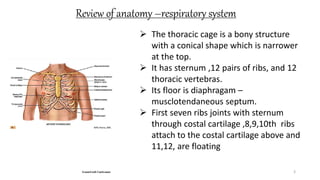
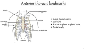
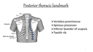
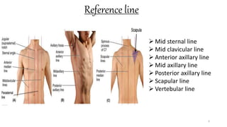
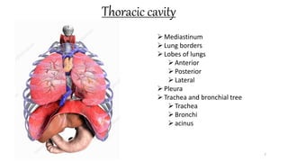
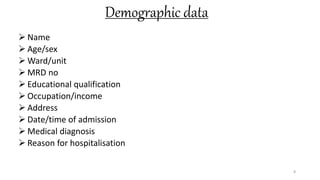
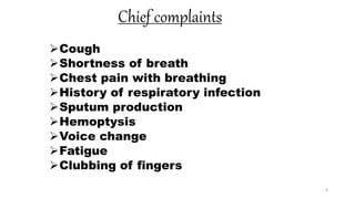
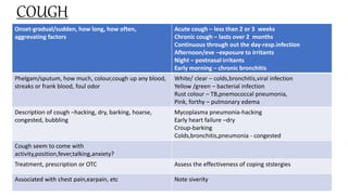
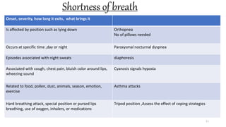
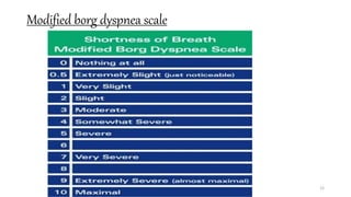
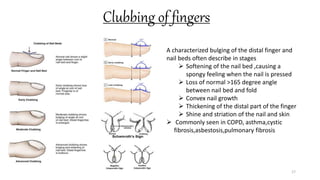
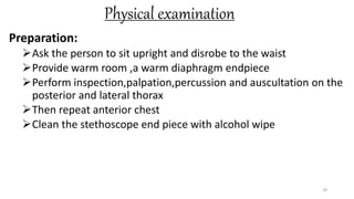
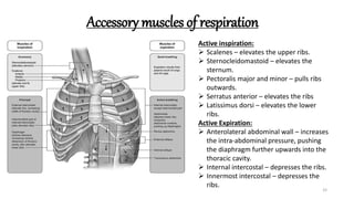
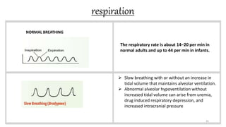
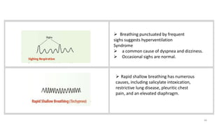
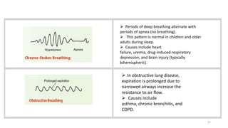
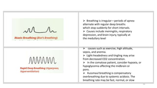
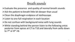
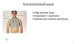
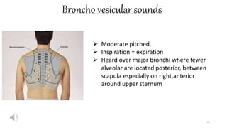
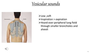
Call Girls Secunderabad 7001305949 all area service COD available Any Time
Respiratory assessment
1. College Of Nursing
Madras Medical College
Chennai-03
PRESENTED BY
EDWIN JOSE.L
MSc(nursing) I YEAR
College of nursing
Madras medical college
Chennai-03
1
2. INTRODUCTION
Correct diagnosis depends on an accurate health history and a
through physical examination.
A respiratory assessment can be done as part of a comprehensive
physical examination or as an examination in itself.
Judgement must be used in determining whether all or part of the
historm and physical examination will be completed based on the
problems
If respiratory distress is severe ,only pertinent information should
be obtained and a through assessment should be deferred until the
patient ‘s condition stabilizes
2
3. Review of anatomy –respiratory system
3
The thoracic cage is a bony structure
with a conical shape which is narrower
at the top.
It has sternum ,12 pairs of ribs, and 12
thoracic vertebras.
Its floor is diaphragam –
musclotendaneous septum.
First seven ribs joints with sternum
through costal cartilage ,8,9,10th ribs
attach to the costal cartilage above and
11,12, are floating
6. Reference line
6
Mid sternal line
Mid clavicular line
Anterior axillary line
Mid axillary line
Posterior axillary line
Scapular line
Vertebular line
8. Demographic data
Name
Age/sex
Ward/unit
MRD no
Educational qualification
Occupation/income
Address
Date/time of admission
Medical diagnosis
Reason for hospitalisation
8
9. Chief complaints
Cough
Shortness of breath
Chest pain with breathing
History of respiratory infection
Sputum production
Hemoptysis
Voice change
Fatigue
Clubbing of fingers
9
10. COUGH
10
Onset-gradual/sudden, how long, how often,
aggrevating factors
Acute cough – less than 2 or 3 weeks
Chronic cough – lasts over 2 months
Continuous through out the day-resp.infection
Afternoon/eve –exposure to irritants
Night – postnasal irritants
Early morning – chronic bronchitis
Phelgam/sputum, how much, colour,cough up any blood,
streaks or frank blood, foul odor
White/ clear – colds,bronchitis,viral infection
Yellow /green – bacterial infection
Rust colour – TB,pnemococcal pneumonia,
Pink, forthy – pulmonary edema
Description of cough –hacking, dry, barking, hoarse,
congested, bubbling
Mycoplasma pneumonia-hacking
Early heart failure –dry
Croup-barking
Colds,bronchitis,pneumonia - congested
Cough seem to come with
activity,position,fever,talking,anxiety?
Treatment, prescription or OTC Assess the effectiveness of coping ststergies
Associated with chest pain,earpain, etc Note siverity
11. Shortness of breath
11
Onset, severity, how long it exits, what brings it
Is affected by position such as lying down Orthopnea
No of pillows needed
Occurs at specific time ,day or night Paroxysmal nocturnal dyspnea
Episodes associated with night sweats diaphoresis
Associated with cough, chest pain, bluish color around lips,
wheezing sound
Cyanosis signals hypoxia
Related to food, pollen, dust, animals, season, emotion,
exercise
Asthma attacks
Hard breathing attack, special position or pursed lips
breathing, use of oxygen, inhalers, or medications
Tripod position ,Assess the effect of coping strategies
13. Chest pain
13
Onset, constant,or does it come and go
Chest pain of thoracic origin occurs with muscle soreness
from coughing or from inflammation of pleura,
Description of pain : burning/stabing
Brought on by respiratory infection, coughing, trauma,
feverdeep breathing, unequal chest inflation
Treatment if any
14. History of respiratory infection
14
Past history of breathing trouble,lung disease like
bronchitis,emphysema,asthma, pneumonia, covid 19
Consider sequalae of these condition
Most people have had some colds,it is meaningfull to
ask about excess number or severity
Any unusually frequent or unusually severe colds
Any family history of allergies,tuberculosis,or asthma
16. hemoptysis
Hemoptysis is defined as the spitting of blood derived from the lungs
or bronchial tubes as a result of pulmonary or bronchial hemorrhage.
The patient’s history should help determine the amount of blood and
differentiate between hemoptysis, pseudo-hemoptysis, and
hematemesis.
Hemoptysis is classified as non-massive or massive based on the
volume of blood loss
considered non-massive if blood loss is less than 200 mL per day
In adults, bronchitis, bronchogenic carcinoma, and pneumonia are the
major causes.
16
17. Clubbing of fingers
17
A characterized bulging of the distal finger and
nail beds often describe in stages
Softening of the nail bed ,causing a
spongy feeling when the nail is pressed
Loss of normal >165 degree angle
between nail bed and fold
Convex nail growth
Thickening of the distal part of the finger
Shine and striation of the nail and skin
Commonly seen in COPD, asthma,cystic
fibrosis,asbestosis,pulmonary fibrosis
18. Past health history:
Frequency of upper respiratory problems including childhood
respiratory disorders
Allergies- medications, pollens, smoke, mold, pet exposure
Past history of lower respiratory problems – asthma, COPD,
pneumonia, covid 19 etc…
History of HIV infection
18
19. Medications:
Prescription and over the counter drugs- antihistamines,
bronchodilators, corticosteroids, cough suppressants,
and antibiotics.
Reason for taking medicines –it’s name, dose, frequency,
length of time taken, it's effects and side effects ,if any
Use of angiotensin converting enzyme (ACE) inhibitors
Use of oxygen,FiO2 ,liter flow, method of administration,
no of hours used per day, effectiveness of therapy
19
20. Surgery or other treatments:
Previous hospitalization, if so the dates, therapy, and current status of the
problem
Use and the response to respiratory treatments such as nebulizer,
humidifier ,airway clearance, high frequency chest oscillation, postural
drainage and percussion
Tobacco usage:
Pack years – is the number of years that a patient has smoked multiplied
by number of pack of cigarettes smoked a day
Second hand smokers
Radon gas
Asbestos
Paddy fields
20
21. Family history
Type of family
Ordinal position in the family
Role in the family
Risk factors among the family members
History of any heredity/genetic disorder,cardiac disease
,etc….
Enquire family history of tuberculosis,bronchiectasis,cystic
fibrosis, lung cancer etc…..
21
22. Soci-economic history
Smoking history
Enquire about passive smoker
Enquire about exposure to birds
Exposure of air pollution
Type of house, waste disposal, drainage facility,
ventilation
Family income
22
24. Functional health patterns
1. Health perception – health management pattern:
Describe your daily activities?
Health status in last several days?
How your breathing problem affects self care abilities?
Have you ever smoked?
Have you had flu vaccination?
What equipments you use to relieve breathing difficulties?
24
25. 2.Nutritional –Metabolic pattern:
Have you recently lost weight? How much? Voluntarily?
Do any particular foods affect your sputum production or
breathing
3. Elimination patterm:
Does your respiratory problem make it difficulty for get to the
toilet?
Are you inactive because of dyspnea which causes constipation?
4.Activity – exercise pattern:
Are you ever short of breath during exercise
What you do when you get shortness of breath ?
Can you walk steps without stopping?
25
26. Sleep rest pattern:
Do breathing problems causes you to awaken during the night
Can you lie flat at night ?how many pillows do you use?
Do you need to sleep upright in a chair?
Do you have morning headache
Do you fall asleep easily during the day?
Cognitive – perceptual pattern:
Do you have any pain associated with breathing?
Pain scale 0-10
Does it hurt more on inspiration?
Self perception- self concept pattern:
Describe how your respiratory problems have changed your life
Do you ever go out without bringing your oxygen ,when and why?
26
27. Role relationship pattern:
Has your respiratory problem caused any difficulty in your work, family or social
relationship
Sexuality –reproductive pattern:
Has your respiratory problem caused a change in your sexual activity?
Do you want to discuss ways to decrease dyspnea during sexual activity?
Coping stress tolerance pattern:
How often do you leave your home?
Would you want to join a support group ?
Does stress have any effect on breathing?
What effects does you respiratory problem have on your emotions?
value –belief pattern:
What do you believe cause your respiratory problems?
Do you think the things you have been told to do for your respiratory problems really
help?if not why?
27
28. Physical examination
Preparation:
Ask the person to sit upright and disrobe to the waist
Provide warm room ,a warm diaphragm endpiece
Perform inspection,palpation,percussion and auscultation on the
posterior and lateral thorax
Then repeat anterior chest
Clean the stethoscope end piece with alcohol wipe
28
29. inspection
NOSE:
Patency – naris is checked for air patency by occluding other naris
Inflammation- inspect with speculum for edema,exudate or bleeding
Deformities – observe for deviation, perforation ,bleeding
observe for polyps
Discharge – assess for color and consistency
purulent/malodorous indicates presence of foregin body
watery discharge – allergies or from cerebrospinal fluid.
blood discharge – from trauma or dryness
thick mucosal discharge – presence of infection
29
30. Mouth and pharynx
Inspects the interior of the mouth for
color,lesions,masses,gum retraction, bleeding, and poor
dentation
Tongue is inspected for symmetry and presence of lesions
Inspect pharynx for exudate, ulceration, swelling or postnasal
drip
Tonsils are noted for colour, symmetry and any enlargement
Assess for gag reflex – indicates the cranial nerves IX and X
are intact
30
31. neck
Inspects for symmetry and presence of tender or swollen areas
The lympnodes are palpated while the patient is sitting erect with the
neck slightly flexed
Patient may have small, mobile,non-tender nodes (shotty nodes) which
are not a sign of a pathological condition
Tender ,hard,or fixed nodes indicates disease.
31
32. Thorax and lungs-inspection
Appearance – evidences of respiratory distress, tachypnea or use
of accessory muscles
Shape – elliptical shape with downward slopping ribs abour 45
degrees relative to the spine
Chest movements – equal ,symmetry, AP diameter < transverse
diameter by a ratio 1:2
32
33. Accessory muscles of respiration
33
Active inspiration:
Scalenes – elevates the upper ribs.
Sternocleidomastoid – elevates the
sternum.
Pectoralis major and minor – pulls ribs
outwards.
Serratus anterior – elevates the ribs
Latissimus dorsi – elevates the lower
ribs.
Active Expiration:
Anterolateral abdominal wall – increases
the intra-abdominal pressure, pushing
the diaphragm further upwards into the
thoracic cavity.
Internal intercostal – depresses the ribs.
Innermost intercostal – depresses the
ribs.
34. Cont….
Barrel chest
34
AP diameter = tranverse diameter with
ratio of 1:1
Ribs are horizontal indtead of normal
downward slope
Seen in normal aging and hyperinflated
lungs such as COPD
Due to overactivity of scalene and
sternocleidomastoid muscle which lifts the
upper ribs and sternum and this overuse
causes remodelling of the chest
35. Pectus excavatum –funnel breast
35
A markedly sunken or concave
appearance of sternum and adjacent
cartilages
Depression begins with 2nd ICS
becoming depressed most at
junction of xiphoid process
Congenital disorder
36. Pectus carinatum-pigeon chest
36
A forward protrusion of
the sternum with ribs
sloping back at either
side and vertical
depression along
costochondral junctions
Congenital disorder
39. Tripod position-dahl’s sign
39
Bilateral, symmetric, slanting regions of
hyperpigmentation on anterior thighs,associated with
tripod position
Seen in COPD
Tripod position – reduced work of breathing and activity
of scalene and sternocleidomastoid muscle if ones leans
forward ,improvement in thoraco abdominal movements.
Dahl’s sign – seen in long term ,chronic respiratory
illness caused by patients spending long periods of time
in the tripod position
40. Harrison’s sulcus (Harrison’s groove)
40
Visible depression of the lower ribs
above the costal margin, at the area of
attachment of the diaphragm.
Seen in rickets, severe asthma in
childhood, cystic fibrosis, pulmonary
fibrosis
Before the bone mineralize and harden
,the downward tension from the
diaphragm and other accessory muscles
used during increased respiratory effort
can bend the ribs inwards over time
41. hoover’s sign
41
Paradoxical inward movement of the lower costal
margins on inspiration
Seen in emphysema, chest hyperinflation – COPD
When the chest becomes hyperinflated, the
diaphragm often becomes stretched, which causes
contration of diaphragm at inspiration results in an
inward movement , bringing the costal margins
with it, as opposed to normal downward movement
42. Pursed lip breathing
42
A breathing practice often taught which includes a
long ,slow expiration against pursed lips
Seen in COPD
Inflammation of the airways leads to destruction of
lung parenchyma,results in reduction in elastic
recoil,fibrosis, and muscle hypertrophy causes
increased airways resistanceand premature airway
closing on expiration or expiratory airflow limitation.
This results in air trapping at end expiration and with
time hyperinflation
43. Tracheal tug
43
Downward displacement of thyroid cartilage during
inspiration
Most common- respiratory
distress/COPD(Campbell’s sign)
Less common – Arch of aorta aneurysm (Oliver’s
sign)
44. Clubbing of fingers
44
A characterized bulging of the distal finger and nail
beds often describe in stages
Softening of the nail bed ,causing a spongy
feeling when the nail is pressed
Loss of normal <165 degree angle between
nail bed and fold
Convex nail growth
Thickening of the distal part of the finger
Shine and striation of the nail and skin
Commonly seen in COPD, asthma,cystic
fibrosis,asbestosis,pulmonary fibrosis
45. respiration
45
NORMAL BREATHING
The respiratory rate is about 14–20 per min in
normal adults and up to 44 per min in infants.
Slow breathing with or without an increase in
tidal volume that maintains alveolar ventilation.
Abnormal alveolar hypoventilation without
increased tidal volume can arise from uremia,
drug induced respiratory depression, and
increased intracranial pressure
46. 46
Breathing punctuated by frequent
sighs suggests hyperventilation
Syndrome
a common cause of dyspnea and dizziness.
Occasional sighs are normal.
Rapid shallow breathing has numerous
causes, including salicylate intoxication,
restrictive lung disease, pleuritic chest
pain, and an elevated diaphragm.
47. 47
Periods of deep breathing alternate with
periods of apnea (no breathing).
This pattern is normal in children and older
adults during sleep.
Causes include heart
failure, uremia, drug-induced respiratory
depression, and brain injury (typically
bihemispheric).
In obstructive lung disease,
expiration is prolonged due to
narrowed airways increase the
resistance to air flow.
Causes include
asthma, chronic bronchitis, and
COPD.
48. 48
Breathing is irregular—periods of apnea
alternate with regular deep breaths
which stop suddenly for short intervals.
Causes include meningitis, respiratory
depression, and brain injury, typically at
the medullary level
causes such as exercise, high altitude,
sepsis, and anemia.
Light-headedness and tingling may arise
from decreased CO2 concentration.
In the comatose patient, consider hypoxia, or
hypoglycemia affecting the midbrain or
pons.
Kussmaul breathing is compensatory
overbreathing due to systemic acidosis. The
breathing rate may be fast, normal, or slow
49. Skin colour
Watch for cyanosis – late sign of hypoxemia
For dark skin patient – observe in conjunctiva, lips,palms, and under the tongue
49
50. Palpation- posterior chest
50
Confirm symmetry chest
expansion by placing warmed
hands sideways on the
posterolateral chest wall with
thumbs pointing together at the
level of T9 or T10 and pinch a
fold of skin
Ask to inhale deeply ,thumbs
should move apart symmetrically
Unequal expansion seen in
atelectasis, lobar pneumonia,
pleural effusion, thoracic
trauma,# ribs, pneumothorax
Pain in deep breathing seen when
the pleura are inflammed
51. Tactile fermitus
51
Fermitus is a palpable vibrations.
Sounds generated from the larynx
are transmitted through patent
bronchi and the lung parenchyma
to the chest wall,where we feel
them as vibrations
Use either the palmar base of the
finger or ulnar edge of one hand
and touch the person’s chest while
the patient repeats the word
“ninety nine” or “blue moon”
Start over the lung apices and
palpate from one side to another
side
Decreased fremitus –odstructed
bronchus,pleural effusion or
thickening, pneumothorax,
emphysema
Increased fremitus – compression
or consolidation of lung tissue
Rhonchal fremitus – palpable with
thick bronchial secretion
Pleural friction fremitus –
inflammation of pleura
52. crepitus
52
Normal Abnormal findings
using the fingers ,gently palpate the
entire chest wall.
This enables you to note any areas of
tenderness,to note skin temperature
and moisture,to detect any superficial
lumps pr masses ,and explore any skin
lesions noted in inspection
Crepitus is a coarse, crackiling
sensation palpable over the skin
surface.
It occurs in subcutaneous emphysema
when air escapes from the lung and
enters the subcutaneous tissue ,as
after open thoracic injury or surgery
53. Percussion-posterior chest
53
Start at the apices and percuss the band
of normally resonant tissue across the
tops of both shoulders
Percuss the interspaces, mark a side to
side comparison all the way down the
lung region
Percuss at 5cm intervals
Avoid the damping effect of scapula and
ribs
54. Percussion –posterior chest
54
Resonance Low pitched, clear, hollow,
sound that predominates in
healthy lung tissue
Hyperresonance Lower pitched ,blooming
sound seen in emphysema or
pneumothorax
Dull Soft, muffled, seen in
pneumonia, pleural effusion,
atelectasis, or tumor
55. Diaphragmatic excursion
55
Percuss to map out the lower lung border
in both expiration and inspiration
Ask the patient to exhale and hold it ,and
percuss down the scapular line until the
sound changes from resonance to dull on
each side and mark the spot
Now ask the patient to take deep breath
and hold it. continue to percuss down
from the first mark and mark the level
where the sound changes from resonance
to dull
Measure the difference
It should be equal bilaterally equal and
measure about 3-5cm
Note for high level of dullness and absence
of excursion and is seen in pleural effusion
or atelectasis in lower lobe
56. Ascultation-posterior chest
56
The passage of air through the tracheobronchial tree
creates a characteristic set of sounds that are audible
through the chest wall.
Breath sounds are changed by obstruction in the
passage ways or by disease in the lung parenchyma,
the pleura,or the chest wall
57. Breath sounds
Evaluate the presence and quality of normal breath sounds
Ask the patient to breath little bit deeper than usual
Clean the diaphragm endpiece of stethoscope
Listen to one full respiration in each location
Do not confuse with background noise with lung sounds
While standing behind the person listen to the following areas
posterior from apices at C7 to T10 and laterally from axilla down
to 7th or 8th rib
57
59. Broncho vesicular sounds
59
Moderate pitched,
Inspiration = expiration
Heard over major bronchi where fewer
alveolar are located posterior, between
scapula especially on right,anterior
around upper sternum
61. Adventitious sounds
crackles (rales)
Non continuous ,explosive popping sounds heared more often on
inspiration can also present on expiration
It may be fine and coarse
Coarse crackles are associated with larger airways and fine crackles are
associated with smaller branches
Seen in asthma,COPD, bronchiectasis, pulmonary edema, pneumonia, lung
cancer, pulmonary fibrosis
61
62. Wheezes
Continual, high pitched musical sounds heared at the end of
inspiration or at the start of expiration
Seen in asthma, COPD, respiratory tract infection
Airway narrowing allows airflow induced oscillation of airway
walls producing acoustic waves
Monophonic wheeze – single notes
Polyphonic wheeze – different tones
62
63. Stridor
Stridor is a continuous, high-frequency, high-pitched musical sound
produced during
airflow through a narrowing in the upper respiratory tract.
Stridor is best heard over the neck during inspiration, but can be biphasic.
Causes of the underlying airway obstruction include tracheal stenosis from
intubation, airway edema after device removal, epiglottitis, foreign body,
and anaphylaxis.
Immediate intervention is warranted.
63
64. pleural rub
A pleural rub is a discontinuous, low-frequency,
grating sound that arises from inflammation and
roughening of the visceral pleura as it slides against
the parietal pleura.
This nonmusical sound is biphasic, heard during
inspiration and expiration, and often best heard in the
axilla and base of the lungs.
64
65. mediastinal crunch
A mediastinal crunch is a series of precordial crackles synchronous with
the heartbeat, not with respiration.
Best heard in the left lateral position, it arises from air entry into the
mediastinum causing mediastinal emphysema (pneumomediastinum).
It usually produces severe central chest pain and may be spontaneous.
It has been reported in cases of tracheobronchial injury, blunt trauma,
pulmonary disease, use of recreational drugs, childbirth, and rapid ascent
from scuba diving.
65
66. bronchophony
Ask the patient to say “ninety-nine.”
Normally the sounds transmitted through the chest wall are muffled and
indistinct.
Louder voice sounds are called bronchophony
Localized bronchophony and egophony are seen in lobar consolidation
from pneumonia.
In patients with fever and cough, the presence of bronchial breath sounds
and egophony more than triples the likelihood of pneumonia.
66
67. Egophony
Ask the patient to say “ee.”
You will normally hear a muffled long ee sound.
If “ee” sounds like “A” and has a nasal bleating quality, an E-to-A
change, or egophony, is present.
Seen in Over consolidation or compression
67
68. Whispered pectoriloquy
Ask the patient to whisper “ninety-nine” or “one-two-three.”
The whispered voice is normally heard faintly and indistinctly, if
at all.
Louder, clearer whispered sounds are called whispered
pectoriloquy
Seen in mild consolidation
68
69. Inspection –anterior chest
69
Shape and configuration Barrel chest has horizontal ribs and costal angle >90 degree
Facial expression
Assess symmetrical chest expansion
Tensed, strained, tired facies and pursed lipped breathing seen
in COPD, Asthma
Unequal expansion occurs in obstructed or collapsed lungs
Assess the level of consciousness
Assess the use of accessory muscle
Cerebral hypoxia may reflected by exercise ,drowsiness or
anxiety ,restlessness and irritability
Seen in airway obstruction and massive atelectasis
Note skin color, nail beds, for cyanosis or unusual pallor.
Explore skin lesion
Clubbing of distal phalanges occurs with COPD because of
growth of vascular connective tissue
Cutaneous angiomas associated with liver disease or PHT
Assess the quality of respirations
Assess for respiratory rate
Assess for chest retraction or bulging
Noisy breathing seen in asthma and COPD
Tachypnea, bradypnea, Cheyne-stroke respiration……
Retraction suggests obstruction and bulging suggests
emphysema or asthma
70. Palpation-anterior chest
70
Palpate symmetric chest
expansion
Place hand on the anterior
lateral wall with thumbs
along the costal margin
and pointing towards
xiphoid process
Ask the person to take a
deep breath
Watch the thumb move
apart symmetrically
Assess tactile fremitus
Palpate anterior chest wall
for tenderness,
lumps,masses
Abnormal costal wide angle
occurs in emphysema
Lag in expansion occurs in
atelectasis, pneumonia,
postoperative guarding
Grating sensation indicates
pleural friction fremitus
71. Percussion-anterior wall
71
As needed, percuss the anterior and
lateral chest, again comparing both
sides.
The heart normally produces an area of
dullness to the left of the sternum from
the 3rd to the 5th interspaces.
Dullness represents airway obstruction
from inflammation or secretions.
Because pleural fluid usually sinks to the
lowest part of the pleural space
(posteriorly in a supine patient), only a
very large effusion can be detected
anteriorly.
The hyperresonance of COPD may obscure
dullness over the heart.
The dullness of right middle lobe
pneumonia typically occurs behind the
right breast.
Unless you displace the breast, you may
miss the abnormal percussion note.
72. Ascultation-anterior chest
Listen to the chest anteriorly and laterally as the patient breathes with
mouth open, and somewhat more deeply than normal.
Compare symmetric areas of the lungs, using the pattern suggested for
percussion and extending it to adjacent areas, if indicated.
Listen to the breath sounds, noting their intensity and identifying any
variations from normal vesicular breathing.
Breath sounds are usually louder in the upper anterior lung fields.
Bronchovesicular breath sounds may be heard over the large airways,
especially on the right
72
74. OXIMETRY
Arterial O2 saturation can be monitored noninvasively
andcontinuously using a pulse oximetry probe on the finger,
toe, ear,forehead, or bridge of the nose.
The abbreviation SpO2is used toindicate the O2 saturation of
hemoglobin as measured by pulseoximetry.
SpO2 and heart rate are displayed on the monitor as digital
readings. Normal SpO2 values are 94% to 99%
74
75. BLOOD STUDIES
Hemoglobin:
Test reflects the amount of hemoglobin available for combination with
oxygen
Normal – 13.5 to18mg/dl (men)
12 to 16 mg/dl (women)
hematocrit
Test reflects ratio of red cells to plasma
Increased hematocrit found in hypoxemia
Normal – 40 to 54% (men)
38 to 47 (women)
75
76. Arterial Blood Gases
ABGs are obtained to determine oxygenation status and acid-base balance.
ABG analysis includes measurement of the PaO2 , PaCO2 (the partial pressure of CO2 in
arterial blood), acidity (pH), bicarbonate (HCO3 ), and SaO2 .
Blood for ABG analysis can be obtained by arterial puncture or from an arterial catheter,
which is usually inserted into the radial or femoral artery.
Both techniques allow only intermittent analysis, but an arterial catheter permits ABG
sampling without repeated arterial punctures.
The normal PaO2 decreases with advancing age.
It varies in relation to the distance above sea level.
At higher altitudes, the barometric pressure is lower, resulting in a lower inspired O2
pressure and a lower PaO2 .
76
77. Sputum studies
Culture and sensitivity
Single sputum specimen is collected ina sterile container
Purpose is to to diagnose bacterial infection ,select antibiotics and evaluate
treatment
Takes 48 -72 hours for results
Gram stain:
Staining of sputum permits classification of bacteria into gram negative and gram
positive types
Results guides therapy until culture and sensitivity results are obtained
Acid fast smear and culture:
Test is to performed to collect sputum for acid fast bacilli
A series of three early morning specimen is used
77
78. cytology
Cytology:
Single sputum specimen is collected in special container with fixative
solution
Purpose is to determine presence of abnormal cells that may indicate
malignant condition
Chest X-ray:
It is most commonly used test for assessment that exposes a patients
respiratory system
Used to assess progressive of disease and response to treatment
The most common views used are the posterior-anterior view and lateral
78
79. Computed tomography
A computed tomography ,which exposes a patients to radiation may be used to
examine cross section of the entire body
Used to evaluates areas that are difficulty to assess by conventional X rays
Common types of CT scan are helical or spiral CT in which contrast dye is usually used
In high resolution CT contrast dye is not used
Spiral CT is most common non invasive imaging procedure used to diagnose pulmonary
embolism
Magnetic resonance imaging:
In a strong magnetic field ,the alignment of spinning nuclei can be changed with a super
imposed radio frequencyand the rate at which they return to alignment with the field
can be measured
The patient is not exposed to radiation
79
80. Ventilation-perfusion scan:
A ventilation perfusion scan is used primarily to check the presence of pulmonary
embolism
But it cannot determine with 100% certainty of the presence of PE
An iv isotope is given and the pulmonary vasculature is outlined and photographed
The patient inhales a radioactive gas (xenon, krypton) which outlines the alveoli
and another photograph is taken
Pulmonary angiography:
Pulmonary angiography is the most specific examination used to confirm the
diagnosis of pulmonary edema
A series of X- ray is taken after radio opaque dye is injected into the pulmonary
artery
This test also detect congenital and acquired lesions of the pulmonary vessels
80
81. Positron emission tomography:
Positron emission tomography scans the use of radio nuclides with short half lives
Used to distinguish benign and malignant solitary pulmonary nodules,because
malignant lung cells have an increased uptake of glucose
Bronchoscopy:
bronchoscopy is a procedure in which the bronchi are visualised through a
fiberoptic tube
Used to obtain biopsy specimen and assess changes resulting from treatment
Small amount (30ml) of sterile saline may be injected through the scope and
withdrawn and examined for cells ,a technique termed as bronchoalveolar lavage
Used to diagnose pneumonia, mucus plug, foregion bodies
Mediastinoscopy:
A scopy is inserted through a small incision in the supra sternal notch and advanced
through mediastinum to inspect and biopsy lymph nodes
The test is used to diagnose carcinoma, non-hodgkins lymphoma, granulomatous
infections, and sarcoidosis
81
82. Lung biopsy:
Lung biopsy may be done
1. Transbronchially
2. Percutaneously or via transthoracic needle aspiration
3. Video assisted thoracic surgery
4. As an open lung biopsy
purpose is to obtain tissue ,cells or secretion for evaluation
Thoracenthesis:
It is the insertion of a large bore needle through the chest wall into pleural
space to obtain specimen for diagnosis ,evaluation ,remove pleural fluids ,or
instil medications into the pleural space
The patient is positioned upright with elbows in an overbed table and feet
supported
The skin is cleansed and a local anesthetic is instilled subcutaneously
A test tube may be inserted to permit further drainage of fluids
82
83. Pulmonary function test:
Pulmonary function test measures lung volumes and airflow
The results pf PFT are used to diagnose pulmonary disease,monitor disease
progression ,evaluate disability and evaluate response to bronchodilators
Airflow is measured by a spirometer and administered by trained personal
The patients inserts a mouth piece ,takes as deep breath as possible and
exhales as hard fast and long as possible
Spirometry may be ordered before and after the administration of
bronchodilator to determine the degree of response
Home spirometry may be used to monitor lung function in person with
asthma or cystic fibrodis
83
84. Exercise testing:
Exercise testing is used to disgnose in determining exercise capacity and
for disability evaluation
A complete exercise test involves walking on a threadmill while expired
oxygen and carbondioxide ,respiratory rate ,heart rate, and heart rhythm
are monitored
A modified test (desaturation test ) may be used to monitor SpO2
Skin test:
Skin test may be performed to test for allergic reactions or exposure to
tuberculosis bacilli or fungai
It involves the intradermal injection of an antigen
A positive result on a TB skin test indicate tha TB is currently active
A negative results indicates patients has exposed to TB
84