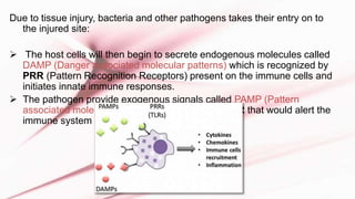
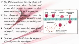
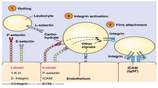
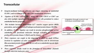
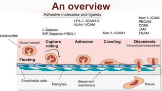
The T cell receptor (TCR) is a protein complex found on the surface of T cells that is responsible for recognizing fragments of antigen bound to MHC molecules. It consists of an alpha and beta chain, with 95% of T cells containing these chains and 5% containing gamma and delta chains instead. Each chain contains a variable region that binds the peptide-MHC complex and a constant region near the cell membrane. The variable regions contain three hypervariable complementarity-determining regions important for antigen recognition. The TCR is associated with CD3 proteins that transmit activation signals into the T cell upon peptide binding. TCR diversity arises from genetic recombination of DNA segments during T cell development.
T-Cell Activation
• Concept of immune response
• T cell-mediated immune response
• B cell-mediated immune response
I. Concept of immune response
• A collective and coordinated response to the introduction of foreign substances in an individual mediated by the cells and molecules in the immune system.
II. T cell-mediated immune response
• Cell-mediated immunity is the arm of the adaptive immune response whose role is to combat infection of intracellular pathogens, such as intracellular bacteria (mycobacteria, listeria monocytogens), viruses, protozoa, etc.
The document discusses major histocompatibility complexes (MHCs), which are glycoproteins found on cell surfaces that present antigen fragments to T cells. It describes MHC Class I and Class II molecules, which present antigens from intracellular and extracellular pathogens, respectively. MHC genes were first identified as important in tissue graft rejection and show high polymorphism between individuals.
This document discusses cellular immune response and the roles of its key components. It describes how antigen presenting cells present antigens to T lymphocytes via MHC molecules, providing the necessary stimulatory and co-stimulatory signals for T cell activation. Activated T cells then differentiate into effector and memory T cells. Effector CD8+ T cells induce apoptosis of infected cells, while effector CD4+ T cells secrete cytokines to activate macrophages. The interactions between these immune cells are regulated by cytokines. The document also discusses antigen presentation pathways, T cell maturation in the thymus, and the roles of superantigens and cytokines in the immune response.
The document discusses cytokines, their properties, classification, receptors, and signaling pathways. It notes that cytokines exhibit pleiotropy, redundancy, synergy and antagonism. Major cytokine families include hematopoietins, interferons, chemokines, and TNF. Cytokine receptors include Ig superfamily, class 1, class 2, TNF and chemokine receptors. Cytokine binding induces receptor dimerization and JAK/STAT pathway activation. Cytokines play essential roles in hematopoiesis and immunity, but imbalances can lead to disease.
B cells are lymphocytes that play a key role in humoral immunity by producing antibodies. B cell development occurs in the bone marrow, progressing from pro-B cells to immature B cells that express IgM, and then to mature B cells that express IgM and IgD. Activation of B cells leads to proliferation and differentiation into plasma cells in secondary lymphoid organs. Defects in B cell development or function can cause immunodeficiencies characterized by poor antibody production and recurrent bacterial infections. Some examples of B cell immunodeficiencies include X-linked agammaglobulinemia caused by mutations in BTK, IgA deficiency, and common variable immunodeficiency.
The complement system is part of the innate immune system and consists of over 30 proteins. It was originally identified in the 1890s by Jules Bordet and Paul Ehrlich as a heat-labile component of serum that enhanced the ability of antibodies to kill bacteria. There are three complement activation pathways: the classical pathway which is initiated by antibody-antigen complexes, the lectin pathway which is activated by mannose-binding lectin, and the alternative pathway which is spontaneously activated by microbial surfaces. Complement activation results in opsonization, inflammation, and formation of the membrane attack complex to kill microbes. Deficiencies in specific complement components can increase susceptibility to certain infections.
Cytokines are cell signaling molecules that aid cell-to-cell communication and stimulate cell movement. They are produced by immune cells and mediate processes like immunity, inflammation, and blood cell production. Cytokines bind to receptors on target cells and alter gene expression through signal transduction pathways. They exhibit properties like redundancy, synergy, and antagonism that allow for coordinated regulation of cellular activity. Diseases have been linked to overproduction or underproduction of cytokines, such as septic shock resulting from excessive cytokine levels during bacterial infection.
The document discusses cytokines, which are proteins that mediate communication between cells of the immune system. It describes the different types of cytokines, including interleukins produced by T-helper cells, lymphokines produced by lymphocytes, and monokines produced by monocytes. The document outlines the roles and functions of specific cytokines like IL-1, IL-2, TNF, IFN-γ and GM-CSF. It also discusses how cytokines are classified based on their structure and roles in innate versus adaptive immunity.
The complement system is a part of the immune system that helps or complements the ability of antibodies and phagocytic cells to clear pathogens from an organism. It is part of the innate immune system, which is not adaptable and does not change over the course of an individual's lifetime.
consists of three pathways: 1. alternative
2. classical
3. lectin pathway
The document provides an overview of the immune system, including its main cells and functions. It discusses how immune cells such as lymphocytes (B cells, T cells, NK cells), neutrophils, macrophages, mast cells, dendritic cells, and others work together to protect the body. The adaptive immune system mounts targeted responses through B cells and T cells, while the innate immune system provides initial defenses using cellular and chemical methods. Key cells include macrophages that phagocytose pathogens and present antigens, B cells that produce antibodies, and T cells that help activate other immune cells and identify and attack infected cells.
This document provides an overview of basic immunology. It begins with an introduction to immunity, the immune system, and immunology. It then discusses the history of immunology, types of immunity including innate and acquired immunity. It describes the tissues and cells involved in immunity. It covers basic aspects like antigens, antibodies, antigen-antibody reactions, and the complement system. It also discusses major histocompatibility complex, cytokines, immune disorders, and immune responses in periodontal pathogenesis.
dendritic cells are part of innate immune system, antigen presenting cells in skin, activation of t cells and inducing and maintaining immune tolerance, 4 types- langerhans cells, dermal dendritic cells, merkel cells, melanocytes
Cell-mediated immunity involves T lymphocytes, macrophages, and natural killer cells. It provides defense against viruses, fungi, and some bacteria through these cells, without involving antibodies. When antigens from invading microbes are presented on antigen-presenting cells like macrophages and dendritic cells, helper T cells are activated and stimulate cytotoxic T cells and B cells. Cytotoxic T cells then directly attack and destroy infected cells. Memory T cells also enhance future immune responses. Overall, cell-mediated immunity protects against intracellular pathogens through cellular immune responses.
The document summarizes key aspects of T cell antigen receptor (TcR) structure and generation of diversity. It describes how TcR were discovered using monoclonal antibodies that recognize unique structures on T cell clones. TcR are heterodimers composed of α and β chains that are similar to antibody structures but do not undergo somatic hypermutation. TcR diversity is generated through combinatorial rearrangement of variable (V), diversity (D), and joining (J) gene segments, as well as junctional diversity from imprecise joining and addition of untemplated nucleotides.
The document summarizes key aspects of major histocompatibility complex (MHC) molecules and T cell receptor (TCR) recognition. It discusses how MHC genes were found to be important in transplant rejection and immune responses. There are three classes of MHC molecules - Class I found on nucleated cells, Class II on antigen presenting cells, and Class III molecules like complement components. The TCR recognizes antigen peptides bound to MHC molecules. Both MHC and TCR are highly polymorphic and recognize antigens in a MHC-restricted manner.
Natural killer (NK) cells are a type of cytotoxic lymphocyte that provides rapid responses to viral infections and tumors. NK cells recognize and destroy stressed cells in the absence of antibodies and MHC molecules through activating receptors that induce apoptosis. NK cell activity is regulated by a balance between activating and inhibitory receptors - inhibitory receptors prevent killing of normal cells that express MHC class I, while activating receptors induce killing of infected or abnormal cells missing MHC I. NK cells help initiate early immune responses by releasing perforin and granzymes to induce apoptosis of virally-infected cells they detect missing MHC class I.
This document discusses cell adhesion molecules (CAMs), which are glycoproteins located on cell surfaces that help cells stick to each other and their surroundings. CAMs are classified into five major families: cadherins, Ig superfamily CAMs, selectins, integrins, and mucins. Cadherins are calcium-dependent and form connections between cells called desmosomes. Selectins help with inflammation and lymphocyte homing. Integrins facilitate cell-cell and cell-extracellular matrix adhesion and are composed of alpha and beta subunits. Malfunctions in CAMs can lead to conditions like breast cancer and leukocyte adhesion deficiency syndrome.
Cytokines are proteins that act as chemical messengers and are produced by immune cells to mediate and regulate immune responses and inflammation. They exist in different classes including interleukins, interferons, chemokines, and tumor necrosis factor. Cytokines act by binding to specific receptors on target cells and triggering signaling pathways that alter gene expression. This allows cytokines to influence diverse biological functions such as immune cell development, activation and communication.
T CELL ACTIVATION AND IT'S TERMINATIONpremvarma064
T cell activation requires two signals: 1) recognition of antigens displayed on antigen-presenting cells by T cell receptors and 2) co-stimulatory signals through molecules like CD28. This leads T cells to proliferate, differentiate into effector and memory cells, and perform effector functions. Proper activation requires interaction between T cells and antigen-presenting cells in lymphoid tissues, where costimulatory molecules are highly expressed. Dysregulation of T cell activation can lead to autoimmunity or susceptibility to infection.
B-cell activation can occur through two routes - dependent or independent of T helper cells. T-helper cells interact with antigen-bound B-cells via CD40/CD40L and B7-CD28 costimulation, releasing cytokines that cause the B-cell to proliferate and differentiate into a plasma cell. The interaction of cytokines and proliferation factors released by T-helper cells provides signals needed for B-cell proliferation and class switching.
T-cell activation requires interaction between the TCR and antigen-bound MHC on an antigen presenting cell, as well as CD28-B7 costimulation, which causes cytokine release and leads to T-cell proliferation, clonal expansion, and differentiation into memory and
NK cells are lymphocytes that constitute 5-10% of cells in human peripheral blood. They play an important role in the innate immune response by killing infected or abnormal cells. NK cells are divided into two main subpopulations, CD56dim NK cells which are highly cytotoxic and mediate antibody-dependent cellular cytotoxicity, and CD56bright NK cells which rapidly produce cytokines and chemokines upon activation. A specialized NK cell population in the uterus, uterine NK cells, help form the placenta during pregnancy by enlarging blood vessels to supply the growing fetus.
This document discusses T cell activation and maturation. It describes the processes of negative and positive selection that screen T cells. T cell activation occurs through engagement of the T cell receptor and co-stimulatory molecules by MHC peptide and co-stimulatory molecules on antigen presenting cells. Binding of the T cell receptor provides the first signal, while co-stimulation provides the second signal required for an effective immune response.
T cells can be categorized into several subsets including helper T cells, cytotoxic T cells, memory T cells, and regulatory T cells. Helper T cells assist other immune cells, cytotoxic T cells destroy infected and tumor cells, memory T cells provide faster responses upon reexposure to pathogens, and regulatory T cells suppress immune activation and prevent autoimmunity. Understanding regulatory T cells in HIV-1 could lead to new immunotherapy or vaccine strategies, but their exact role in HIV-1 pathogenesis requires further study.
The document discusses the major histocompatibility complex (MHC) in mammals. It notes that MHC acts as antigen presenting receptors and are involved in cell-cell interaction, antigen presentation, and recognition of self and non-self molecules. MHC is found on chromosome 6 in humans and is referred to as the HLA complex. MHC molecules are divided into three main classes - Class I MHC present antigens to cytotoxic T cells, Class II MHC present antigens to helper T cells, and Class III MHC genes encode complement components and cytokines. The structures and functions of Class I and Class II MHC molecules are described in detail.
The document discusses induced innate immunity and the roles of cytokines and chemokines. It can be summarized as follows:
1. Induced innate immunity begins 4-96 hours after exposure to pathogens and involves the recruitment of defense cells through cytokines and chemokines produced in response to pathogen recognition.
2. Cytokines are small proteins released by cells in response to stimuli that induce responses by binding to receptors, acting in autocrine, paracrine, or endocrine manners. Chemokines are a class of cytokines that induce cell migration.
3. Key cytokines and chemokines involved in innate immunity include interleukin-1, interleukin-6, interleukin-12, TNF-α, and interleukin-8. They
Cell adhesion molecules help cells stick to each other and their surroundings through proteins. There are several types of cell adhesion molecules including immunoglobulin super family CAMs, integrins, selectins, and cadherins. Cadherins like E-cadherin form adherens junctions between cells and link to actin through catenins. Changes in cell adhesion can lead to diseases such as cancer where reduced adhesion allows cancer cells to invade other tissues. Cell adhesion molecules are important for tissue development and function.
Immunity :
It is defined as the resistance exhibited by the host against any
foreign antigen including microorganisms.
Plays a major role in prevention of infectious diseases.
The immune system is a network designed for the homeostasis of large molecules (oligomers) and cells based on specific recognition processes.
It is the collection of cells, tissues and molecules that function to defend us against infectious microbes
Intercellular connections and molecular motorsAnwar Siddiqui
This document summarizes a physiology seminar on intercellular connections and molecular motors. It discusses various cell adhesion molecules like cadherins, selectins, immunoglobulin superfamily molecules, and integrins that mediate cell-cell and cell-matrix adhesion. It also describes different types of intercellular junctions such as tight junctions, desmosomes, and hemidesmosomes. Finally, it provides an overview of molecular motors like kinesin, dynein, and myosin that transport cargo within cells and generate forces through ATP hydrolysis.
Cell junctions are specialized contact sites that hold cells together and attach cells to the extracellular matrix. They are classified into three main groups: tight junctions, gap junctions, and adherens junctions. Tight junctions form continuous seals around cells to control permeability and prevent diffusion between cells. Gap junctions allow small molecules and ions to pass directly between cells to facilitate cell-cell communication. Adherens junctions, such as desmosomes and hemidesmosomes, anchor cells to other cells or the extracellular matrix. Cell adhesion molecules like cadherins, integrins, and immunoglobulin superfamily proteins mediate cell-cell and cell-matrix adhesion through homophilic or heterophilic binding interactions.
Acute inflammation is the immediate and early response to harmful stimuli, characterized by vascular changes that increase blood flow and permeability, allowing plasma proteins and cells to enter tissues. This forms an exudate containing antibodies, leukocytes, and other factors that work to dilute, destroy, and remove the cause of injury. The cardinal signs of inflammation - heat, redness, swelling, pain, and loss of function - result. Acute inflammation is usually short-lived and resolves once the stimulus has been dealt with.
This document summarizes the key processes involved in acute inflammation. It describes how inflammation is initiated by increased blood flow and vascular permeability, allowing leukocytes and proteins to exit blood vessels and enter infected or injured tissues. The roles of different leukocytes, especially neutrophils and macrophages, in recognizing, phagocytosing, and destroying pathogens are discussed. Critical adhesion molecules and chemokines involved in leukocyte recruitment are also outlined.
2. inflammation cellular events dr ashutosh kumarDrAshutosh Kumar
The document discusses the process of acute inflammation and the cellular events involved. It describes how leukocytes are recruited to the site of injury through a process called chemotaxis. It then outlines the steps of phagocytosis - adherence, ingestion, digestion. It discusses the mechanisms used by phagocytes to kill and degrade microbes, including both oxygen-dependent and oxygen-independent processes. Finally, it briefly mentions some genetic defects and acquired conditions that can cause defects in leukocyte function.
The document describes a study examining the role of the FUT7 gene during leukocyte adhesion and extravasation. The study used CRISPR/Cas9 to knockout the FUT7 gene in HL-60 cells. A microfluidic flow chamber was used to compare cell rolling and adhesion of FUT7 knockout HL-60 cells to wild type HL-60 cells on P-selectin and E-selectin coatings under flow. A transmigration assay compared the ability of the two cell types to migrate across activated HUVEC monolayers under static conditions. Preliminary results showed FUT7 plays a dominant role in P-selectin mediated adhesion but a smaller role in E-selectin binding. FUT7 knockout did not
series of events which takes place at the time of acute inflammation includes two different kinds of, one at the vascular level and other one is at the cellular level. which works as the primary level of immunity protection and leads to the phagocytosis of the pathogenic microbes. The presence of foreign bodies such as bacteria within the bodies provokes a protective inflammatory response...characterized by redness, swelling, warmth and the pain at the site of infection. These signs are due to increased blood flow, increased capillary permeability and the escape of fluid and cells into the tissue spaces. The increased permeability is due to several chemical mediators of which histamines, prostaglandins and leukotriens are the most important ones.
White blood cells (leukocytes) play an important role in immune responses and tissue maintenance. There are two main types - polymorphonuclear granulocytes which have lobed nuclei and cytoplasmic granules, and mononuclear agranulocytes which lack granules and have large nuclei. Neutrophils, eosinophils, basophils, lymphocytes and monocytes are the major leukocyte types and each plays distinct roles in immune function and response to pathogens. Leukocytes are produced through myeloid and lymphoid stem cell lineages in the bone marrow.
The document summarizes the key steps and processes involved in acute inflammation. It describes how immune cells like macrophages recognize pathogens or damage and release inflammatory mediators. These mediators cause vasodilation, increased permeability, and the classic signs of inflammation - redness, heat, swelling and pain. The document then details the leukocyte adhesion cascade by which immune cells migrate to the site of inflammation, and the processes of chemotaxis, phagocytosis, intracellular killing, and extracellular release involved in the immune response.
This document provides an overview of myeloid leucopoiesis, or the formation of white blood cells from myeloid progenitor cells. It discusses the sites where white blood cell formation occurs, the growth factors involved in differentiation and proliferation, and the maturation process for granulocytes and monocytes. The key functions of neutrophils, eosinophils, basophils and monocytes/macrophages are also summarized, including their roles in phagocytosis and the innate immune response.
Cell adhesion molecules and mechanisms of cell adhesionKunaal Agrawal
This document discusses the extracellular matrix and cell adhesion molecules that help connect cells to each other and their surroundings. It covers the main components of the extracellular matrix, including collagens, elastins, proteoglycans, and hyaluronic acid. It also examines various cell adhesion molecules like integrins, cadherins, selectins, and the immunoglobulin superfamily that allow cell-cell and cell-matrix binding through interactions with extracellular matrix proteins or receptors on other cells. These adhesion complexes are important for tissue structure and function.
1. During acute inflammation, leukocytes marginate and roll along vessel walls before firmly adhering and transmigrating between endothelial cells into tissues.
2. Once in tissues, leukocytes migrate toward sites of infection or injury via chemotaxis guided by chemical gradients of chemoattractants.
3. The adhesion and migration of leukocytes is regulated by cell surface adhesion molecules including selectins, immunoglobulins, integrins, and mucins that bind counterparts on endothelial cells.
The document summarizes the biochemistry of the vascular system. It describes how blood is pumped through arteries and returns via veins, exchanging nutrients and waste through capillaries. It focuses on the endothelium and adhesion molecules that allow immune cells to exit blood vessels at sites of infection. Selectins mediate initial attachment and rolling of leukocytes along endothelium. Integrins and cellular adhesion molecules then firm attachment and transmigration. The endothelium regulates vascular tone and homeostasis through nitric oxide, prostacylandins, and other factors.
The presentation may give you an idea abouth the disease, its pathophysiology, signs, symptoms, diagnosis, treatment....Thanks toall the websites which helped me to make this presentation.
The tryptophan operon regulates the biosynthesis of tryptophan in E. coli through transcriptional attenuation and repression. It contains five genes encoding the enzymes needed to synthesize tryptophan. When tryptophan levels are high, the tryptophan repressor binds to the operator site, preventing transcription. Additionally, a regulatory region can form a terminator stem-loop structure to halt transcription if tryptophan tRNA levels are high during translation of the leader mRNA sequence. However, if tryptophan levels are low, the terminator structure does not form and transcription of the operon proceeds.
This presentation gives information about the antimetabolites and suicide inhibitors that we are frequently using. Their mechanism and various examples are also given
This document discusses antigens and their classification. It defines antigens as substances that can induce an immune response. Antigens are classified as either exogenous (external) or endogenous (internal) antigens. Exogenous antigens enter the body from the external environment, while endogenous antigens are further divided into xeno-genic, allogenic, and autologous antigens based on their origin. The document also discusses the properties of immunogens and antigens, as well as factors that contribute to immunogenicity.
The document discusses the structure of proteins at various levels of organization:
- Proteins are composed of amino acids linked together by peptide bonds to form polypeptide chains. The sequence and interactions of these chains determine the protein's structure.
- There are four levels of protein structure - primary, secondary, tertiary, and quaternary. Secondary structure includes alpha helices and beta sheets formed by hydrogen bonding between amino acids in the chain. Tertiary structure describes the overall 3D shape formed by interactions between amino acid side chains. Quaternary structure involves the interaction of multiple polypeptide chains.
- Protein structure enables proteins to perform their diverse functions through processes like enzyme catalysis, oxygen transport, and providing structure
Colloids are substances microscopically dispersed throughout another substance. The dispersed particles range in size from 1-100 nm. Colloids exhibit properties between true solutions and suspensions due to their intermediate particle size. They are able to pass through filters but not semipermeable membranes. Common examples include milk, fog, mayonnaise and paints. Colloids can be classified based on factors like the physical state of the phases, the interaction between the phases, the size and nature of dispersed particles, and the electrical charge on particles. They are purified using techniques like dialysis, electrodialysis, and ultrafiltration which separate colloidal particles from dissolved substances.
Mitochondria are double-membrane organelles found in the cytoplasm of eukaryotic cells that produce energy through aerobic respiration. They originated from endosymbiotic bacteria and contain their own DNA. Mitochondria have an outer and inner membrane, intermembrane space, cristae folds in the inner membrane that increase surface area, and a matrix space containing enzymes. They produce ATP through oxidative phosphorylation to power cellular activities, making them the powerhouses of the cell. Mitochondrial DNA is maternally inherited and encodes proteins essential for mitochondrial function.
Immersive Learning That Works: Research Grounding and Paths ForwardLeonel Morgado
We will metaverse into the essence of immersive learning, into its three dimensions and conceptual models. This approach encompasses elements from teaching methodologies to social involvement, through organizational concerns and technologies. Challenging the perception of learning as knowledge transfer, we introduce a 'Uses, Practices & Strategies' model operationalized by the 'Immersive Learning Brain' and ‘Immersion Cube’ frameworks. This approach offers a comprehensive guide through the intricacies of immersive educational experiences and spotlighting research frontiers, along the immersion dimensions of system, narrative, and agency. Our discourse extends to stakeholders beyond the academic sphere, addressing the interests of technologists, instructional designers, and policymakers. We span various contexts, from formal education to organizational transformation to the new horizon of an AI-pervasive society. This keynote aims to unite the iLRN community in a collaborative journey towards a future where immersive learning research and practice coalesce, paving the way for innovative educational research and practice landscapes.
The binding of cosmological structures by massless topological defectsSérgio Sacani
Assuming spherical symmetry and weak field, it is shown that if one solves the Poisson equation or the Einstein field
equations sourced by a topological defect, i.e. a singularity of a very specific form, the result is a localized gravitational
field capable of driving flat rotation (i.e. Keplerian circular orbits at a constant speed for all radii) of test masses on a thin
spherical shell without any underlying mass. Moreover, a large-scale structure which exploits this solution by assembling
concentrically a number of such topological defects can establish a flat stellar or galactic rotation curve, and can also deflect
light in the same manner as an equipotential (isothermal) sphere. Thus, the need for dark matter or modified gravity theory is
mitigated, at least in part.
Current Ms word generated power point presentation covers major details about the micronuclei test. It's significance and assays to conduct it. It is used to detect the micronuclei formation inside the cells of nearly every multicellular organism. It's formation takes place during chromosomal sepration at metaphase.
When I was asked to give a companion lecture in support of ‘The Philosophy of Science’ (https://shorturl.at/4pUXz) I decided not to walk through the detail of the many methodologies in order of use. Instead, I chose to employ a long standing, and ongoing, scientific development as an exemplar. And so, I chose the ever evolving story of Thermodynamics as a scientific investigation at its best.
Conducted over a period of >200 years, Thermodynamics R&D, and application, benefitted from the highest levels of professionalism, collaboration, and technical thoroughness. New layers of application, methodology, and practice were made possible by the progressive advance of technology. In turn, this has seen measurement and modelling accuracy continually improved at a micro and macro level.
Perhaps most importantly, Thermodynamics rapidly became a primary tool in the advance of applied science/engineering/technology, spanning micro-tech, to aerospace and cosmology. I can think of no better a story to illustrate the breadth of scientific methodologies and applications at their best.
ESR spectroscopy in liquid food and beverages.pptxPRIYANKA PATEL
With increasing population, people need to rely on packaged food stuffs. Packaging of food materials requires the preservation of food. There are various methods for the treatment of food to preserve them and irradiation treatment of food is one of them. It is the most common and the most harmless method for the food preservation as it does not alter the necessary micronutrients of food materials. Although irradiated food doesn’t cause any harm to the human health but still the quality assessment of food is required to provide consumers with necessary information about the food. ESR spectroscopy is the most sophisticated way to investigate the quality of the food and the free radicals induced during the processing of the food. ESR spin trapping technique is useful for the detection of highly unstable radicals in the food. The antioxidant capability of liquid food and beverages in mainly performed by spin trapping technique.
The technology uses reclaimed CO₂ as the dyeing medium in a closed loop process. When pressurized, CO₂ becomes supercritical (SC-CO₂). In this state CO₂ has a very high solvent power, allowing the dye to dissolve easily.
Travis Hills of MN is Making Clean Water Accessible to All Through High Flux ...Travis Hills MN
By harnessing the power of High Flux Vacuum Membrane Distillation, Travis Hills from MN envisions a future where clean and safe drinking water is accessible to all, regardless of geographical location or economic status.
Authoring a personal GPT for your research and practice: How we created the Q...Leonel Morgado
Thematic analysis in qualitative research is a time-consuming and systematic task, typically done using teams. Team members must ground their activities on common understandings of the major concepts underlying the thematic analysis, and define criteria for its development. However, conceptual misunderstandings, equivocations, and lack of adherence to criteria are challenges to the quality and speed of this process. Given the distributed and uncertain nature of this process, we wondered if the tasks in thematic analysis could be supported by readily available artificial intelligence chatbots. Our early efforts point to potential benefits: not just saving time in the coding process but better adherence to criteria and grounding, by increasing triangulation between humans and artificial intelligence. This tutorial will provide a description and demonstration of the process we followed, as two academic researchers, to develop a custom ChatGPT to assist with qualitative coding in the thematic data analysis process of immersive learning accounts in a survey of the academic literature: QUAL-E Immersive Learning Thematic Analysis Helper. In the hands-on time, participants will try out QUAL-E and develop their ideas for their own qualitative coding ChatGPT. Participants that have the paid ChatGPT Plus subscription can create a draft of their assistants. The organizers will provide course materials and slide deck that participants will be able to utilize to continue development of their custom GPT. The paid subscription to ChatGPT Plus is not required to participate in this workshop, just for trying out personal GPTs during it.
The debris of the ‘last major merger’ is dynamically youngSérgio Sacani
The Milky Way’s (MW) inner stellar halo contains an [Fe/H]-rich component with highly eccentric orbits, often referred to as the
‘last major merger.’ Hypotheses for the origin of this component include Gaia-Sausage/Enceladus (GSE), where the progenitor
collided with the MW proto-disc 8–11 Gyr ago, and the Virgo Radial Merger (VRM), where the progenitor collided with the
MW disc within the last 3 Gyr. These two scenarios make different predictions about observable structure in local phase space,
because the morphology of debris depends on how long it has had to phase mix. The recently identified phase-space folds in Gaia
DR3 have positive caustic velocities, making them fundamentally different than the phase-mixed chevrons found in simulations
at late times. Roughly 20 per cent of the stars in the prograde local stellar halo are associated with the observed caustics. Based
on a simple phase-mixing model, the observed number of caustics are consistent with a merger that occurred 1–2 Gyr ago.
We also compare the observed phase-space distribution to FIRE-2 Latte simulations of GSE-like mergers, using a quantitative
measurement of phase mixing (2D causticality). The observed local phase-space distribution best matches the simulated data
1–2 Gyr after collision, and certainly not later than 3 Gyr. This is further evidence that the progenitor of the ‘last major merger’
did not collide with the MW proto-disc at early times, as is thought for the GSE, but instead collided with the MW disc within
the last few Gyr, consistent with the body of work surrounding the VRM.
3. LEUKOCYTES
• The cellular components of blood include erythrocytes,
leukocytes and platelets.
• Leukocytes are a heterogeneous group of nucleated cells.
• Normal human blood contains 4000-10000 leukocytes/µl.
• Leukocytes are classified onto five:
Neutrophils (40-75%)
Eosinophils (1-6%)
Granulocytes
Basophils (less than
1%)
Monocytes (2-10%)
Agranulocytes
6. Due to tissue injury, bacteria and other pathogens takes their entry on to
the injured site:
The host cells will then begin to secrete endogenous molecules called
DAMP (Danger associated molecular patterns) which is recognized by
PRR (Pattern Recognition Receptors) present on the immune cells and
initiates innate immune responses.
The pathogen provide exogenous signals called PAMP (Pattern
associated molecular patterns) recognized by PRR that would alert the
immune system to the presence of pathogen.
7. The APC present near the injured site will
also phagocytose these bacteria and
present their peptide fragments on their
membrane to activate the T lymphocytes.
Non phagocytic granulocytes near the
injured tissue will release histamine which
increases the blood vessel permeability and
smooth muscle activity.
Cytokines are also secreted by neutrophils,
eosinophils, macrophage, activated T
cells….
Cytokines secreted will direct the movement
of leukocytes to the injured area.
8. LEUKOCYTE EXTRAVASATION
• Also called Diapedesis.
• It is the movement of leukocytes out of the circulatory system and
towards the site of tissue damage or infection.
• Chemokines or chemoattractant cytokines are a family of pro
inflammatory mediators produced at the inflammatory site which induces
the mechanism of extravasation.
9. Leukocyte extravasation is orchestrated by the
combined action of cellular adhesion receptors and
chemotactic factors, and involves radical
morphological changes in both leukocytes and
endothelial cells.
10. Recruitment of Leukocytes to Inflammation Site
Multistep Model
1. Tethering (Capture)
2. Rolling
3. Activation
4. Firm Adhesion
5. Transendothelial
migration
11. TETHERING AND ROLLING
The generated inflammatory chemicals, bind to seven
helix receptors on endothelial cells and stimulate the
fusion of cytoplasmic vesicles called Weibel-Palade
bodies with the plasma membrane.
This exposes, P-Selectin formerly stored in the
vesicle membranes , on the cell surface facing the
blood.
Selectins bind mucins that are constitutively exposed
on the surface of neutrophils, tethering them to the
surface.
The bond forms and break rapidly, allowing the
neutrophils to roll along the surface of the
endothelium at rates greater then 10µm/s as the
blood wall pushes them along.
12. Interaction between Selectin and carbohydrate ligand present on the leucocytes.
• WBC and Platelets use selectins to interact with
vascular endothelial cells.
• Selectins are a group of transmembrane molecules,
expressed on the surface of leukocytes and activated
endothelial cells.
• Selectins contain an N-terminal extracellular
domain with structured homology to Ca-dependent
lectins, followed by a domain homologous to
epidermal growth factor and 2-9 consensus repeats
similar to sequences found in complementary
regulatory proteins.
13. • All selectin ligands are transmembrane glycoproteins
which present oligosaccharide structures to the
selectin.
• All three selectins recognize glycoproteins or
glycolipids containing the tetrasaccharide Sialyl-
Lewis.
• This tetrasaccharide is found on all circulating
myeloid cells and is composed of sialic acid, galactose,
fucose and N-acetyl galactosamine.
The low affinity nature of selectins is
what allows the characterestic rolling
action of leukocytes.
14.
15.
16. Activation & Firm Adhesion
• During the process of leukocyte rolling the contact of
leukocytes with the luminal endothelial surface allows
leukocytes to effectively ‘sense’ the endothelial
surface-bound chemokines.
• These surface-deposited chemokines are presented to
rolling leukocytes.
• By interaction with the G-protein coupled chemokine
receptors on leukocytes chemokines induce
intracellular signals leading to inside-out integrin
activation and firm leukocyte adhesion.
17. Binding of chemokines to their receptors on leukocytes
results in the inside-out signalling activation of leukocyte β1-
integrins and the β2-integrins LFA-1 and Mac-l, that
mediate firm arrest of leukocytes.
Neutrophils use both LFA-1 and Mac-l for adhesion.
The endothelial counter-ligands for leukocyte integrins are
members of the immunoglobulin superfamily and include
(ICAM)-1–5, (VCAM)-1 and mucosal addressin cell adhesion
molecule-1 (MAdCAM-1).
LFA-1 and Mac-1 bind to endothelial ICAMs such as ICAM-
1 and ICAM-2.
ICAM-1 and ICAM-2 are constitutively expressed, and
ICAM-1 expression is further increased after endothelial
activation.
In contrast, endothelial VCAM-1 is recognized by β-1
integrin receptors predominantly found on lymphocytes and
monocytes .
18.
19. Transmigration
Transmigration of leukocytes through the vascular endothelium can
take place in a paracellular or transcellular manner.
Actin rearrangement ocuurs in leukocytes, pseudopods are formed and
then they begin to squeeze through the endothelial cells moving
towards higher cocentration of chemokines.
20. Paracellular
• Leukocytes encounter multiple endothelial cell junctional
molecules and molecular complexes during paracellular
transmigration.
• CD99 and CD31 (platelet endothelial cell adhesion molecule-1
(PECAM-1)) are expressed on endothelium and are enriched at
cell-cell lateral junctions.
• These molecules also are expressed on most leukocyte types.
• Both molecules interact through homophilic interactions, that is,
CD99 on one endothelial cell binds to the same molecule on
adjacent endothelial cells.
• Transmigrating leukocytes must cross endothelial tight junctions
(TJ) and adherens junctions (AJ), which contain numerous
proteins involved in selective permeability, growth control, and
cell-cell adhesion.
• The tight junction and its subjacent adherens junction (AJ)
constitute the apical junctional complex (AJC).
21. • Adherens junction include VE-Cadherin and tight junctions that incorporate members of the junctional adhesion
molecule(JAM) family, endothelial cell selective adhesion molecule(ESAM) and claudins.
• A number of other adhesion molecules are also enriched at borders of adjacent endothelial cells such as PECAM 1,
CD99,ICAM 2.
• These multiple junctional endothelial cell adhesion molecules are thought to recycle in a variety of intracellular
compartments or vesicles including the membranous lateral border recycling compartment (LBRC).
• LBRC supports leukocyte TEM through efficient recruitment of key molecules to sites of leukocyte diapedesis.
• These molecules will undergo various mechanisms and causes the loosening of the junctions between endothelial
cells to enable leukocyte damage.
22. Transcellular
Integrin-mediated leukocyte adhesion can trigger clustering of endothelial
ICAM-1, and recruitment of VCAM-1 into membranous structures.
Ligation of ICAM-1 and VCAM-1, along with CD9, CD151 or CD47 can
also elicit multiple signaling events in endothelial cells postulated to reduce
endothelial barrier properties.
This includes increased intracellular Ca2+, reactive oxygen species (ROS)
generation, and activation of p38 mitogen-activated protein kinase (MAPK).
In addition, ICAM-1 ligation can result in tyrosine phosphorylation of key
endothelial cell junctional molecules through activation of endothelial
proline-rich tyrosine kinase 2 (Pyk2) and Src kinase.
These responses can couple to the triggering of RhoA (a small GTPase
involved in regulation of actin cytoskeletal networks) and its downstream
Rho-associated protein kinase (ROCK), as well as to endothelial myosin light-
chain kinase.
These responses finally result in the formation of intracellular channels
through which the leukocytes enter.