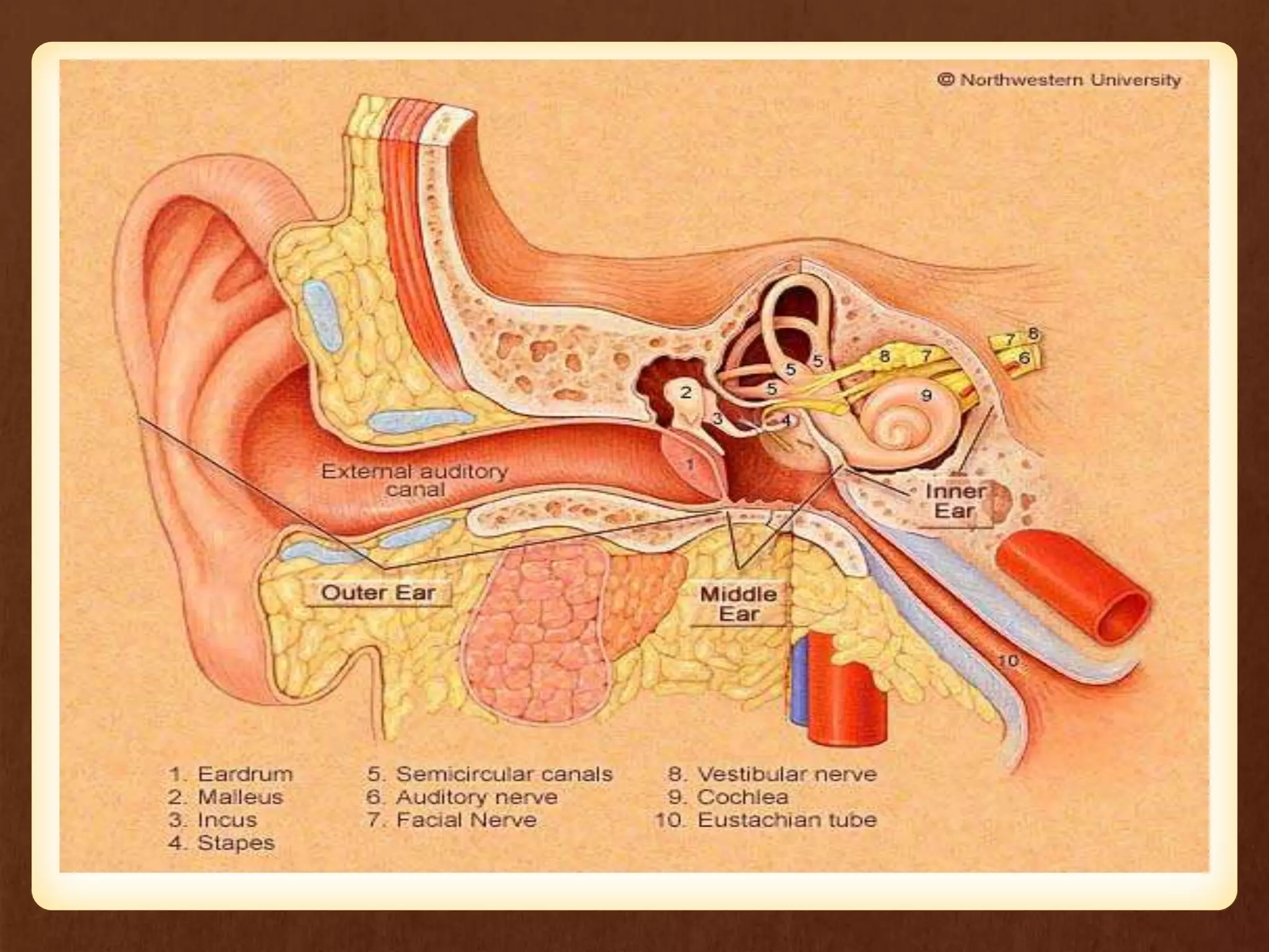
DEPARTMENT OF
MEDICAL SURGICAL NURSING
LEARNING OBJECTIVES
At the end of this lecture, students should be able to :
• define otosclerosis
• list out the causes
• explain the pathophysiology
• enumerate the diagnostic measures
• describe the management
Introduction
• primary metabolic bone disease of the otic
capsule and ossicles
• It causes fixation of the ossicles (stapes)
• It results in conductive or mixed hearing loss.
• It is genetically-mediated via autosomal
dominant transmission
Definition
• Otosclerosis or “hardening of the ear” result from
the formation of an abnormal spongy bone , like
bone growth along the stapes in the middle ear.
• With the new bone growth , the stapes become
immobile prevents transmission as sound vibration
into the ear, leading to conductive hearing loss.
• Otosclerosis usually affect the both ears.
Etiology
Many theories have been proposed such as
• Hereditary, endocrine,
• metabolic, Infectious lesion
• Vascular
• Measles
• autoimmune
Pathophysiology
• Due to etiological causes
• Formation of new bone, or re growth at the area of stepes
• limited to the vibration of the bone
• Conducting hearing loss
Clinical Manifestations
• Hearing loss
• Dizziness
• Tinnitus
• Roaring
• Buzzing the ear
• Vertigo
• headache & earache
Diagnostic evaluation
• History
• Physial examination
• Tuning fork test
• Audiometry test
• Tympanocentesis – fluid for middle ear send for culture
• CT scan – collection of fluidin ear & mastoid region ,
abscess formation
• MRI – evaluation of tumor & soft tissue
• AUDIOGRAPHY – to assess hearing loss
Diagnosis
• Slowly progressive
• Tinnitus is associated with 75% patients
• The age of onset of hearing loss is young
Diagnosis
• low-volume speech.
• Paracusis of Willis.
• Two-thirds of patients will report a family history of
hearing loss.
• Women with pregnancy worse her hearing
Complications
• Complete deafness
• Nerve damage
• Infection, dizziness, pain, or blood clot in the ear
after surgery
Management
• Otosclerosis may slowly get worse. The condition
may not require treatment until you having severe
hearing problems.
• Medications such as fluoride, calcium, or vitamin D
may help to slow the hearing loss, but the benefits
have not yet been proved.
• No known medical treatment exists for this form of
deafness, but amplification with a hearing aid may
be helpful.
• Administer analgesics such as –
• IBUPROFEN
• OXYCODONE
• ACETAMENOPHEN (PCM)
• Hearing aid may be used to treat the hearing loss
General measures :
• Avoidance of noise full environment
• Side lying position
• Continuous applications of medications
• High protein diet
Surgical interventions
• Stapedectomy - The removal of
portion of the sclerotic stepes
footplate of stapes or complete
removal of the stapes and the
implant with prosthesis to
maintain suitable conduction.
• Stapedotomy - Modern surgery called stapedotomy
is performed by drilling a small hole in the stapes
footplate with micro drill or laser, and the insertion
of a piston like prosthesis.
Non-surgical interventions
• Amplification: hearing aide
• Patients who do not want to undergo
surgery for otosclerosis
• patients who are not fit for surgery.
Non-surgical interventions
• Medical treatment:
• Usual dose is about 20-120mg of fluoride a
day
• Efficacy of the treatment can be evaluated
2 years later.
Nursing assessment
1. History of onset & progression of symptoms
2. Extend of hearing loss via audiometry
3. Rinne/weber test – to evaluate loss of air
conduction
4.Past medical history of leasons and infection
5.Family history of otosclerosis
• Nursing diagnosis :
• Impaired hearing activity related to disease
condition
• Acute pain related to disease process
• Risk for injury related to hearing loss
• Imbalance nutritional status less than body
requirement related to vertigo
• Anxiety related to surgical intervention and
prognosis
• Knowledge deficit related to surgical intervention
• Interventions
• Assess the general condition of patient
• Provide side lying position
• Remove sound or noise from the patient area
• Reassure patient that dizziness is tempory
• Maintain fluid balance of the patient
• Prepare patient for surgical intervention
• Explain the surgical procedure and get inform
consent
• Explain the use of hearing device
• Assess the sign of bleeding , drainage at the site of
surgery
• Provide medication as per doctors prescription
• Health education should be given to patient
regarding diet like use of calcium rich diet, high
protein diet, use of hearing device, medication and
followup
• Evaluation
• Evaluate the patient condition and hearing activity
of the patient
• Ask the patient for regular follow up
Summary
So far we have discussed about definition, etiology, clinical
manifestations, investigations, and managements of
otosclerosis.
Bibliography
• Lewis et al, Medical Surgical Nursing, Mosby
Elsevier,7th edition.
• Joyce.M.Black et al, Medical Surgical Nursing,
Saunders publication.
• Brunner and Siddhartha, Medical Surgical Nursing,
Lippincott Williams and Wilkins.
Thank You

Otosclerosis

  • 1.
  • 2.
    LEARNING OBJECTIVES At theend of this lecture, students should be able to : • define otosclerosis • list out the causes • explain the pathophysiology • enumerate the diagnostic measures • describe the management
  • 3.
    Introduction • primary metabolicbone disease of the otic capsule and ossicles • It causes fixation of the ossicles (stapes) • It results in conductive or mixed hearing loss. • It is genetically-mediated via autosomal dominant transmission
  • 4.
    Definition • Otosclerosis or“hardening of the ear” result from the formation of an abnormal spongy bone , like bone growth along the stapes in the middle ear. • With the new bone growth , the stapes become immobile prevents transmission as sound vibration into the ear, leading to conductive hearing loss. • Otosclerosis usually affect the both ears.
  • 8.
    Etiology Many theories havebeen proposed such as • Hereditary, endocrine, • metabolic, Infectious lesion • Vascular • Measles • autoimmune
  • 9.
    Pathophysiology • Due toetiological causes • Formation of new bone, or re growth at the area of stepes • limited to the vibration of the bone • Conducting hearing loss
  • 10.
    Clinical Manifestations • Hearingloss • Dizziness • Tinnitus • Roaring • Buzzing the ear • Vertigo • headache & earache
  • 11.
    Diagnostic evaluation • History •Physial examination • Tuning fork test • Audiometry test • Tympanocentesis – fluid for middle ear send for culture • CT scan – collection of fluidin ear & mastoid region , abscess formation • MRI – evaluation of tumor & soft tissue • AUDIOGRAPHY – to assess hearing loss
  • 12.
    Diagnosis • Slowly progressive •Tinnitus is associated with 75% patients • The age of onset of hearing loss is young
  • 13.
    Diagnosis • low-volume speech. •Paracusis of Willis. • Two-thirds of patients will report a family history of hearing loss. • Women with pregnancy worse her hearing
  • 14.
    Complications • Complete deafness •Nerve damage • Infection, dizziness, pain, or blood clot in the ear after surgery
  • 15.
    Management • Otosclerosis mayslowly get worse. The condition may not require treatment until you having severe hearing problems. • Medications such as fluoride, calcium, or vitamin D may help to slow the hearing loss, but the benefits have not yet been proved. • No known medical treatment exists for this form of deafness, but amplification with a hearing aid may be helpful.
  • 16.
    • Administer analgesicssuch as – • IBUPROFEN • OXYCODONE • ACETAMENOPHEN (PCM) • Hearing aid may be used to treat the hearing loss General measures : • Avoidance of noise full environment • Side lying position • Continuous applications of medications • High protein diet
  • 17.
    Surgical interventions • Stapedectomy- The removal of portion of the sclerotic stepes footplate of stapes or complete removal of the stapes and the implant with prosthesis to maintain suitable conduction.
  • 18.
    • Stapedotomy -Modern surgery called stapedotomy is performed by drilling a small hole in the stapes footplate with micro drill or laser, and the insertion of a piston like prosthesis.
  • 20.
    Non-surgical interventions • Amplification:hearing aide • Patients who do not want to undergo surgery for otosclerosis • patients who are not fit for surgery.
  • 21.
    Non-surgical interventions • Medicaltreatment: • Usual dose is about 20-120mg of fluoride a day • Efficacy of the treatment can be evaluated 2 years later.
  • 22.
    Nursing assessment 1. Historyof onset & progression of symptoms 2. Extend of hearing loss via audiometry 3. Rinne/weber test – to evaluate loss of air conduction 4.Past medical history of leasons and infection 5.Family history of otosclerosis
  • 23.
    • Nursing diagnosis: • Impaired hearing activity related to disease condition • Acute pain related to disease process • Risk for injury related to hearing loss • Imbalance nutritional status less than body requirement related to vertigo • Anxiety related to surgical intervention and prognosis • Knowledge deficit related to surgical intervention
  • 24.
    • Interventions • Assessthe general condition of patient • Provide side lying position • Remove sound or noise from the patient area • Reassure patient that dizziness is tempory • Maintain fluid balance of the patient • Prepare patient for surgical intervention • Explain the surgical procedure and get inform consent
  • 25.
    • Explain theuse of hearing device • Assess the sign of bleeding , drainage at the site of surgery • Provide medication as per doctors prescription • Health education should be given to patient regarding diet like use of calcium rich diet, high protein diet, use of hearing device, medication and followup
  • 26.
    • Evaluation • Evaluatethe patient condition and hearing activity of the patient • Ask the patient for regular follow up
  • 27.
    Summary So far wehave discussed about definition, etiology, clinical manifestations, investigations, and managements of otosclerosis.
  • 28.
    Bibliography • Lewis etal, Medical Surgical Nursing, Mosby Elsevier,7th edition. • Joyce.M.Black et al, Medical Surgical Nursing, Saunders publication. • Brunner and Siddhartha, Medical Surgical Nursing, Lippincott Williams and Wilkins.
  • 29.