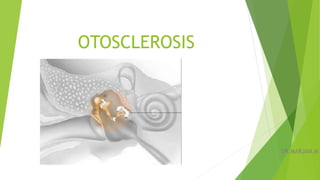
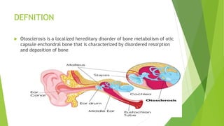
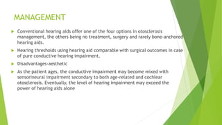
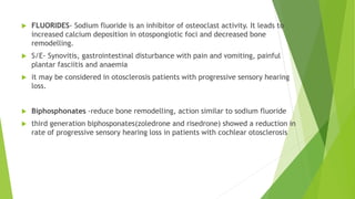
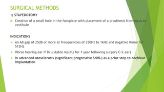
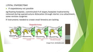
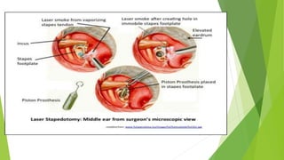
OTOSCLEROSIS
DR.MARJAN.M
DEFNITION
 Otosclerosis is a localized hereditary disorder of bone metabolism of otic
capsule enchondral bone that is characterized by disordered resorption
and deposition of bone
TYPES
 CLINICAL OTOSCLEROSIS - lesions that affect the stapes, stapediovestibular joint or
round window membrane thus cause conductive hearing loss.
 COCHLEAR OTOSCLEROSIS -lesions involving the cochlear endosteum without
affecting the stapes or the stapediovestibular joint, thus causing pure sensorineural
hearing loss.It is very rare
 HISTOLOGIC OTOSCLEROSIS refers to histopathological lesions of temporal bone that
do not affect the stapes, stapediovestibular joint or cochlear endosteum, and thus
remain asymptomatic during life
 Prevalence of clinically apparent otosclerosis is 0.3-0.5% and histological is 10 times
more.
EPIDEMIOLOGY
 More common in Caucasians, less common in Southeast Asians and Native
Americans, rare in Africans
 1.4-2 times higher in women as compared to males.
 Eventually involve both ear in 85-90% patients
PATHOLOGY
 Normal temporal bone -embryonic cartilage rests”globuli interossei”
 The earliest indication of otosclerotic process is RESORPTION OF ENCHONDRAL BONE
AROUND BLOOD VESSELS, with consequent enlargement of perivascular spaces
followed by deposition of immature (woven) bone by osteoblast ,this unslable matrix
is seen histologically as change in extracellular staining pattern- BLUE MANTLE.
 This immature bone continues remodelling mediated by osteoblasts -
OTEOSPONGIOSIS
 Later, more mature bone, sclerotic, dense, irregularly woven, poorly vascularised
(lamellar bone), is deposited- OSTEOSCLEROSIS,
 Active otosclerotic foci - increased vascularity and increased bone turnover &
Inactive (sclerotic) foci consisting dense mineralized bone can sxist together.
 The connective tissue stroma in otosclerotic foci consists of fibroblasts and
osteocytes, while there is complete absence of acute inflammatory cells.
 Electron microscopy studies usually describe osteoclasts in the centres but not
in the periphery of otosclerotic foci, thus possibly indicating a lesser role in
bone resorption in the advancing front.
ORIGIN AND DISTRIBUTION
 The most common site of involvement is the cochlear wall anterior to the oval
window,fistula ante fenestrum(96%), followed by the round window niche(30%) and
the cochlear apex(12%).
 Less frequent sites- foci posterior to the oval window, walls of the internal
auditory canal, around the cochlear duct and the semicircular canals, the entire
footplate and middle ear ossicles.
 Invasion of labyrinthine spaces and the vestibular aqueduct are rare, while invasion
of the internal auditory canal or the facial nerve canal have not been reported.
AETIOLOGY
1)GENETIC PREDISPOSITION
 50% hereditary
 AD with incomplete penetration of 20-40%
 OTSC1 (chromosome 15q25-26), OTSC2 (chromosome 7q34-36), OTSC3 (chromosome
6p21.3-22.3), OTSC4 (chromosome 16q21-23.2), OTSC5 (chromosome 3q22-24) and
OTSC7 (chromosome 6q13-16.1) are monogenetic otosclerosis loci- rare
 mutations and altered expression of the SERPINF1 gene in patients with familial
otosclerosis.-PEDF which is a known regulator of bone density
 Genes associated with this complex form of otosclerosis include COL1A1, TGFB1 and
RELN.
2) VIRAL INFECTION
 ultrastructural & immunohistochemical evidence of MEASLES VIRUS proteins and
antigenicity in active otosclerotic lesions
 increased expression of specific measles virus receptor CD46 isoforms in otosclerotic
footplates
 lower levels of anti-measles virus IgG were found in serum from otosclerosis patients
with virus-positive footplates
 significant decrease in otosclerosis among the vaccinated population
3) AUTOIMMUNE DISEASE
 antibody production to type II collagen or a closely related antigen that is
abundantly present in the regions of predilection.
 Criticism- ubiquitous presence of type II collagen, in relapsing polychondritis
extremely high titres of circulating antibodies against type II collagen are seen, with
involvement of multiple organ-sites, but without evidence of otosclerosis.
4) CYTOKINES
 ratio of two cytokines, osteoprotegerin (OPG),RANK and RANK-L (receptor activator
nuclear-kb ligand)
 OPG(fibroblasts of spiral ligament) is a competitive inhibitor of RANK-L and inhibit
bone remodelling
 Transforming growth factor β1 (TGF-β1) and bone morphogenetic protein (BMP)-
BMP receptors 1B and 2- play a pivotal role in bone formation as well as the healing
cascade of bone
5) HORMONAL FACTORS
 More in females, initiates or accelerates during pregnancy and lactation
 angiotensin II, TNF-α, RAAS may play a role in the regulation of bone remodelling.
DIAGNOSIS
Based on history , physical examination and audiometric testing
Defnitive diagnosis only during surgery
HISTORY
• painless progressive hearing loss, insidous in onset, in early adulthood- third or fourth
decade, 90% before 50 years
• CHL/ rarely SNHL, U/L or B/L, symmetrical or asymmetrical
• Paracussis villi
• Tinnitus- cochlear and active lesions
• 10-30% vestibular symptoms- can be BPPV, other paroxysmal vertigo attacks, dizziness or
unsteadines
• Monotonous well odulated soft speech
• A history of deterioration of the hearing during pregnancy esp 50% of those with bilateral
and 25% of those with unilateral otosclerosis
1)OTOSCOPY
 Normal, mobile TM
 A ‘flamingo flush’ or Schwartz sign, a red blush of the
tympanic membrane over the promontory, is said to be
due to the vascularity of an active otosclerotic focus,
rarely seen.
 Proper examination of external ear(infection,exostosis),
middle ear (fluid,TSP,retraction pockets, COM,TSP)
2) TUNING FORK TEST
 Negative Rinne, Weber lateralised to effected ear, ABC normal or decreased
in SNHL
 A negative 512kHz is a prerequisite for surgery(30-45dB loss)
3) PTA
 Conductive, mixed or pure SNHL, according to
the magnitude and site of the disease
 In early disease there will be AB gp loss
greatest in low frequencies- due to anterior
foci resulting in posterior footplate
displacement.
 Max CHL from stapes foot plate is 55-60dB
 Presence of Cahart's notch- not
pathognomic(pseudo loss, an audiometric
artifact- related to resnance of EAC & ME in
face of fixed ossicles)
 Patients with normal bone-conduction
thresholds, the air–bone gap will usually have
the classical notch at 2kHz Carhart notch
4)Speech Audiometry
Normal decrimination score except in those with cochlear involvement
5)ACOUSTIC REFLEX
will be absent in OTOSCLEROSIS
helps to r/o mobile third window like SSCD- can present similarly as low
freequencyCHL
but will be supranormal bone conduction in low freequencies,acoustic
reflex present
6)IMAGING
not routinely done
MANAGEMENT
 Conventional hearing aids offer one of the four options in otosclerosis
management, the others being no treatment, surgery and rarely bone-anchored
hearing aids.
 Hearing thresholds using hearing aid comparable with surgical outcomes in case
of pure conductive hearing impairment.
 Disadvantages-aesthetic
 As the patient ages, the conductive impairment may become mixed with
sensorineural impairment secondary to both age-related and cochlear
otosclerosis. Eventually, the level of hearing impairment may exceed the
power of hearing aids alone
 FLUORIDES- Sodium fluoride is an inhibitor of osteoclast activity. It leads to
increased calcium deposition in otospongiotic foci and decreased bone
remodelling.
 S/E- Synovitis, gastrointestinal disturbance with pain and vomiting, painful
plantar fasciitis and anaemia
 it may be considered in otosclerosis patients with progressive sensory hearing
loss.
 Biphosphonates -reduce bone remodelling, action similar to sodium fluoride
 third generation biphosponates(zoledrone and risedrone) showed a reduction in
rate of progressive sensory hearing loss in patients with cochlear otosclerosis
SURGICAL METHODS
1) STAPEDOTOMY
 Creation of a small hole in the footplate with placement of a prosthesis from incus to
vestibule
INDICATIONS
 An AB gap of 25dB or more at freequencies of 250Hz to 1kHz and nagative Rinne for
512Hz
 Worse hearing ear if B/L(stable results for 1 year following surgery C/L ear)
 In advanced otosclerosis (significant progressive SNHL) as a prior step to cochlear
implantation
CONTRAINDICATIONS
 Infected ME, or EE.
 TM perforation
 Only hearing ear
 Threatened hearing in C/L ear
 Menier's disease
 SNHL and old age are not contraindications
2)TOTAL STAPEDECTOMY
 If stapedotomy not possible
eg:floating footplate, comminuted # of stapes,footplate inadvertently
removed during suprastructure dislocation through nterior crus attachment,
some revision surgeries
If instruments needed to create small fenestra are lacking
Post operative Care
 Discharged after few hours of surgery
 Asked to keep ears dry, to avoid strenous activities,nose blowing, sneeze with open mouth
 Oral antibiotics for 1 week
 If unabsorbable packing , pack removal after 1 week
 Audiometric evaluation after6-8 weeks
Results
 Closure of AB gap to less than 10dB, and incidence of profound SNHL not more than 1%
INTRAOPERATIVE PROBLEMS AND COMPLICATIONS
 Tears in TM flap- due to elevation of flap in a limited segment, not in broad front or
elevating TM without annulus.Repaired by medially placed tragal perichondrium or
fascia graft,smll tears covered by gel foam.
 Subluxation of the Incus- during curettage of annulus, separation of IS joint,
manipulation of oval window and crimping.if disarticulation- best to remove incus and
use a malleus attachment prosthesis.
 Overhanging of Facial Nerve - if prolapsed nerve abuts the promontery inferior to
the oval window, surgery should not be completed.
 Obliterative otosclerosis of Oval Window-fisrst saucerise and thin out the
obstructing bone
 Otosclerosis involving Round Window- if complete can cause persistance of CHL after
surgery
 Persistant Stapedial Artery- Normally seen as a small vessel running accross the the
footplate.If persists, it is from ICA to either replace Middle meningeal artery or to branch
into three arteries accompanying V CN.( 1in 5000- 10,000).Usually in the anterior half of
footplate, so fenestra should be made in posterior half.
 Malleus Ankylosis-incidence 0.5%.Pnematic otoscopy reduced mobility of
umbo,manubrium, lateral process of manubrium,confirmed by Laser Doppler
Vibriometry.Reconstruction by malleus attachment prosthesis by removing incus and head
of malleus.
 Perilymph Gushers and Oozers-Fenestration may be followed by fluid egress from the
vestibule to middle ear. a gusher is a strong and forceful flow originating from the defect
in cibrose area of fundus of internal auditary canal.Should be immediately packed
withtissue graft or cotton pledget
 X linked stapes gushers syndrome should be suspected in male patients with childhood
onset of hearing impairment.
 Floating or Depressed Footplate- A footplate that is irretrievably depressed into the
vestibule/ - no safe way to extract it.Fenestration by laser is advised in such conditions.In
case of floating footplate- fenestration can be made by laser. If footplate is too thick, a
small bur hole is created inferior to the annular ligament and footplate is elevated with a
small hook and later sealed off with tissue graft
POST OPERATIVE COMPLICATIONS
1. Facial palsy- immediate paralysis is due to LA( completely recover) or intraoperative trauma(
if persists for more than 3 hrs).
 If surgeon is sure about the integrity of the nerve, no intervention needed except for eye
protection and a course of systemic steroids.
 If not sure about the nerve status,exploration is required.Repair with or without cable graft
may be required.
 Delayed facial n palsy- 5 to 20 days later will resolve in 1-2 months
2.Chorda tympani dysfunction-
 A severed nerve will cause temporary hypogeusia and dysguesia, will
recover by 3-6 months.
 A dried out nerve recovers its function quickly
 A stretched nerve cause more disturbing symptoms like metallic
taste, unpleasant taste, altered taste, so better to sacrifice it.
3.Otitis media--
 AOM immediate post-op(6 weeks) is a a risk for suppurative
labrynthitis and meningitis.
 Admit the patient and start iv antibiotics, change according to
culture.
 Removal of any ear packing.
 Start systemic steroids to decrease inner ear damage
4..VERTIGO
 during surgery indicates air entering vestibule”Pneumo labrynth”
while suctioning of oval window which resolve by 24-48 hrs
 Or insult to membraneous labyrinth
 Blood causes chemical irritation and will last for days.
 Delayed vertigo is rare and is due to due BPPV.
 Perilymphatic fistula can cause vertigo early or late post operative
period
5. Reparative granuloma-
 is a mass of exuberant granulation tissue developing in reaction to surgery,
foreign body(surgical glove powder)gelfoam or to perilymph.
 Manifest on 5-15 th day
 Associated labrynthitis symptoms of dizzineess, tinnitus, hearing loss and
nystagmus develop after a period of hearing gain.
 Otoscopy reveals edema, thickening and hyperemia of skin flaps & TM.
 Audiometry shows mixed hearing loss & decreased speech discrimination score
 Immediate re exploration shoud be done- Granulation tissue alon with the
prosthesis are removed & fenestra is sealed off wit tissue graft.
 Systemic streoids are started
 Vestibular symptoms resolve in weeks to months.
6.Sensorineural HL
 <5dB loss- immediately postop is due to mild serous labrynthitis
 Eary loss at highr frequencies indicate surgical trauma
 Delayed SNHL- PLF
 Delayed fluctuating low freequency- post-traumatic hydrops
7.Conductive HL
Immediate - 1)malfunctioning prosthesis
2)Unrecognised malleus fixation
3)Unrecognised round window obliteration
4)MEE
5)unrecognised SSCD
Revision surgery may be considered after months
appearing late
1)erosion of incus at site of prosthesis attachment-fluctuating loss,
improved intermittently by Valsalva or changing head position
2)malpositioned prosthesis
3)bony or fibrous regrowth at the oval window
4) round window obliteration
REVISION STAPES SURGERY
 Delayed or immediate postoperative CHL of at least 20dBin the speech
frequencies
 Otosclerotic regrowth
 Incus necrosis
 Better result wen there was an immediate improvement after primary
stapedectomy
 Results are less and complications are more.
THANK YOU
Otosclerosis

Otosclerosis

  • 1.
  • 2.
    DEFNITION  Otosclerosis isa localized hereditary disorder of bone metabolism of otic capsule enchondral bone that is characterized by disordered resorption and deposition of bone
  • 3.
    TYPES  CLINICAL OTOSCLEROSIS- lesions that affect the stapes, stapediovestibular joint or round window membrane thus cause conductive hearing loss.  COCHLEAR OTOSCLEROSIS -lesions involving the cochlear endosteum without affecting the stapes or the stapediovestibular joint, thus causing pure sensorineural hearing loss.It is very rare  HISTOLOGIC OTOSCLEROSIS refers to histopathological lesions of temporal bone that do not affect the stapes, stapediovestibular joint or cochlear endosteum, and thus remain asymptomatic during life  Prevalence of clinically apparent otosclerosis is 0.3-0.5% and histological is 10 times more.
  • 4.
    EPIDEMIOLOGY  More commonin Caucasians, less common in Southeast Asians and Native Americans, rare in Africans  1.4-2 times higher in women as compared to males.  Eventually involve both ear in 85-90% patients
  • 5.
    PATHOLOGY  Normal temporalbone -embryonic cartilage rests”globuli interossei”  The earliest indication of otosclerotic process is RESORPTION OF ENCHONDRAL BONE AROUND BLOOD VESSELS, with consequent enlargement of perivascular spaces followed by deposition of immature (woven) bone by osteoblast ,this unslable matrix is seen histologically as change in extracellular staining pattern- BLUE MANTLE.  This immature bone continues remodelling mediated by osteoblasts - OTEOSPONGIOSIS  Later, more mature bone, sclerotic, dense, irregularly woven, poorly vascularised (lamellar bone), is deposited- OSTEOSCLEROSIS,
  • 6.
     Active otoscleroticfoci - increased vascularity and increased bone turnover & Inactive (sclerotic) foci consisting dense mineralized bone can sxist together.  The connective tissue stroma in otosclerotic foci consists of fibroblasts and osteocytes, while there is complete absence of acute inflammatory cells.  Electron microscopy studies usually describe osteoclasts in the centres but not in the periphery of otosclerotic foci, thus possibly indicating a lesser role in bone resorption in the advancing front.
  • 7.
    ORIGIN AND DISTRIBUTION The most common site of involvement is the cochlear wall anterior to the oval window,fistula ante fenestrum(96%), followed by the round window niche(30%) and the cochlear apex(12%).  Less frequent sites- foci posterior to the oval window, walls of the internal auditory canal, around the cochlear duct and the semicircular canals, the entire footplate and middle ear ossicles.  Invasion of labyrinthine spaces and the vestibular aqueduct are rare, while invasion of the internal auditory canal or the facial nerve canal have not been reported.
  • 8.
    AETIOLOGY 1)GENETIC PREDISPOSITION  50%hereditary  AD with incomplete penetration of 20-40%  OTSC1 (chromosome 15q25-26), OTSC2 (chromosome 7q34-36), OTSC3 (chromosome 6p21.3-22.3), OTSC4 (chromosome 16q21-23.2), OTSC5 (chromosome 3q22-24) and OTSC7 (chromosome 6q13-16.1) are monogenetic otosclerosis loci- rare  mutations and altered expression of the SERPINF1 gene in patients with familial otosclerosis.-PEDF which is a known regulator of bone density  Genes associated with this complex form of otosclerosis include COL1A1, TGFB1 and RELN.
  • 9.
    2) VIRAL INFECTION ultrastructural & immunohistochemical evidence of MEASLES VIRUS proteins and antigenicity in active otosclerotic lesions  increased expression of specific measles virus receptor CD46 isoforms in otosclerotic footplates  lower levels of anti-measles virus IgG were found in serum from otosclerosis patients with virus-positive footplates  significant decrease in otosclerosis among the vaccinated population 3) AUTOIMMUNE DISEASE  antibody production to type II collagen or a closely related antigen that is abundantly present in the regions of predilection.  Criticism- ubiquitous presence of type II collagen, in relapsing polychondritis extremely high titres of circulating antibodies against type II collagen are seen, with involvement of multiple organ-sites, but without evidence of otosclerosis.
  • 10.
    4) CYTOKINES  ratioof two cytokines, osteoprotegerin (OPG),RANK and RANK-L (receptor activator nuclear-kb ligand)  OPG(fibroblasts of spiral ligament) is a competitive inhibitor of RANK-L and inhibit bone remodelling  Transforming growth factor β1 (TGF-β1) and bone morphogenetic protein (BMP)- BMP receptors 1B and 2- play a pivotal role in bone formation as well as the healing cascade of bone 5) HORMONAL FACTORS  More in females, initiates or accelerates during pregnancy and lactation  angiotensin II, TNF-α, RAAS may play a role in the regulation of bone remodelling.
  • 11.
    DIAGNOSIS Based on history, physical examination and audiometric testing Defnitive diagnosis only during surgery HISTORY • painless progressive hearing loss, insidous in onset, in early adulthood- third or fourth decade, 90% before 50 years • CHL/ rarely SNHL, U/L or B/L, symmetrical or asymmetrical • Paracussis villi • Tinnitus- cochlear and active lesions • 10-30% vestibular symptoms- can be BPPV, other paroxysmal vertigo attacks, dizziness or unsteadines • Monotonous well odulated soft speech • A history of deterioration of the hearing during pregnancy esp 50% of those with bilateral and 25% of those with unilateral otosclerosis
  • 12.
    1)OTOSCOPY  Normal, mobileTM  A ‘flamingo flush’ or Schwartz sign, a red blush of the tympanic membrane over the promontory, is said to be due to the vascularity of an active otosclerotic focus, rarely seen.  Proper examination of external ear(infection,exostosis), middle ear (fluid,TSP,retraction pockets, COM,TSP)
  • 13.
    2) TUNING FORKTEST  Negative Rinne, Weber lateralised to effected ear, ABC normal or decreased in SNHL  A negative 512kHz is a prerequisite for surgery(30-45dB loss)
  • 14.
    3) PTA  Conductive,mixed or pure SNHL, according to the magnitude and site of the disease  In early disease there will be AB gp loss greatest in low frequencies- due to anterior foci resulting in posterior footplate displacement.  Max CHL from stapes foot plate is 55-60dB  Presence of Cahart's notch- not pathognomic(pseudo loss, an audiometric artifact- related to resnance of EAC & ME in face of fixed ossicles)  Patients with normal bone-conduction thresholds, the air–bone gap will usually have the classical notch at 2kHz Carhart notch
  • 15.
    4)Speech Audiometry Normal decriminationscore except in those with cochlear involvement 5)ACOUSTIC REFLEX will be absent in OTOSCLEROSIS helps to r/o mobile third window like SSCD- can present similarly as low freequencyCHL but will be supranormal bone conduction in low freequencies,acoustic reflex present 6)IMAGING not routinely done
  • 16.
    MANAGEMENT  Conventional hearingaids offer one of the four options in otosclerosis management, the others being no treatment, surgery and rarely bone-anchored hearing aids.  Hearing thresholds using hearing aid comparable with surgical outcomes in case of pure conductive hearing impairment.  Disadvantages-aesthetic  As the patient ages, the conductive impairment may become mixed with sensorineural impairment secondary to both age-related and cochlear otosclerosis. Eventually, the level of hearing impairment may exceed the power of hearing aids alone
  • 17.
     FLUORIDES- Sodiumfluoride is an inhibitor of osteoclast activity. It leads to increased calcium deposition in otospongiotic foci and decreased bone remodelling.  S/E- Synovitis, gastrointestinal disturbance with pain and vomiting, painful plantar fasciitis and anaemia  it may be considered in otosclerosis patients with progressive sensory hearing loss.  Biphosphonates -reduce bone remodelling, action similar to sodium fluoride  third generation biphosponates(zoledrone and risedrone) showed a reduction in rate of progressive sensory hearing loss in patients with cochlear otosclerosis
  • 18.
    SURGICAL METHODS 1) STAPEDOTOMY Creation of a small hole in the footplate with placement of a prosthesis from incus to vestibule INDICATIONS  An AB gap of 25dB or more at freequencies of 250Hz to 1kHz and nagative Rinne for 512Hz  Worse hearing ear if B/L(stable results for 1 year following surgery C/L ear)  In advanced otosclerosis (significant progressive SNHL) as a prior step to cochlear implantation
  • 19.
    CONTRAINDICATIONS  Infected ME,or EE.  TM perforation  Only hearing ear  Threatened hearing in C/L ear  Menier's disease  SNHL and old age are not contraindications
  • 20.
    2)TOTAL STAPEDECTOMY  Ifstapedotomy not possible eg:floating footplate, comminuted # of stapes,footplate inadvertently removed during suprastructure dislocation through nterior crus attachment, some revision surgeries If instruments needed to create small fenestra are lacking
  • 22.
    Post operative Care Discharged after few hours of surgery  Asked to keep ears dry, to avoid strenous activities,nose blowing, sneeze with open mouth  Oral antibiotics for 1 week  If unabsorbable packing , pack removal after 1 week  Audiometric evaluation after6-8 weeks Results  Closure of AB gap to less than 10dB, and incidence of profound SNHL not more than 1%
  • 23.
    INTRAOPERATIVE PROBLEMS ANDCOMPLICATIONS  Tears in TM flap- due to elevation of flap in a limited segment, not in broad front or elevating TM without annulus.Repaired by medially placed tragal perichondrium or fascia graft,smll tears covered by gel foam.  Subluxation of the Incus- during curettage of annulus, separation of IS joint, manipulation of oval window and crimping.if disarticulation- best to remove incus and use a malleus attachment prosthesis.  Overhanging of Facial Nerve - if prolapsed nerve abuts the promontery inferior to the oval window, surgery should not be completed.  Obliterative otosclerosis of Oval Window-fisrst saucerise and thin out the obstructing bone
  • 24.
     Otosclerosis involvingRound Window- if complete can cause persistance of CHL after surgery  Persistant Stapedial Artery- Normally seen as a small vessel running accross the the footplate.If persists, it is from ICA to either replace Middle meningeal artery or to branch into three arteries accompanying V CN.( 1in 5000- 10,000).Usually in the anterior half of footplate, so fenestra should be made in posterior half.  Malleus Ankylosis-incidence 0.5%.Pnematic otoscopy reduced mobility of umbo,manubrium, lateral process of manubrium,confirmed by Laser Doppler Vibriometry.Reconstruction by malleus attachment prosthesis by removing incus and head of malleus.
  • 25.
     Perilymph Gushersand Oozers-Fenestration may be followed by fluid egress from the vestibule to middle ear. a gusher is a strong and forceful flow originating from the defect in cibrose area of fundus of internal auditary canal.Should be immediately packed withtissue graft or cotton pledget  X linked stapes gushers syndrome should be suspected in male patients with childhood onset of hearing impairment.  Floating or Depressed Footplate- A footplate that is irretrievably depressed into the vestibule/ - no safe way to extract it.Fenestration by laser is advised in such conditions.In case of floating footplate- fenestration can be made by laser. If footplate is too thick, a small bur hole is created inferior to the annular ligament and footplate is elevated with a small hook and later sealed off with tissue graft
  • 26.
    POST OPERATIVE COMPLICATIONS 1.Facial palsy- immediate paralysis is due to LA( completely recover) or intraoperative trauma( if persists for more than 3 hrs).  If surgeon is sure about the integrity of the nerve, no intervention needed except for eye protection and a course of systemic steroids.  If not sure about the nerve status,exploration is required.Repair with or without cable graft may be required.  Delayed facial n palsy- 5 to 20 days later will resolve in 1-2 months
  • 27.
    2.Chorda tympani dysfunction- A severed nerve will cause temporary hypogeusia and dysguesia, will recover by 3-6 months.  A dried out nerve recovers its function quickly  A stretched nerve cause more disturbing symptoms like metallic taste, unpleasant taste, altered taste, so better to sacrifice it. 3.Otitis media--  AOM immediate post-op(6 weeks) is a a risk for suppurative labrynthitis and meningitis.  Admit the patient and start iv antibiotics, change according to culture.  Removal of any ear packing.  Start systemic steroids to decrease inner ear damage
  • 28.
    4..VERTIGO  during surgeryindicates air entering vestibule”Pneumo labrynth” while suctioning of oval window which resolve by 24-48 hrs  Or insult to membraneous labyrinth  Blood causes chemical irritation and will last for days.  Delayed vertigo is rare and is due to due BPPV.  Perilymphatic fistula can cause vertigo early or late post operative period
  • 29.
    5. Reparative granuloma- is a mass of exuberant granulation tissue developing in reaction to surgery, foreign body(surgical glove powder)gelfoam or to perilymph.  Manifest on 5-15 th day  Associated labrynthitis symptoms of dizzineess, tinnitus, hearing loss and nystagmus develop after a period of hearing gain.  Otoscopy reveals edema, thickening and hyperemia of skin flaps & TM.  Audiometry shows mixed hearing loss & decreased speech discrimination score  Immediate re exploration shoud be done- Granulation tissue alon with the prosthesis are removed & fenestra is sealed off wit tissue graft.  Systemic streoids are started  Vestibular symptoms resolve in weeks to months.
  • 30.
    6.Sensorineural HL  <5dBloss- immediately postop is due to mild serous labrynthitis  Eary loss at highr frequencies indicate surgical trauma  Delayed SNHL- PLF  Delayed fluctuating low freequency- post-traumatic hydrops 7.Conductive HL Immediate - 1)malfunctioning prosthesis 2)Unrecognised malleus fixation 3)Unrecognised round window obliteration 4)MEE 5)unrecognised SSCD Revision surgery may be considered after months
  • 31.
    appearing late 1)erosion ofincus at site of prosthesis attachment-fluctuating loss, improved intermittently by Valsalva or changing head position 2)malpositioned prosthesis 3)bony or fibrous regrowth at the oval window 4) round window obliteration
  • 32.
    REVISION STAPES SURGERY Delayed or immediate postoperative CHL of at least 20dBin the speech frequencies  Otosclerotic regrowth  Incus necrosis  Better result wen there was an immediate improvement after primary stapedectomy  Results are less and complications are more.
  • 33.