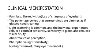
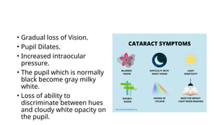
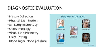
Cataract
Department of Medical Surgical Nursing
Introduction
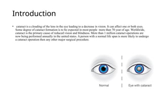
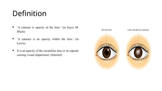
• cataract is a clouding of the lens in the eye leading to a decrease in vision. It can affect one or both eyes.
Some degree of cataract formation is to be expected in most people more than 70 year of age. Worldwide,
cataract is the primary cause of reduced vision and blindness. More than 1 million cataract operations are
now being performed annually in the united states. A person with a normal life span is more likely to undergo
a cataract operation then any other major surgical procedure.
Definition
 ‘A cataract is opacity of the lens.’ (in Joyce M.
Black)
 ‘A cataract is an opacity within the lens.’ (in
Lewis)
 It is an opacity of the crystalline lens or its capsule
causing visual impairment. (Internet)
Risk factors
• Aging
• Associated Ocular Conditions (Retinitis pigmentosa,
Myopia, Retinal detachment and retinal surgery, Infection)
• Nutritional Factors (Reduced levels of antioxidants, Poor
nutrition, Obesity)
• Physical Factors (Dehydration, Blunt trauma, perforation of
the lens, foreign body,)
• Systemic Diseases and Syndromes (Diabetes mellitus, Down
syndrome, Disorders related to lipid metabolism, Renal
disorders)
TYPES OF CATARACT
1. Congenital cataract - A congenital cataract is clouding of
the lens of eyes, that is present at birth.
2. Acquired cataract – Are caused by diseases or
medications. Diseases that are linked with the development
of cataracts include Glaucoma and Diabetes. Medication like
the use of steroid (prednisone) and other medication can
sometime lead to cataracts.
3. Sanile cataract – sanile cataract is a age related, vision-
impairing disease characterized by gradual progressive
thickening of the lens of the eye.
4. Traumatic cataract – traumatic cataract develop after an
injury to the eye, but it can take several years for this to
happen.
5. Radiation cataract- radiation cataracts can form after a
person undergoes radiation treatment for cancer.
Pathophysiology
Due to causes
Reduction in oxygen uptake and a reduction in initial increase
in water content followed by dehydration of the lens .
Sodium and calcium contents are
increased ,Potassium ,ascorbic acid and proteins are decreased
The protein in the lens undergoes numerous age related changes .
Yellowing from formation of fluorescent components and
molecular changes .
OPACIFICATION OF THE LENS
CLINICAL MENIFESTATION
• Pain less, Blurred vision(loss of sharpness of eyesight).
• The patient perceives that surroundings are dimmer, as if
glasses need cleaning.
• Light scattering is common, and the individual experiences
reduced contrast sensitivity, sensitivity to glare, and reduced
visual acuity.
• Abnormal color perception.
• Photophobia(light sensitivity).
• Nystagmus(involuntary eye movement ).
• Gradual loss of Vision.
• Pupil Dilates.
• Increased intraocular
pressure.
• The pupil which is normally
black become gray milky
white.
• Loss of ability to
discriminate between hues
and cloudy white opacity on
the pupil.
DIAGNOSTIC EVALUATION
• History Collection
• Physical Examination
• Slit Lamp Microscopy
• Opthalmoscopy
• Visual Field Perimetry
• Glare Testing
• blood sugar, blood pressure
Complication
• Postoperative infection
• Bleeding
• Macular edema
• Wound leaks
• Complete Vision loss
Medical Management
• No nonsurgical treatment cures cataracts.
• In the early stages of cataract development, glasses, contact
lenses, strong bifocals, or magnifying lenses may improve
vision.
• Reducing glare with proper light and appropriate lighting
can facilitate reading.
• Mydriatics can be used as short-term treatment to dilate the
pupil and allow more light to reach the retina, although this
increases glare(cause difficult to drive at night).
• Lifestyle adjustment.
• Reassurance
• Preoperative Medications- Eye drops may include the
dilating agent such as tropicamide (mydriacyl) to facilitate
the surgery.
• A cycloplegic cyclopentolate (cyclogyl) may also be
administered to paralyze the ciliary muscles.
Surgical Management
• Intracapsular Cataract
Extraction(ICCE)
• Extracapsular cataract
Surgery (ECCE)
• Phacoemulsification
• Lens Replacement
Preoperative care
• To reduce the risk for retrobulbar hemorrhage (after retrobulbar
injection), any anticoagulation therapy that the patient is receiving is
withheld, if medically appropriate.
• Aspirin should be withheld for 5 to 7 days, nonsteroidal anti-
inflammatory medications (NSAIDs) for 3 to 5 days, and warfarin
(Coumadin) until the prothrombin time of 1.5 is almost reached.
• Dilating drops are administered every 10 minutes for four doses at least
1 hour before surgery. Additional dilating drops may be administered in
the operating room (immediately before surgery) if the affected eye is
not fully dilated.
• Antibiotic, corticosteroid,and NSAID drops may be administered
prophylactically to prevent postoperative infection and inflammation.
Postoperative care
• After recovery from anesthesia, the patient receives verbal
and written instruction regarding how to protect the eye,
administer medications, recognize signs of complications,
and obtain emergency care.
• The nurse also explains that there is minimal discomfort
after surgery and instructs the patient to take a mild
analgesic agent, such as acetaminophen,as needed.
• Antibiotic, anti-inflammatory, and corticosteroid eye drops
or ointments are prescribed postoperatively.
• Position Patient on back or unoperated side to prevent
pressure in operated eye.
• Keep siderails up as necessary for protection.
• Place call light within reach.
• The nurse instruct the patient to be careful to prevent so up
water from entering the operative eye during face or hair
washing.
• Stress avoidance of action the increases I.O.P.
NURSING DIAGNOSIS
• 1.Disturbed sensory perception(visual) related to lens
extraction and replacement and use of eye patch
• 2.Anxiety related to lack of knowledge
• 3.Risk for injury related to blurred vision
• 4.Acute pain related to trauma to the incision and increased
IOP
• 5.Risk for infection related to trauma to the incision.

cataract and its management .pptx

  • 1.
  • 2.
    Introduction • cataract isa clouding of the lens in the eye leading to a decrease in vision. It can affect one or both eyes. Some degree of cataract formation is to be expected in most people more than 70 year of age. Worldwide, cataract is the primary cause of reduced vision and blindness. More than 1 million cataract operations are now being performed annually in the united states. A person with a normal life span is more likely to undergo a cataract operation then any other major surgical procedure.
  • 3.
    Definition  ‘A cataractis opacity of the lens.’ (in Joyce M. Black)  ‘A cataract is an opacity within the lens.’ (in Lewis)  It is an opacity of the crystalline lens or its capsule causing visual impairment. (Internet)
  • 5.
    Risk factors • Aging •Associated Ocular Conditions (Retinitis pigmentosa, Myopia, Retinal detachment and retinal surgery, Infection) • Nutritional Factors (Reduced levels of antioxidants, Poor nutrition, Obesity) • Physical Factors (Dehydration, Blunt trauma, perforation of the lens, foreign body,) • Systemic Diseases and Syndromes (Diabetes mellitus, Down syndrome, Disorders related to lipid metabolism, Renal disorders)
  • 6.
    TYPES OF CATARACT 1.Congenital cataract - A congenital cataract is clouding of the lens of eyes, that is present at birth. 2. Acquired cataract – Are caused by diseases or medications. Diseases that are linked with the development of cataracts include Glaucoma and Diabetes. Medication like the use of steroid (prednisone) and other medication can sometime lead to cataracts.
  • 7.
    3. Sanile cataract– sanile cataract is a age related, vision- impairing disease characterized by gradual progressive thickening of the lens of the eye. 4. Traumatic cataract – traumatic cataract develop after an injury to the eye, but it can take several years for this to happen. 5. Radiation cataract- radiation cataracts can form after a person undergoes radiation treatment for cancer.
  • 8.
    Pathophysiology Due to causes Reductionin oxygen uptake and a reduction in initial increase in water content followed by dehydration of the lens . Sodium and calcium contents are increased ,Potassium ,ascorbic acid and proteins are decreased
  • 9.
    The protein inthe lens undergoes numerous age related changes . Yellowing from formation of fluorescent components and molecular changes . OPACIFICATION OF THE LENS
  • 10.
    CLINICAL MENIFESTATION • Painless, Blurred vision(loss of sharpness of eyesight). • The patient perceives that surroundings are dimmer, as if glasses need cleaning. • Light scattering is common, and the individual experiences reduced contrast sensitivity, sensitivity to glare, and reduced visual acuity. • Abnormal color perception. • Photophobia(light sensitivity). • Nystagmus(involuntary eye movement ).
  • 11.
    • Gradual lossof Vision. • Pupil Dilates. • Increased intraocular pressure. • The pupil which is normally black become gray milky white. • Loss of ability to discriminate between hues and cloudy white opacity on the pupil.
  • 12.
    DIAGNOSTIC EVALUATION • HistoryCollection • Physical Examination • Slit Lamp Microscopy • Opthalmoscopy • Visual Field Perimetry • Glare Testing • blood sugar, blood pressure
  • 13.
    Complication • Postoperative infection •Bleeding • Macular edema • Wound leaks • Complete Vision loss
  • 14.
    Medical Management • Nononsurgical treatment cures cataracts. • In the early stages of cataract development, glasses, contact lenses, strong bifocals, or magnifying lenses may improve vision. • Reducing glare with proper light and appropriate lighting can facilitate reading. • Mydriatics can be used as short-term treatment to dilate the pupil and allow more light to reach the retina, although this increases glare(cause difficult to drive at night).
  • 15.
    • Lifestyle adjustment. •Reassurance • Preoperative Medications- Eye drops may include the dilating agent such as tropicamide (mydriacyl) to facilitate the surgery. • A cycloplegic cyclopentolate (cyclogyl) may also be administered to paralyze the ciliary muscles.
  • 16.
    Surgical Management • IntracapsularCataract Extraction(ICCE) • Extracapsular cataract Surgery (ECCE) • Phacoemulsification • Lens Replacement
  • 17.
    Preoperative care • Toreduce the risk for retrobulbar hemorrhage (after retrobulbar injection), any anticoagulation therapy that the patient is receiving is withheld, if medically appropriate. • Aspirin should be withheld for 5 to 7 days, nonsteroidal anti- inflammatory medications (NSAIDs) for 3 to 5 days, and warfarin (Coumadin) until the prothrombin time of 1.5 is almost reached. • Dilating drops are administered every 10 minutes for four doses at least 1 hour before surgery. Additional dilating drops may be administered in the operating room (immediately before surgery) if the affected eye is not fully dilated. • Antibiotic, corticosteroid,and NSAID drops may be administered prophylactically to prevent postoperative infection and inflammation.
  • 19.
    Postoperative care • Afterrecovery from anesthesia, the patient receives verbal and written instruction regarding how to protect the eye, administer medications, recognize signs of complications, and obtain emergency care. • The nurse also explains that there is minimal discomfort after surgery and instructs the patient to take a mild analgesic agent, such as acetaminophen,as needed. • Antibiotic, anti-inflammatory, and corticosteroid eye drops or ointments are prescribed postoperatively.
  • 20.
    • Position Patienton back or unoperated side to prevent pressure in operated eye. • Keep siderails up as necessary for protection. • Place call light within reach. • The nurse instruct the patient to be careful to prevent so up water from entering the operative eye during face or hair washing. • Stress avoidance of action the increases I.O.P.
  • 21.
    NURSING DIAGNOSIS • 1.Disturbedsensory perception(visual) related to lens extraction and replacement and use of eye patch • 2.Anxiety related to lack of knowledge • 3.Risk for injury related to blurred vision • 4.Acute pain related to trauma to the incision and increased IOP • 5.Risk for infection related to trauma to the incision.